
Abstract
Leiomyomas arising outside of the uterus and gastrointestinal tract are uncommon, though these benign soft tissue neoplasms have been reported in the lower extremity and foot. To our knowledge, the sonographic appearance of a lower extremity leiomyoma has not been described in the literature. This report involves a case of leiomyoma of the foot and its sonographic imaging features. MR correlative imaging and histopathology are also provided.
Keywords
Introduction
Leiomyomas arising outside of the uterus and gastrointestinal tract are uncommon, though these benign soft tissue neoplasms have been reported in the lower extremity and foot. To our knowledge, the sonographic appearance of a lower extremity leiomyoma has not been described in the literature. This report involves a case of leiomyoma of the foot and its sonographic imaging features. MR correlative imaging and histopathology are also provided.
Clinical Case
A healthy 50-year-old male presented for evaluation of a slowly enlarging but non-painful right foot mass. The patient described a 10-year history of a mass affecting the lateral aspect of the right foot, with a slow increase in size over time. There was no antecedent trauma. The patient reported some discomfort while doing yoga, but was otherwise asymptomatic and could comfortably wear a shoe. About 4 to 5 years prior to the current presentation, the patient brought this mass to the attention of a physician, and the question of a ganglion cyst was raised. No further treatment was sought at that time. On current physical exam, a 3 × 4-cm mass overlying the lateral aspect of the right foot was palpated. The mass was subcutaneous and firm, but non-tender and without overlying erythema.
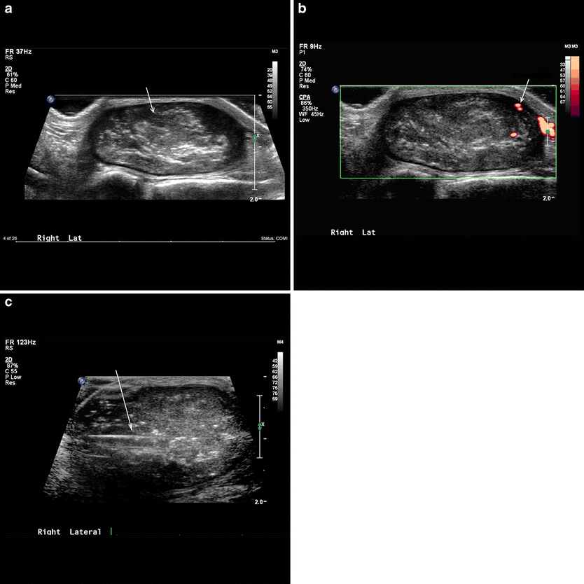
Ultrasound demonstrated a well-marginated, 2.8 × 1.4-cm ellipsoid, heterogeneous and mildly hyperechoic soft tissue mass on the dorso-lateral aspect of the right ankle. The soft tissue mass demonstrated a thickened capsule which showed increased vascularity on power Doppler imaging. Punctate calcifications, internal septations, and vascularity were also demonstrated within the mass (Fig. 1a, b). The sonographic features, though not entirely specific, raised the possibility of a fat-containing mass, such as a lipoma that had undergone previous hemorrhage with subsequent dystrophic calcification. Under ultrasound guidance, a Monopty 12-gauge 10-cm biopsy gun (Bard) was advanced into the soft tissue mass and two core specimens were obtained (Fig. 1c). The specimens were placed in formalin and sent to pathology.
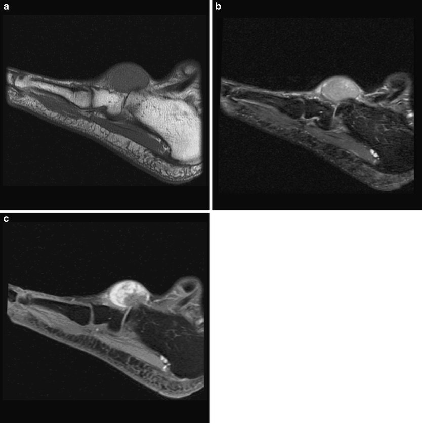
Magnetic resonance imaging of the right foot was also performed and demonstrated a well-defined subcutaneous mass on the dorso-lateral aspect of the foot with its epicenter at the level of the calcaneo-cuboid joint (Fig. 2a–c). The mass insinuated itself between the extensor digitorum tendons on the dorsum of the foot and the peroneus brevis tendon on the lateral aspect of the foot. On T1-weighted MR images, the mass was isointense to muscle, becoming hyperintense on T2-weighted fat suppressed images (Fig. 2a, b). Following the administration of contrast agent, the mass demonstrated moderate to marked inhomogeneous enhancement (Fig. 2c).
The differential diagnosis for a solid, enhancing lesion of the foot includes benign neoplasms such as schwannoma, neurofibroma, desmoid tumor, and localized pigmented villonodular synovitis (giant cell tumor of the tendon sheath). The duration of this lesion and its slow growth suggested a non-aggressive process. Nonetheless, a low-grade malignancy, such as a well-differentiated liposarcoma or a low-grade myxomatous tumor could not be excluded. The options of core-needle biopsy, incisional biopsy, excisional biopsy, marginal excision, and wide excision were evaluated, and a final decision was made to proceed with ultrasound-guided core-needle biopsy.
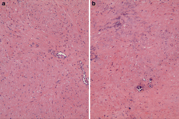
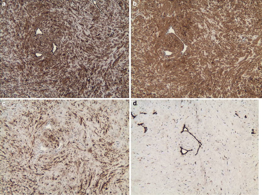
The specimens consisted of two cores of glistening white and focally yellow soft tissue, each measuring approximately 1.6 cm in length and 0.2 cm in diameter. Histological examination revealed a tumor of low cellularity composed of bland spindle cells with a haphazard arrangement, exhibiting discrete eosinophilic cytoplasm and blunt nuclei with occasional perinuclear vacuolation with interspersed capillary vessels lined by flat endothelial cells (Fig. 3a). Scattered dystrophic calcifications were present in one core (Fig. 3b). Supplemental immunohistochemical studies showed the tumor cells to stain diffusely positive with variable intensity for vimentin, smooth muscle actin, and desmin consistent with a histological diagnosis of leiomyoma (Fig. 4a–c). Capillary vessels stained positive for CD31 and the sparse pattern of vascularity ruled out angioleiomyoma (Fig. 4d).
The patient elected to forgo surgical excision.
Discussion
Soft tissue masses of the foot and ankle are relatively rare and include tumor-like lesions and benign and malignant neoplasms. Only approximately 8% of all benign soft tissue lesions and 5% of malignant soft tissue tumors occur in this location [1]. The spectrum of soft tissue neoplasms in the foot does not necessarily parallel those of other regions of the body, reflecting the unique proportion of the various soft tissue elements [2]. The majority of biopsy proven soft tissue lesions in the foot and ankle are, however, benign.
Non-tumoral lesions of the foot and ankle include ganglion cysts, epidermoid cysts, rheumatoid nodules, and also synovial proliferative diseases such as intra-articular localized and diffuse pigmented villonodular synovitis and giant cell tumor of the tendon sheath [3]. Benign tumors include hemangiomas, neurogenic tumors, lipomas, and deep fibromatosis (extra-abdominal desmoid tumor), the last one classified as benign but with a well-known potential for local recurrence and aggressiveness [3]. Because the clinical manifestations are often non-specific, clinicians must maintain a high index of suspicion and consider a diagnostic biopsy when presented with soft tissue masses of the foot [2]. Primary malignancies to consider include liposarcoma, low-grade myxomatous sarcoma, synovial sarcoma, clear cell sarcoma, malignant fibrous histiocytoma, and leiomyosarcoma.
Leiomyomas are quite unusual outside of the uterus and gastrointestinal tract. When in the soft tissues, leiomyomas are usually small and cutaneous or subcutaneous in location. Rarely, leiomyomas may be encountered in the deep soft tissues. These deep soft tissue lesions are much larger than their superficial counterparts and are more likely to be confused with leiomyosarcoma [4]. Soft tissue leiomyomas can be subdivided into three distinct groups, although others have been described. The most common form, the cutaneous leiomyoma, arises from the erector pili muscles of the skin and the deep dermis of the scrotum, labia major, and nipple. These lesions are quite small and often present as clustered papules measuring on the order of several millimeters each. The cutaneous leiomyomas are considered dermatologic lesions and are rarely evaluated radiographically [4].
The second group of soft tissue leiomyomas, the angioleiomyomas (also known as angiomyomas or vascular leiomyomas), are differentiated by their subcutaneous location and histology, which is characterized by a conglomeration of thick-walled vessels associated with smooth muscle tissue. These are solitary lesions and most commonly present in the adult, with two thirds occurring in the fourth through sixth decades. These lesions are typically small (<2 cm) and located in the extremities, with the lower extremity most frequently involved [4]. The foot is a common location and patients may present with symptoms related to footwear. Pain or tenderness may or may not be present. These lesions are slow-growing and may be present for 10 to 15 years prior to presentation [4].
The third group, leiomyomas of the deep soft tissues, may be located in the deep soft tissues of the extremities or the retroperitoneum. The retroperitoneal variety is more commonly found in females, possibly reflecting an origin from hormonally sensitive smooth muscle akin to uterine leiomyomas [4].
Case reports describing superficial leiomyomas in the lower extremity and their MRI imaging characteristics have been reported [5, 6]. In this case report, we have been given the opportunity to describe the sonographic appearance of a leiomyoma in the foot. Our pedal leiomyoma appeared heterogeneous and mildly hyperechoic under ultrasound, with scattered areas of punctate calcification, internal septations, and vascularity. This sonographic appearance may not be dissimilar from the most commonly encountered leiomyomas, those arising within the uterine myometrium. Though uterine leiomyomas typically appear as well-defined hypoechoic masses under ultrasound, heterogeneity induced by fibrous elements, necrosis, and calcification is not uncommon. The sonographic features of our pedal leiomyoma remained non-specific. Differentiation from other benign or malignant neoplasms, especially those of low-grade malignant potential, was not possible based solely on sonographic features. Biopsy was thus recommended for definitive evaluation.
The advantage of ultrasound-guided core-needle biopsy is minimal invasiveness coupled with the ability to provide the pathologist with adequate tissue in order to correctly make the diagnosis. This technique is particularly useful in low-grade myxomatous tumors because other methods to acquire tissue can potentially spread the tumor and hasten recurrence.