
Abstract
Introduction
The term epithelioid hemangioendothelioma (EH) was first identified and described in 1982 by Weiss and Enzinger [15] for a rare endothelial vascular neoplasm in soft tissues with a histologic appearance and clinical course between hemangioma and angiosarcoma. EH of the bone is extremely rare, with a 1 % incidence among all vascular bone tumors, and usually involves the skull, spine, femur, tibia, and feet of adults [1,5,8]. EH involving bones of the hand is exceptional, with only four cases previously reported [10,14].
We report a case of HE involving the pisiform bone. To our knowledge, this is the first case that has been reported.
Case Report
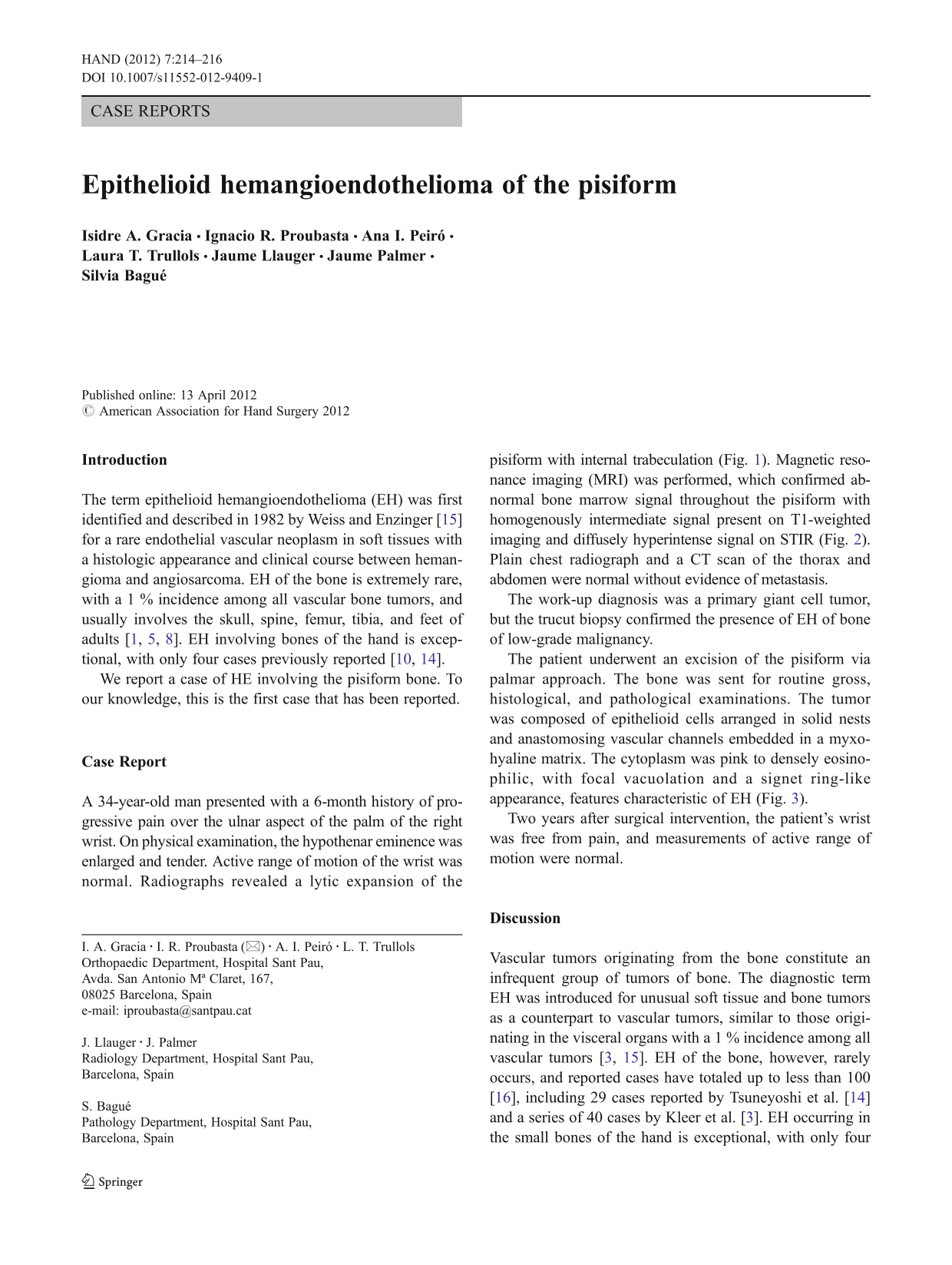
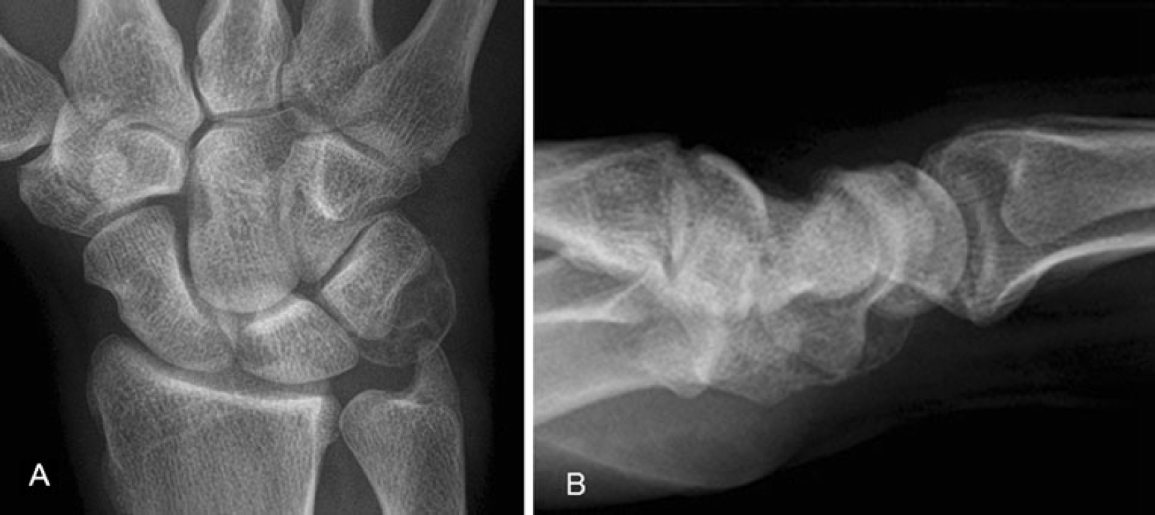
A 34-year-old man presented with a 6-month history of progressive pain over the ulnar aspect of the palm of the right wrist. On physical examination, the hypothenar eminence was enlarged and tender. Active range of motion of the wrist was normal. Radiographs revealed a lytic expansion of the pisiform with internal trabeculation (Fig. 1). Magnetic resonance imaging (MRI) was performed, which confirmed abnormal bone marrow signal throughout the pisiform with homogenously intermediate signal present on T1-weighted imaging and diffusely hyperintense signal on STIR (Fig. 2). Plain chest radiograph and a CT scan of the thorax and abdomen were normal without evidence of metastasis.
Preoperative radiographic findings. Posteroanterior (
MRI of the wrist. On coronal proton density-weighted image with fat suppression (DP-SPAIR), the lesion showed a hyperintense and heterogeneous signal (
The work-up diagnosis was a primary giant cell tumor, but the trucut biopsy confirmed the presence of EH of bone of low-grade malignancy.
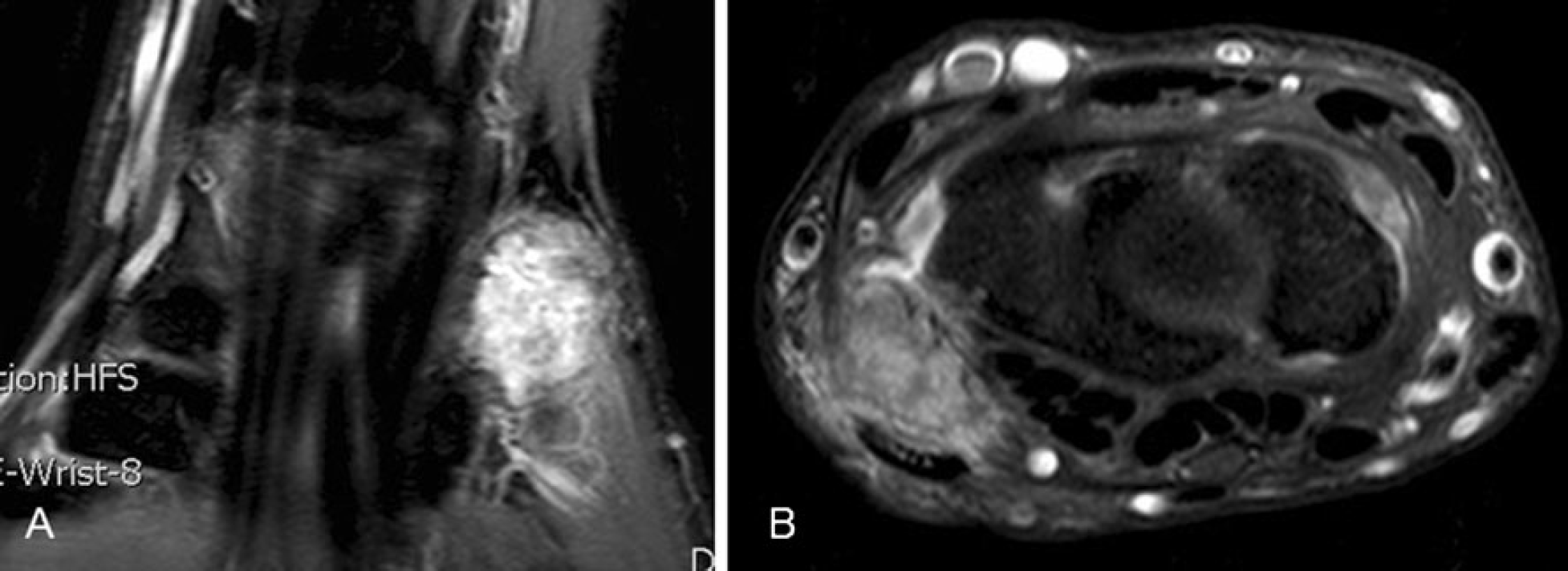
The patient underwent an excision of the pisiform via palmar approach. The bone was sent for routine gross, histological, and pathological examinations. The tumor was composed of epithelioid cells arranged in solid nests and anastomosing vascular channels embedded in a myxohyaline matrix. The cytoplasm was pink to densely eosinophilic, with focal vacuolation and a signet ring-like appearance, features characteristic of EH (Fig. 3).
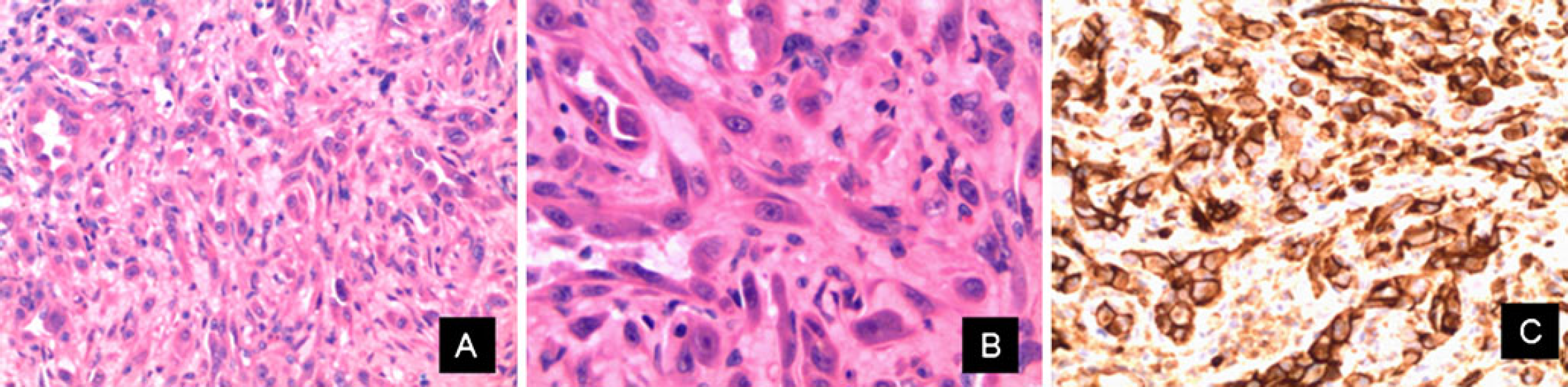
Histopathology.
Two years after surgical intervention, the patient's wrist was free from pain, and measurements of active range of motion were normal.
Discussion
Vascular tumors originating from the bone constitute an infrequent group of tumors of bone. The diagnostic term EH was introduced for unusual soft tissue and bone tumors as a counterpart to vascular tumors, similar to those originating in the visceral organs with a 1 % incidence among all vascular tumors [3,15]. EH of the bone, however, rarely occurs, and reported cases have totaled up to less than 100 [16], including 29 cases reported by Tsuneyoshi et al. [14] and a series of 40 cases by Kleer et al. [3]. EH occurring in the small bones of the hand is exceptional, with only four cases previously reported [10,14]. In this regard, Tsuneyoshi et al. [14] reported 14 patients with EH of bone, including a 16-year-old patient with EH occurring in the metacarpal bone. Kleer et al. [11] described 40 patients with EH of bone, including two patients with EH occurring in the small bones of the hand. Finally, Kitagawa et al. [10] reported a 12-year-old girl with a rare phalangeal EH lesion with a 10-year postsurgical follow-up evaluation after the resection of the phalanx.
In EH, lesions can appear in a multicentric manner with an incidence of over 50 % and below 62 %, particularly with a predilection for bones of the lower extremities in one anatomic region [14,16]. The multicentric type of EH seems to take a benign course in affected regions, but is more indolent than the solitary type [12,16]. Because of the potential for multifocal bone or visceral involvement, patients with EH should be thoroughly evaluated with CT of the chest and abdomen, bone scintigraphy, and a skeletal survey.
EH can occur at almost any age [15], most frequently between the ages of 20 and 30, with a male predilection of approximately 2:1 [1,8]. Generally, patients complain of pain and swelling in the affected area, as in our case.
Radiographs and CT typically reveal a lytic lesion without matrix mineralization, and osseous expansile remodeling may be seen. These lesions can be localized in the cortical or medullary bone. A honeycomb appearance on the radiograph is sometimes noted [12]. Periosteal reaction is rare, but soft tissue extension is common. Joint invasion is also a common feature [12].
There is no specific pattern of signal intensity at MR imaging. Most frequently, EH has low to intermediate signal intensity on T1-weighted images and high signal intensity on T2-weighted images, with homogeneous enhancement after the injection of gadolinium-based contrast material. Interestingly, serpentine vascular structures are not typically seen at MR imaging of EH of bone, findings that would suggest the diagnosis [8]. However, this phenomenon is likely related to the small size of the vascular channels and the hypercellularity seen at pathologic analysis [6]. The differential diagnosis for EH of bone should include angiomatosis, Langerhans cell histiocytosis (LCH), angiosarcoma, infection, myeloma, metastasis, and lymphoma [6]. It is for this reason that the biopsy is a mandatory previous treatment. Because of its low incidence, the variable clinical course and indistinct malignancy characteristics, the choice of treatment is not well established. In this regard, there are various treatment strategies for the treatment of EH of bone: (1) surgery (curettage, resection, or amputation), (2) radiotherapy, (3) radiofrequency treatment, (4) chemotherapy, and (5) various combinations (curettage followed by radiation or chemotherapy, resection followed by radiation or chemotherapy, or resection followed by radiotherapy and chemotherapy) [14,16]. At present, treatment plans should be tailored to the individual patient in correlation with the extension and location of the disease. However, based on our case and previously reported series, full resection is the treatment of choice of EH of bone [2,4,7,9,13,14,16,17].