
Abstract
Carpal instability includes a broad spectrum of osseous and ligamentous injuries which have been subclassified into greater and lesser arc injuries, in addition to combinations of both (Mayfield et al. J Hand Surg [Am] 5:226–241, 1980; Yaeger et al. Skeletal Radiol 13(2):120–30, 1985). The injuries typically occur from a fall on the outstretched hand with the wrist in ulnar deviation, hyperextension, and intercarpal supination (Yaeger et al. Skeletal Radiol 13(2):120–30, 1985). The force classically propagates from the radial to the ulnar side of the wrist resulting in a fracture (greater arc) or dislocation (lesser arc) pattern with the extent of the injury occurring in an orderly pattern depending upon the degree of hyperextension and the duration and magnitude of the force (Mayfield et al. J Hand Surg [Am] 5:226–241, 1980; Yaeger et al. Skeletal Radiol 13(2):120–30, 1985). Multiple variations occur, including transradial styloid fractures as well as fractures through carpal bones surrounding the lunate (Mayfield et al. J Hand Surg [Am] 5:226–241, 1980; Yaeger et al. Skeletal Radiol 13 (2):120–30, 1985; Kozin SH. J Am Acad Orthop Surg 6 (2): 114–20, 1998. Although carpal dislocations have been noted for many years, the mechanisms and classification have only been recently clarified. We report a case of a complex dislocation involving the entire proximal carpal row without an associated fracture. While this type of complex carpal dislocation has been previously described, to our knowledge, it has never been reported without a fracture of the forearm, wrist, or hand.
Case Report
A 44-year-old female with known schizophrenia presented to a level 1 trauma center following an unwitnessed fall from a second story window. The patient's daughter reported finding her mother down, but conscious. In the trauma bay, the patient was found to have a Glascow Coma Scale of 14 with confusion and multiple injuries including a subdural hematoma, T5, T12, and L1 burst fracture, distal sacral fracture, a left navicular foot fracture, and left proximal carpal dislocation.
Figures 1 and 2 are the posterior-anterior (PA) and lateral views of the left wrist which demonstrate traumatic volar dislocation of the proximal carpal row, with respect to the radius. The distal carpal row has also migrated proximally along with mild subluxation of the ulna at the distal radialulnar joint. No fractures of the left wrist or hand are detected. All of the carpometacarpal joints along with the forearm osseous structures remain intact.
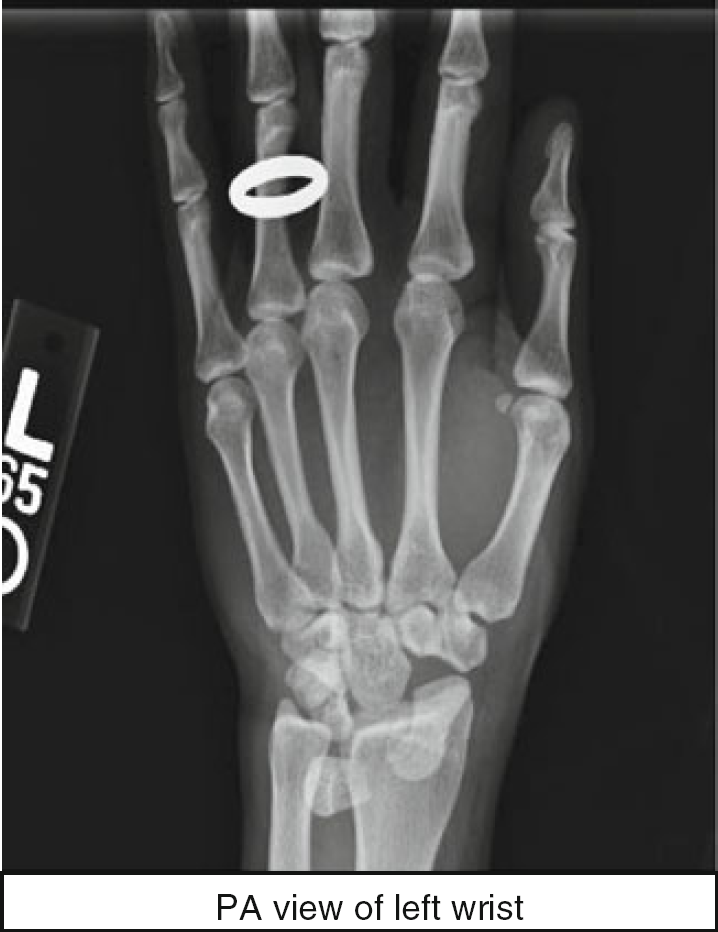
PA view of the left wrist
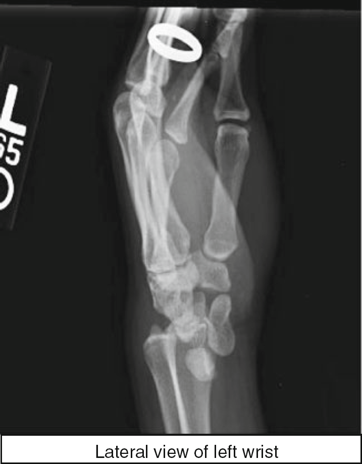
Lateral view of the left wrist
The patient underwent a closed reduction of the left carpal dislocation in the trauma bay. Subsequent postreduction films demonstrated that the lunate was still volarly displaced in relation to the radius (Figs. 3 and 4) with interval reduction of the scaphoid and triquetrum.
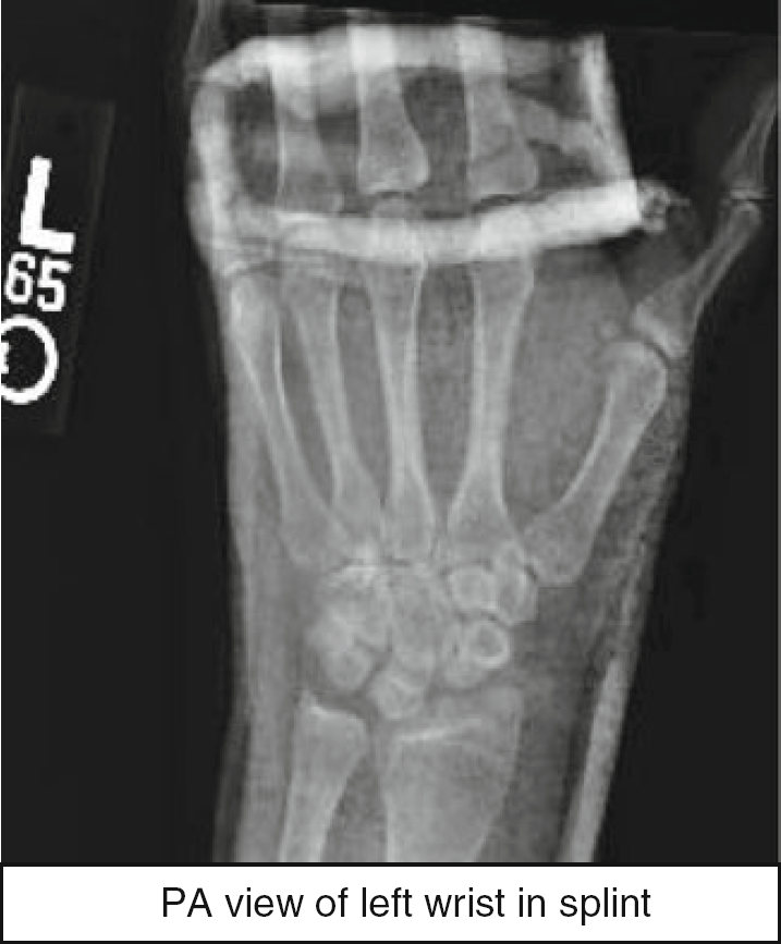
PA view of the left wrist in splint
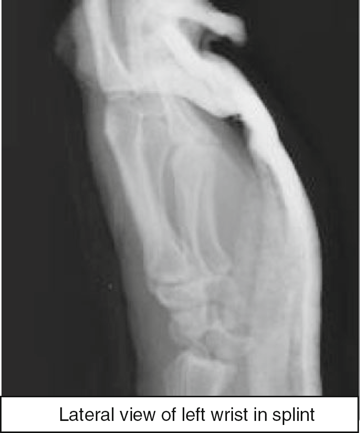
Lateral view of the left wrist in splint
The scaphoid and triquetrum, however, did align with the radius and ulna respectively. The patient had no symptoms of compression neuropathy at this time.
The patient was taken to the operating room where an open reduction of the lunate and repair of perilunate ligaments was performed. A volar approach was used initially, which allowed for reduction of the lunate and repair of the volar capsule. Following carpal tunnel release, the median nerve and flexor tendons were retracted radially to expose the floor of the carpal tunnel. The rent in the volar wrist capsule was localized through which the lunate was visible. The lunate was easily reduced with manual pressure on the volar side of the bone.
At this point, a dorsal incision was made over the radiocarpal joint, the tendons were retracted, and the wrist capsule was incised transversely for identification of the proximal carpal row. The dorsal scapholunate ligament and lunotriquetral ligaments were both found to be avulsed. There were still ligamentous structures attached to the lunate; however, the triquetral and scaphoid attachments of the ligament had been avulsed from bone with small articular fragments. These fragments were too small for fixation and were excised. A mini suture anchor (Depuy Mitek, Raynham, MA) was placed into the proximal pole of the scaphoid and used to secure the lunate insertion of the scapholunate ligament. A second suture anchor was then drilled into the triquetrum and used to secure the lunotriquetral ligament. With these two ligaments repaired, fluoroscopic images in AP and lateral projections were obtained which revealed anatomic alignment of the carpus (Figs. 5 and 6).
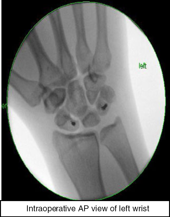
Intraoperative AP view of the left wrist
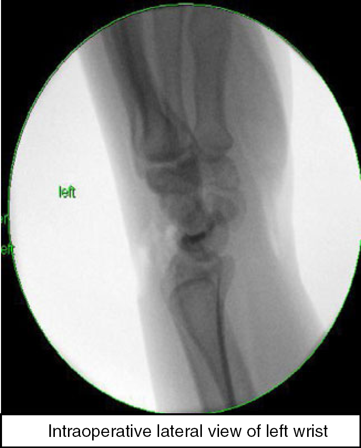
Intraoperative lateral view of the left wrist
The decision was made to forgo percutaneous pin fixation across the carpal bones, given the concern about the patient's compliance with wound care and follow-up appointments. The patient was immobilized in short arm fiberglass. She tolerated this procedure well, was treated for her other injuries, and was discharged from the hospital 4 days later.
Discussion
Most carpal dislocations are caused by hyperextension secondary to a fall on the outstretched hand. The proposed mechanism is that the primary dislocation occurs at the midcarpal joint, with the capitate usually displaced dorsal to the lunate [3,6]. As the scaphoid bridges both the rows, it may either fracture or rotate when the capitate dislocates dorsally, leading to ligamentous instability and a perilunate dislocation [3,9]. Following the groundbreaking 1980 study of Mayfield et al. [7], the manifestation of the most severe form of this injury was believed to be lunate dislocation into the carpal canal. However, this case presents a more complex carpal dislocation in which not only the lunate but the entire proximal carpal row has dislocated. Carpal instabilities have been characterized by Dobyns et al. as dissociative, which disrupt joints within a carpal row, or as nondissociative, which have dislocations or subluxations between carpal rows [4]. This case represents both dissociative and nondissociative proximal carpal dislocation, with complete disruption of the midcarpal, scapholunate, and lunotriquetral articulations.
While a proximal carpal bone dislocation is in itself exceedingly rare, this case is even more unusual in that it resulted in no associated fractures. Both the mechanism of injury and complex dislocation of carpal bones suggest a more traumatic outcome to the wrist, however, this was solely a variation of a lesser arc injury.
Capo et al. reported a similar case of a nondissociative complex carpal dislocation with ligamentous disruption between the proximal and distal carpal rows [1]. However, unlike our case, their patient also sustained an associated fracture at the base of the first metacarpal of the same hand [1]. A reported case of proximal carpal dislocation without fracture was reported back in 1982; however, this was isolated to the scaphoid and lunate bones, not the entire carpal row [2]. In 1966, a complex carpal dislocation without fracture was reported, but this was isolated to the radiocarpal articulation, as there was no instability demonstrated between the distal and proximal rows [8].
Repair of carpal dislocation via open reduction can be performed through the dorsal approach or, as in our case, a combined dorsal and volar one [5]. The volar approach provided direct access for lunate reduction, carpal tunnel release, and volar capsule repair. The dorsal access allowed for repair of the scapholunate and lunotriquetral ligaments. While hyperextension of the wrist upon high impact axial load is speculated, the exact nature of the mechanism that resulted in this rare injury is uncertain, providing a topic of discussion for future investigations.
Footnotes
The authors declare that they have no conflicts of interest, commercial associations, or intent of financial gain regarding this research.