
Abstract
Injuries to the scapholunate ligament are common, especially among young active individuals. Surgeons are faced with a difficult problem because of imperfect surgical outcomes and the high demands of this patient population. Here, we review the current concepts and newest literature on scapholunate ligament injuries as well as the classification and treatment options for each stage of scapholunate instability. Emphasis is on stages in which reconstructive rather than salvage procedures can be performed. The natural history is poorly understood; it is unknown which and how many scapholunate injuries lead to wrist arthritis (SLAC wrist). Partial injuries are rare and in small studies did well with arthroscopic treatment. Complete injuries are graded based on the acuity of the injury, the presence and reducibility of scapholunate malalignment, and, finally, cartilage status. In acute injuries, anatomic repair usually leads to satisfactory results, and many authors augment the repair with a capsulodesis technique. In chronic injuries, the presence of static malalignment usually leads to inferior outcomes. Various techniques have been devised and improved over the years. These techniques appear to provide a more anatomic reconstruction, with less loss of motion; motion is 60–80 % of the contralateral side and grip strength averages 65–90 %. Once there is cartilage loss, the surgeon only has salvage procedures to choose from, tailored to the degree of arthritis.
Keywords
Introduction
Scapholunate interosseous ligament (SLIL) injuries are common, affecting a wide range of patients. Since young, active individuals are particularly prone to these injuries a reconstructive technique that withstands the high demands placed on these wrists is required. Often, scapholunate (SL) ligament injuries are not diagnosed or treated during the acute phase of injury when direct repair of the ligament would be possible. Therefore surgeons are left to choose from reconstructive techniques that will best reproduce and maintain the natural carpal kinematics and load transmission while providing satisfactory range of motion and strength.
In this article, we provide a review of the pertinent anatomy and biomechanics, the natural history of SLIL injuries, classification schemes, and treatment tailored to each stage of instability.
Anatomy and Biomechanics
The SLIL is a true intra-articular ligament bathed in synovial fluid which contributes to its poor healing potential [50]. Sokolow noted that the ligament can be divided both anatomically and histologically into three parts: dorsal, intermediary, and volar. The volar section contains obliquely oriented collagen fascicles. With a tensile strength of 150 N, it is responsible for controlling rotational motion. The volar section is not visible from a dorsal wrist approach when the scapholunate ligament is intact. It is highly innervated and believed to have a major proprioceptive role [31]. The intermediate section of the ligament is the weakest segment with maximum tensile strength of only 25–50 N. This section has very little connective tissue. It is composed of fibrocartilaginous tissue completely devoid of neurovascular bundles. Because of its poor strength and low blood supply, it is commonplace to see perforating degenerative tears as well as avulsion of the ligament off of the scaphoid. The dorsal component is the strongest part. It has a tensile strength of 300 N and primarily controls flexion and extension. The edges blend with the radiocarpal articular capsule dorsally, the scaphotriquetral and dorsal intercarpal ligaments posteriorly, and the intermediate portion of the ligament proximally [50].
Many of the adjacent carpal ligaments play a role as secondary stabilizers of the scapholunate complex, particularly the scaphotrapezial–trapezoidal (STT) and radioscaphocapitate (RSC) ligaments. [5, 6, 46–48] Selective sectioning studies have established that the SLIL is the primary stabilizer. There are no statistical changes in carpal motion during flexion–extension or radio-ulnar deviation with selective sectioning of the STT or RSC alone, whereas isolated SLIL sectioning destabilizes the scaphoid and lunate throughout the motion cycles. [47] Subsequent loss of the RSC and STT ligaments results in angular changes of the carpal bones throughout a larger portion of the motion cycle [46], with an increase in angular and translational motion of the scaphoid and a decrease in lunate motion. [59] This indicates that the secondary stabilizers are responsible for stabilizing at the extremes of motion. In a cadaveric model, the effects of scaphoid rotatory subluxation were examined; progressive rotatory subluxation decreased the contact surface area, i.e., 20° of subluxation caused a 77 % reduction in the contact surface area and shifted the forces toward the dorsal lip of the scaphoid fossa of the radius. The biomechanical data correlate well with the carpal changes seen at long-term follow-up after SL ligament injuries: arthritis at the radioscaphoid joint, but not at the radiolunate joint.
The so-called dart-throwing motion occurs mainly through the mid-carpal joint while the scaphoid and lunate essentially do not move [12, 59]. This provides a rationale for rehabilitation after surgery for scapholunate ligament injuries by allowing early wrist motion while protecting the repair, but has not been validated in clinical studies yet.
Natural History
Selecting the optimal treatment and advising patients about operative intervention requires knowledge of the natural history of SLIL injuries. To date, the natural history is essentially unknown, largely because the majority of injuries go undetected in the acute stage or patients believe they simply suffered a sprain. We therefore do not know the true rate of symptomatic instability and/or arthritis development after SLIL injury.
Watson, in a widely quoted study, retrospectively analyzed 4,000 wrist radiographs in a cross-sectional fashion and identified 210 cases of wrist arthritis. Fifty-seven percent of the arthritic wrists demonstrated the characteristic scapholunate advanced collapse (SLAC) pattern. The arthritis began at the radial styloid and scaphoid junction and progressed to include the radioscaphoid articular surface. As the SL interval widened, the head of the capitate migrated proximally into the widened interval, resulting in arthritic changes at the capitate–lunate joint [55]. This study, however, did not prove that scapholunate ligament injury inevitably led to arthritis, nor did it elucidate the natural history.
Harrington et al. [23] postulated that the natural history of SLIL injury was deformity with dorsal intercalated segment instability (DISI), which consists of lunate extension, loss of capitate–lunate collinear relationship, and progression to arthritis. No evidence to support that is provided. Sebald et al. [45] analyzed the case reports of 57 patients with wrist pain, 48 of which showed DISI. He grouped the patients according to the length of time from injury and assessed radiographic results by group. Sebald et al. observed increasing signs of DISI in the patients as the length of time increased, but did not prospectively follow injuries to the scapholunate ligament to provide evidence that “the natural history of the condition begins with ligament damage, which leads to collapse deformities and then to degenerative arthritis.”
Geissler developed a useful arthroscopic classification system for scapholunate ligament injuries which is used by many authors (Table 1). We will introduce it at this time since it is relevant to a natural history study. O'Meeghan et al. [37] reported on the evolution of Geissler grade I and II scapholunate injuries (without static gap or DISI deformity) over a 7-year period on average. At the final follow-up, only one of the 11 patients had arthritis. Two others with arthroscopic but no radiographic evidence of early degenerative changes showed no progression on radiographs. None required salvage fusion procedures. The authors noted that none of the wrists demonstrated widening of the SL interval or DISI deformity, indicating that there was not complete dissociation of the scaphoid and lunate. O'Meeghan proposed conservative management of minor SL ligament injuries; however, it should be noted that all patients reported some level of pain, decreased grip strength, and decreased range of motion (ROM). Activities such as grocery shopping and using a screwdriver were frequently difficult, and the majority of the patients changed occupations and adapted their lifestyles. Younger, more active individuals may find this unacceptable.
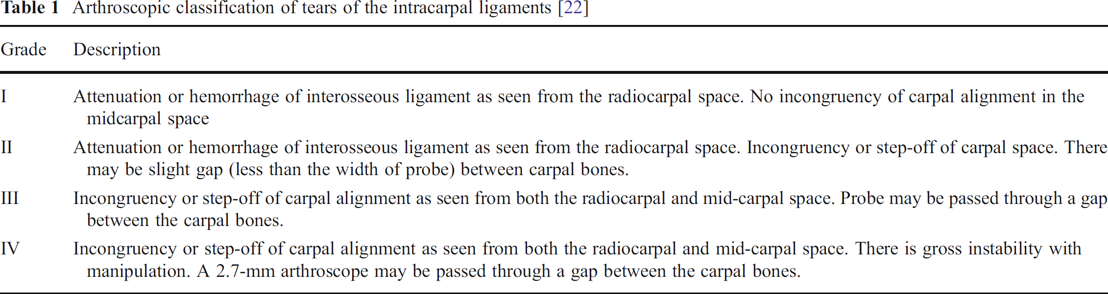
Arthroscopic classification of tears of the intracarpal ligaments [22]
Classification of Scapholunate Ligament Injuries
Garcia-Elias et al. [21] proposed five prognostic factors to consider when evaluating scapholunate ligament injuries.
What is the integrity of the dorsal SL ligament? Most partial tears include only the volar and interosseous components. Watson's scaphoid shift test would only be positive if the dorsal component is completely disrupted.
What is the healing potential of the disrupted ligaments? Intra-substance tears have low healing potential. Osseous avulsions may heal well with anatomic reattachment.
What is the status of the secondary scaphoid stabilizers (STT, SC, and palmar STT capsule)? Loss of these stabilizers causes rotary subluxation of the scaphoid and static deformity. This would require a more extensive stabilizing procedure.
What is the reducibility of the carpal malalignment? If finger traction alone can realign the joint, then it is easily reducible and could be fixed with a soft tissue reconstruction. If reduction is not possible after soft tissue releases, these injuries will require a selective arthrodesis procedure.
What is the cartilage status? In the face of arthritis, the operative options are reduced to salvage procedures, such as a proximal row carpectomy (PRC) or partial wrist fusion, most common scaphoidectomy with four corner fusion. The prerequisite for a PRC is intact cartilage on the capitate (i.e., SLAC wrist stages I and II).
Scapholunate injuries can be divided into six stages by answering yes or no to each of the above questions (Table 2) [21].
Staging of scapholunate dissociation injuries [21]
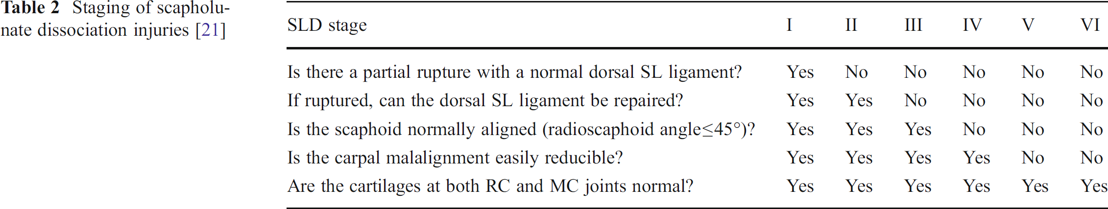
Stage 1: Partial scapholunate ligament injury
Stage 2: Complete disruption with repairable ligament
Stage 3: Complete disruption with irreparable ligament but normal alignment
Stage 4: Complete disruption with irreparable ligament and reducible rotary subluxation of the scaphoid
Stage 5: Complete disruption with irreducible malalignment and intact cartilage
Stage 6: Chronic SLIL disruption with cartilage loss (SLAC)
This classification provides a logical framework utilizing anatomic and biologic factors for treating SLIL injuries.
Radiographic Diagnosis of SLIL Injuries
The radiographic diagnosis of SLIL injuries is somewhat controversial, especially for stages 1–3. Once static malalignment is present (i.e., SL angle >65° on a lateral view and scaphoid ring sign on the AP view indicating vertical rotatory subluxation of the scaphoid), there is little disagreement about staging the injury (stage 4). The surgeon has to ensure that there are no early arthritic changes, which would start at the radial styloid tip (stage 6). Stage 5 is an intraoperative determination.
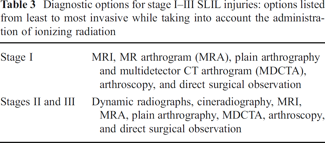
Multiple options exist for the diagnosis of stage 1–3 SLIL injury (Table 3), each with its own set of advantages and disadvantages. There are several studies comparing the sensitivity, specificity, and positive and negative predictive values of radiographic techniques using arthroscopy as the gold standard. A complete review is out of the scope of this article, but since accurate staging of SLIL injuries is critical, we discuss the value of diagnostic imaging below.
Diagnostic options for stage I—III SLIL injuries: options listed from least to most invasive while taking into account the administration of ionizing radiation
In general, most studies show a superiority of magnetic resonance arthrography (MRA) versus plain MRI [36, 43, 45, 54, 61]. It should be noted though that plain MRI is comparable to MRA for complete tears in some studies [30, 36]. More recent literature shows that multidetector CT arthrogram (MDCTA) is superior to MRA for partial ligament injuries [36, 44]. The diagnostic accuracy of plain MRI varies widely depending on multiple factors. Earlier literature showed sensitivity rates of 11–33 % [61]. Modern techniques with dedicated wrist coils on a 3-T magnet have sensitivity values of 89 % [30]. In conclusion, the technique of choice is somewhat dependent from institution to institution and the clinical setting (partial versus complete injury). We employ dynamic radiographs and plain MRI for stages 1–3. In cases where radiographs suggest no injury, we obtain an MRI if the pain is out of proportion for a simple sprain, there is discrete tenderness at the SL interval, and/or there is pain with Watson's maneuver. If complete SLIL injury is suspected on plain radiographs, we obtain an MRI when the patient is considering surgical treatment. If the diagnosis is still not clear, arthroscopy is the next step for diagnostic/therapeutic purposes. We acknowledge the value of MRA, but do not employ it in our algorithm.
In societies or medical systems without easy access to MRI, one may elect to operate solely on the basis of the history, physical exam, and dynamic radiographs and/or cineradiography since arthroscopy is ultimately the golden standard. This approach has low appeal to more sophisticated medical systems since patients often wish to have a reasonable preoperative diagnosis (i.e., partial versus complete injury and preoperative counseling about the risks/benefits/outcomes and anticipated postoperative course).
We will now review the management of scapholunate ligament injuries according to the aforementioned six stages.
Treatment Tailored to Each Stage
Stage 1: Partial Scapholunate Ligament Injury
There are few studies that address partial scapholunate ligament injuries. A case report in 1979 described a patient with clicking and pain in the dorsal wrist. Dynamic radiographs as well as an arthrogram did not show evidence of SL dissociation. Intraoperatively, there was scarring of the dorsal SLIL component which caused catching of the scaphoid proximal pole on the dorsal edge of the lunate [56]. Excision of the scarred portion of the ligament resulted in relief of pain and resolution of symptoms. The authors believed the problem to be a partial SLIL tear which spontaneously healed, although integrity of the SL interosseous and volar fibers was not actually documented.
In the 1990s, two studies by Ruch and Poehling [42] and Weiss et al. [57] reported on the management of partial injuries with arthroscopic debridement leading to relief of symptoms. The incomplete tears occurred in the weaker intermediate component of the ligament in one series [42]. The displaced flap of tissue became impinged, causing pain and crepitus similar to that found with torn menisci. The rest of the ligament continued to provide mechanical stability. Simple debridement of the excess tissue provided excellent relief of symptoms in 13 of 14 patients [42]. Weiss et al. [57] reported complete resolution or only occasional symptoms in 66 % of patients with complete SL versus 85 % of patients with partial SL tears.
Darlis et al. [13] reported on arthroscopic debridement combined with electrothermal shrinkage in Geissler I and II tears; 14 of 16 patients had good results at 19 months mean follow-up. Patients maintained full range of motion in the flexion extension arc as well as 78 % of their grip strength.
Arthroscopic debridement of partial SL Geissler II and III tears in children and adolescents after failure of nonoperative treatment led to very favorable outcomes in 24 of 32 patients in one series. Mayo wrist scores improved from 66.3 to 91.6 at an average follow-up of 43 months. Eight patients needed a subsequent operation for continued pain. Both patients with Geissler III injuries required a reoperation [19].
In summary, arthroscopic treatment without reconstruction or repair attempts seemed to have better outcomes in Geissler I and II injuries. Higher grade injuries in our review of the literature led to poorer outcomes. Follow-up was limited and the series were small.
Stage 2: Complete Disruption with Repairable Ligament
Acute injuries to the scapholunate ligament are defined as occurring within a 2- to 3-week period from injury to surgery. Within this time frame, the ligament itself is often still repairable, allowing for the success of direct ligament repair [2, 3, 32] with or without supplementary capsulodesis procedures. The SLIL almost always avulses off of the scaphoid, sometimes with an osseous fragment. Primary repair techniques utilize bone anchors with attached sutures to reattach the SLIL to the scaphoid, while in the past [29, 38] transosseous tunnels were used. Intraoperatively, the lunate is flexed while the wrist and scaphoid were extended to restore the capitolunate and scapholunate alignment. This is held with strategically placed clamps and/or pins. The SLIL is secured with horizontal mattress sutures [2, 3].
Bicker reported on their results for this technique utilizing a Mitek bone anchor. At follow-up, 10 of 12 patients were determined to have stable SL ligaments, as shown by normal rotational behavior at the scaphoid and lunate with a stable angle and interval that did not demonstrate diastasis with loading. Eight of 12 patients were classified as having good or excellent results, with no apparent correlation between radiographic and functional findings.
Cohen, Minami, Pomerance, Szabo, and Linscheid [9, 29, 33, 40, 51] described a combination of direct ligament repair augmented with a capsulodesis procedure. Cohen performed the dorsal capsular augmentation by attaching a capsular flap to the distal scaphoid with a small bone anchor. This technique predicted a loss of 15° of wrist flexion.
Minami and Kaneda [32] reported on 12 direct SLIL repairs at a mean follow-up of 5 years and reevaluated those 3 years later [33]. The authors found deterioration of the clinical outcome as well as an increase in the SL angle from 50° to 60°. This prompted them to add a modified dorsal capsulodesis to their technique. The authors documented better maintenance of the SL angle with addition of the capsulodesis. Early repair within 8 weeks of injury also had better results with improved SL angles, decreased loss of flexion or grip strength, and overall improved patient satisfaction [33]. It should be noted that many of these patients had incurred an SLIL injury as part of a perilunate dislocation and that the results must be viewed with caution in view of the diagnosis heterogeneity.
Szabo [51] reported a case of direct repair with the dorsal intercarpal ligament capsulodesis technique for an open trans-styloid perilunate fracture dislocation. Good results were achieved at 1 year, with patients returning to unrestricted work without pain and with a SL angle of 49° and SL interval of 2 mm or less.
Pomerance[40] reported on 17 patients with isolated SLIL injury at 66 months average follow-up. Patients involved in strenuous activities have decreased motion, lower outcome scores, and recurrence of instability radiographically at extended follow-up. This raised the question of whether the SL ligament actually healed while the capsulodesis protected and augmented the repair. The author concluded that the direct repair technique with dorsal capsulodesis should be considered as a temporizing measure to minimize the sequelae of the SLIL disruption in patients placing high demands on the wrist [40]. An example of SL ligament repair and dorsal intercarpal ligament (DIC) capsulodesis augmentation is shown in Fig. 1.
Recently, a technique for arthroscopic repair of the volar scapholunate ligament was reported by del Piñal et al. [15]. An isolated volar injury of the SL volar fibers is sometimes difficult to diagnose preoperatively. Since that portion of the ligament is the weakest, the widespread application of this technique remains questionable.
Longer follow-up is required to determine the durability and efficacy of scapholunate ligament repairs in the acute setting. Capsulodesis alone without biologic healing of the SLIL is not expected to withstand the repetitive stresses that lead to stretching of the ligament in a cadaveric model [39]. This is perhaps reflected in the clinical outcomes with longer follow-up in Pomerance's study [40]. The ideal treatment of acute SL ligament injuries is still elusive. It is not unreasonable for surgeons to select a technique that includes repair of the SL ligament and also a reconstruction as in cases of irreparable ligament, which are reviewed in the next section.
Stage 3: Complete Disruption with Irreparable Ligament, but Normal Alignment
For this stage, the ideal theoretical procedure is reconstruction of the SL ligament only. The secondary scaphoid stabilizers are still intact and prevent rotatory subluxation. In this stage, the bone–tendon–bone grafts championed by Weiss have the most theoretic appeal.
Consistently good results were reported by Weiss [58] for patients with dynamic stability harvesting a bone–retinaculum–bone graft from the dorsal distal radius. However, he noted that the results for the static deformity group were very inconsistent, with wide ranges of values for the SL angle and SL interval, 45–87° and 3–11 mm, respectively. The retinacular fibers used in the graft were oriented 90° from the direction that provided the highest resistance to stretch. Harvey and Weiss investigated other possible graft sites, attempting to replicate the strength and stiffness of the original SLIL [24]. The metacarpal–carpal BTB grafts from the base of the second or third metacarpals correlated well to the stiffness and strength of the SLIL. The dorsal retinaculum previously used was much weaker than the SLIL. This was believed to be due to differences in the cross-sectional area between the sample sizes and not actually the mechanical properties of the retinaculum [24]. Vascularized bone grafts have been postulated to lead to better healing at the scaphoid and lunate, but not yet proven to be of superior clinical benefit.
Another option described in patients with Geissler grade III or IV tears is aggressive arthroscopic debridement down to bleeding bone and pinning [14]. Three of 11 patients required revision procedures and were deemed failures. Of the remaining eight, only six reported good or excellent Mayo scores. There appeared to be an association between Geissler grade III tears and better final results, but the sample size was not large enough to make the results statistically significant [14]. Range of motion did not greatly decrease from preoperative values. Within the set of eight, none of the patients progressed to static instability or DISI at the final follow-up of 33 months. However, the authors still deemed the results of the study to be “suboptimal” and suggested considering it only in patients who refused to undergo anything more than an arthroscopic procedure [14].
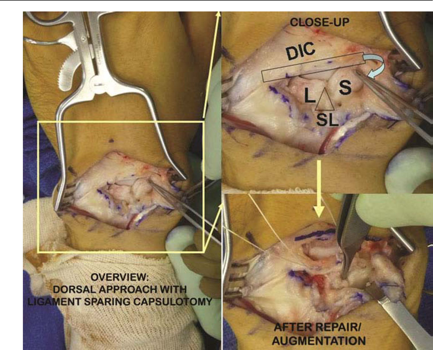
Intraoperative picture of SL repair augmented with DIC capsulodesis. This patient had a subacute SL injury (2 months out from injury) with dynamic instability and poor quality SL ligament. A dorsal approach with ligament-sparing capsulotomy is displayed in the large window. In the close-up view, the scaphoid and lunate are labeled (S and L, respectively). The SLIL is avulsed off of the scaphoid. After reduction of the deformity and fixation with K-wires, the SL ligament was repaired. The proximal one half of the DIC is detached from its radial attachment and attached onto the scaphoid with a suture anchor as described by Pomerance. The scapholunate interval was closed (picture courtesy of David B. Drake)
Many surgeons resort to the procedures listed in the next section because of the results for the techniques described above, especially the bone–ligament–bone grafts which have not been replicated, and because these techniques may be somewhat unpredictable.
Stage 4: Complete Disruption with Irreparable Ligament and Reducible Rotary Subluxation of the Scaphoid
Chronic reducible injuries in which the ligament is irreparable with or without subluxation are the subject of heavy debate. Multiple techniques for treatment are described, including the Blatt capsulodesis [4], Mayo capsulodesis [34], scapholunate arthrodesis [25], Brunelli tenodesis with flexor carpi radialis tendon [5], the Brunelli technique modified by Van Den Abbeele [53], the tri-ligament tenodesis by Garcia-Elias [21], and the RASL procedure with Herbert screw [41]. Of these techniques, the two that showed the most promise and that subsequently have led to the most variations were the Blatt capsulodesis and the Brunelli tenodesis.
Scapholunate fusions have a historic interest only due to the highest nonunion rate of all partial wrist fusions (62.5 %) and non-union correlation with poorer functional outcomes [49]. Attempts at scapholunate ligament reconstruction with a tendon graft through drill holes in the scaphoid and lunate were also historically abandoned because of unsatisfactory results, inability to hold the reduction, and complications such as tunnel fractures and rapid cartilage degeneration [17, 38].
Blatt's dorsal capsulodesis technique, first described in 1987, utilized a long, rectangular, proximally based capsular flap about 1–1.5 cm wide rotated and then attached to the dorsal distal surface of the scaphoid just distal to the center of rotation [4]. This technique was found initially to have excellent results, reducing pain without a significant decrease in range of motion. However, since that time, the results have been mixed.
Lavernia et al., in 1992 [28], looked at 21 patients treated for scapholunate instability with an average of 17 months from injury to operation. The operative treatments were based on the pathology visualized during the procedure. Fourteen underwent SL repair combined with dorsal capsulodesis with drill holes through the scaphoid, four underwent only a SL repair, and three underwent only a dorsal capsulodesis. All patients were pooled into the same results. At 3 years, there was no progression to carpal collapse. There was a loss of 11° of wrist flexion on average, including those patients that did not undergo capsulodesis. Radiographically, the SL angle decreased from 62° to 57°. The stage of injury was heterogeneous; hence, the results should be interpreted with caution.
Wyrick et al. [60] used a similar capsulodesis technique, but with Mitek suture anchors. They reported reduction of the SL interval and angle in the immediate postoperative period, but on final follow-up at an average of 30 months, the reductions were not maintained [60]. The final results of SL angle 72° and 3-mm SL interval were not all that different from the original preoperative values (78° and 4 mm, respectively). The final range of motion was 60 % of the unaffected side, and 60 % of patients had pain with activities of daily living. Initially, the differences between the results in this study and those of Lavernia were attributed to the greater preoperative deformities in the patients in Wyrick's study. It was suspected that Lavernia may have in fact been treating patients with dynamic instability since the average SL angle preoperatively was 62°, only 2° outside the normal range.
Subsequent studies by Moran et al. [34] and Deshmukh et al. [16] supported Wyrick's findings. At both short-term and intermediate follow-up, the range of motion for the flexion extension arc was decreased to 60–70 % of the unaffected side. Moran et al. reported that pain was not completely resolved in the majority of patients. They did not find any statistical differences between the preoperative and postoperative values of the SL angle and interval. At an average follow-up of 54 months, there was a larger progression of carpal malalignment in patients presenting with static instability versus dynamic instability. Despite the continued progression of carpal malalignment only 4 of the 31 patients progressed to a SLAC wrist by final follow-up. Gajendran et al. [20] reported that there was a high patient satisfaction rate (58 %) an average of 5 years out from a dorsal intercarpal ligament capsulodesis despite the fact that 8 of 15 patients showed arthritic changes due to the lack of maintenance of proper carpal kinematics. There did not appear to be a strong correlation between the radiographic findings and clinical outcomes [20, 34].
Palmer et al. [38] described multiple techniques of tenodesis mainly using ECRL or ECRB, but also palmaris, plantaris, FCR, abductor pollicis longus, extensor digiti quinti, and extensor carpi ulna as the donor. Later, Linscheid and Dobyns [29] refined their technique by adding a volar incision to imbricate the STT ligament. Most of these techniques have been abandoned in favor of the Brunelli tenodesis due to fractures, cartilage degeneration, and concerns for AVN of the lunate and scaphoid proximal pole.
Figure 2 describes the three most common tenodesis techniques reported in the literature. The tenodesis technique described by Brunelli crosses the radiocarpal joint and there is a loss of flexion similar to Blatt's capsulodesis technique [5, 6]. Brunelli reported loss of 30–60 % of flexion at 6 months to 2 years postoperatively.
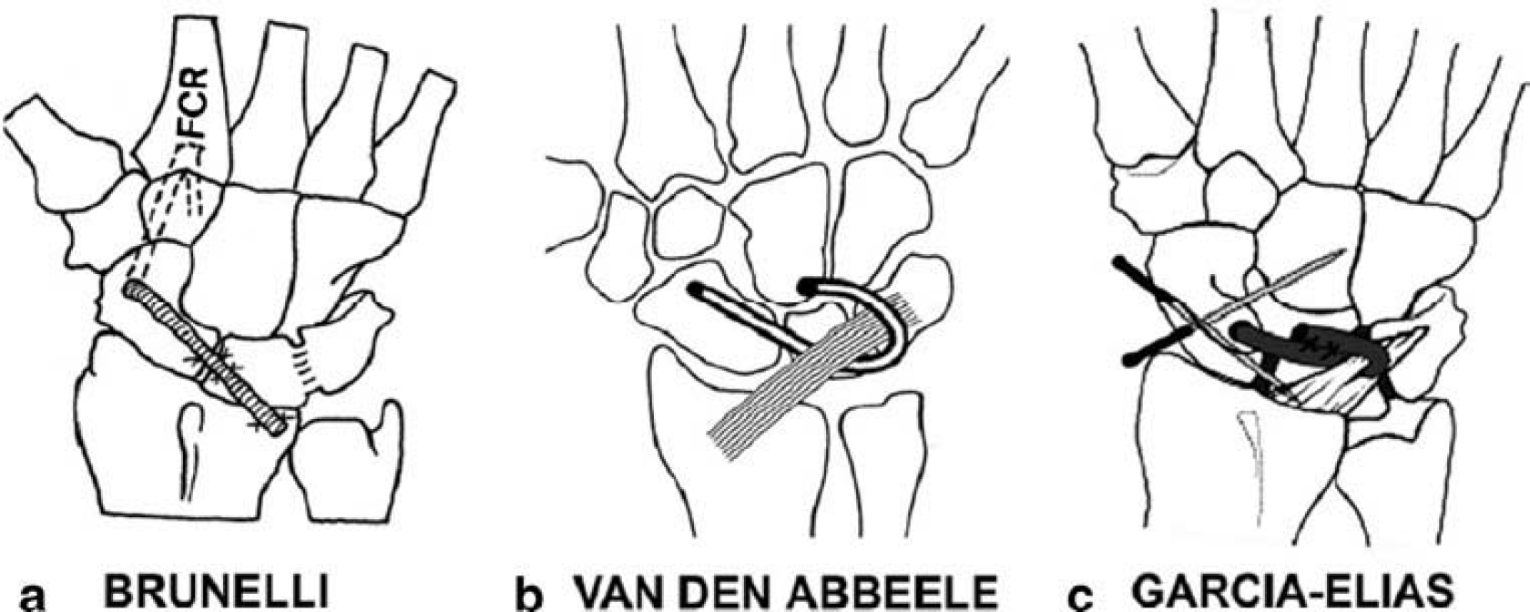
The three most common tenodesis techniques.
Van den Abbeele et al. [53] and Garcia-Elias et al. [21] subsequently modified the original technique to avoid crossing the radiocarpal joint (Fig. 2). Van den Abbeele et al. [53] reported a loss of only 9° of palmar flexion at 9 months, which was a significant improvement from the original technique. Subsequent studies with longer follow-up periods could not replicate Van den Abbeele's range of motion results. Chabas et al. [8] reported on the maintenance of carpal kinematics with the modified Brunelli tenodesis at ˜3 years follow-up. The average wrist motion was 50° extension and 41° flexion (75 and 73 % of the uninvolved wrists, respectively). Grip strength was 78 % of the uninvolved wrists. On radiographs, the mean static SL distance was 2.4 mm (2.8 mm before surgery). There was no widening of the SL gap compared to the immediate postoperative gap. The SL angle improved from a mean preoperative value of 61° to 53° immediately after surgery and rose again to 62° at the final follow-up. The gradual wear on the tendon as well as the stretching in the sagittal plane was attributed to the mechanical and histological differences between the FCR and the SLIL.
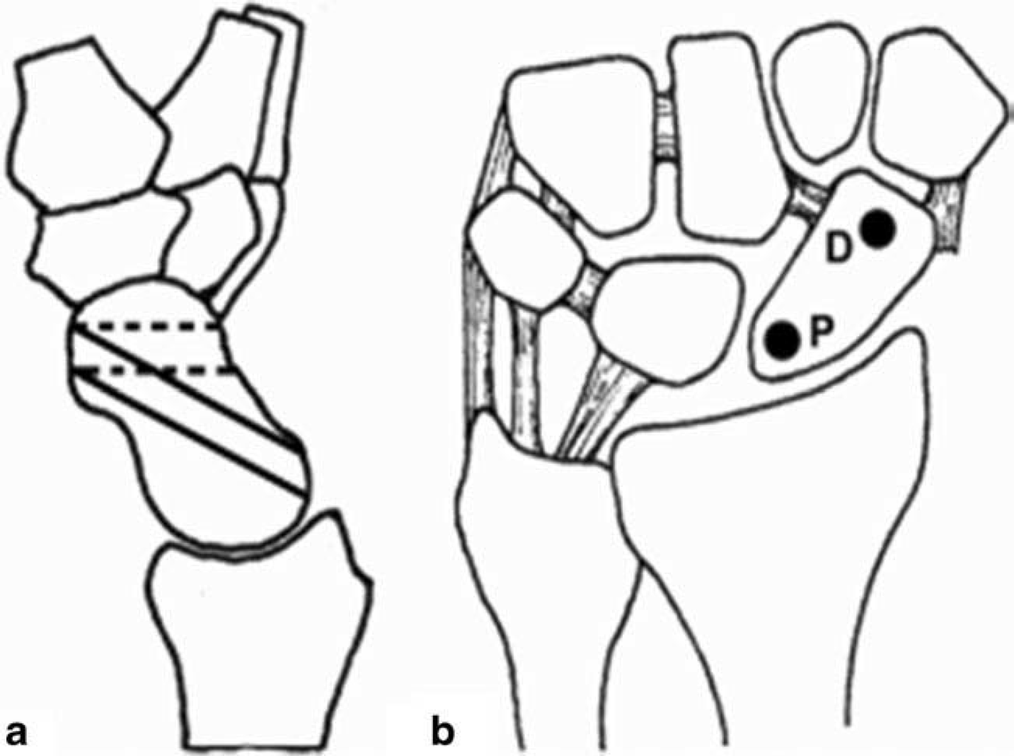
Figure 3 shows the two different tunnel placements studied by Howlett for the modified Brunelli tenodesis technique. He concluded that from the biomechanical point of view, a tunnel that exited distally in the scaphoid was more effective than a tunnel exiting in the proximal pole [26]. This intrinsically makes sense since a more distal tunnel would be more effective at controlling volar flexion of the scaphoid.
Garcia-Elias et al. [21] reviewed 38 patients at an average of 46 months with the reconstruction technique he developed. Pain relief at rest was obtained in 28 patients—eight patients complained of mild discomfort during strenuous activity and two patients had pain in most activities of daily life. The average ranges of motion were flexion, 74 %; extension, 77 %; radial deviation, 78 %; and ulnar deviation, 92 %, versus the contralateral side. Grip strength relative to the contralateral normal side was 65 %. They noted a patient satisfaction rate of 74 %.
This technique employed by other surgeons led to good clinical results in 12 of 14 patients at an average of 10.5 months postoperatively. All but one patient had static deformity. Dorsiflexion was 53°, flexion 35°, and grip strength averaged 80 % of the contralateral side. Radiographic deterioration of the results was noted already at short-term follow-up with SL angle of 73° and SL interval of 3.3 mm versus 73° and 3.6 mm preoperatively [27]. Talwalkar et al. [52] reported on 162 patients after a tri-ligament tenodesis at an average of 4 years postoperatively. This is the largest single patient cohort in the literature. Seventy-seven patients (62 %) had little to no pain. Average dorsiflexion was 80 %, flexion was 69 %, and grip strength averaged 80 % of the unaffected side. There was no difference in outcome between patients with dynamic instability and static instability at the time of presentation [52].
There is only one clinical study comparing the capsulodesis (Mayo technique) versus tenodesis (Van Den Abbeele modification). The two groups were very similar, but the tenodesis patients had more severe preoperative deformity. Moran et al. [35] reported comparable results in wrist motion and grip strength between the two techniques with no statistical difference. The techniques studied were modified to not cross the radiocarpal joint, yet there was still a significant loss of flexion. The tenodesis patients had 63 % and the capsulodesis patients had 64 % of the range of motion of the unaffected side; grip strength was 87 and 91 % of the contralateral side, respectively. Average Mayo wrist scores were 74 in the tenodesis group and 77 in the capsulodesis group [35].
The RASL procedure (reduction and association of the scaphoid and lunate) with a Herbert screw did not reproduce normal wrist mechanics, but improved them by allowing a small degree of motion at the scapholunate interval like the ligament would [41]. The authors reported no cases of screw breakage or cutout from the bone, although movement at the joint caused concerns for this technique. Since the Herbert screw did not directly contact any of the other bones, this technique could be converted into a salvage procedure, if necessary—either a proximal row carpectomy or wrist arthrodesis [41]. This procedure has yet to gain popularity at this time over the modified Brunelli tenodesis and the modified capsulodesis techniques. Formal results have not been published. Arthroscopic RASL was reported in two small retrospective series, with good results on short-term follow-up [1, 7].
Dunn and Johnson [18] reported an interesting technique in a cadaveric model for complete scapholunate dissociations with loss of the volar secondary stabilizers. Using a dual approach, the dorsal scapholunate ligament was repaired directly; the palmar radioscapholunate and radioscaphocapitate ligaments were reconstructed using a free flexor carpi radialis tendon autograft and Mitek mini suture anchors. This technique restored anatomic relationships of the scaphoid and lunate while preserving passive motion in the cadaveric model. The results of this intriguing technique have not been reported in the clinical scenario.
An exciting new development is an arthroscopically assisted, minimally invasive technique for the tri-ligament tenodesis using a distally based FCR tendon strip, passing it through the scaphoid with an oblique tunnel, dorsally across the SL interval to reconstruct the dorsal SLIL fibers, and then in a dorsal-to-volar direction through the lunate. The drill holes are 3 mm in diameter and the graft is secured in both tunnels with tenodesis screws, as described by Corella [10, 11]. The advantages of this technique are its minimally invasive nature and safety as assessed in a cadaveric model. Furthermore, it does not remove the dorsal capsular attachments to the carpus, as required with traditional open-exposure techniques. It has not been tested biomechanically, however, and clinical results have yet to be reported.
The surgeon faced with scapholunate instability and scaphoid rotatory subluxation can choose from a plethora of options which consist of variations of the basic techniques of capsulodesis and tenodesis. No technique is perfect, but with the passage of time, reconstructive techniques have attempted to better replicate the anatomy of the SLIL complex while preserving wrist motion. Interpretation of the results is limited by the heterogeneity of diagnosis in some series. It is not possible to determine at this time whether more anatomic and minimally invasive reconstructions will stand the test of time. It would be interesting to see the results of the techniques described by Dunn and Corella in the clinical setting. Range of motion can be expected to be 60–80 % of the contralateral side, with more flexion loss; grip strength averages 65–90 % of the contralateral side.
Stage 5: Complete Disruption with Irreducible Malalignment and Intact Cartilage
This stage poses an even more difficult task for the treating surgeon and can only be diagnosed intraoperatively. The surgical tactic includes aggressive soft tissue release and mobilization of scar/adhesions around the scaphoid and lunate to convert it to a reducible deformity. In this scenario, experienced surgeons may feel that an attempt at a non-salvage ligament reconstruction procedure is futile with a high failure probability and thus prefer to perform a salvage procedure as the initial treatment. Results for this isolated stage have not been published and these cases are uncommon.
Stage 6: Chronic SLIL Disruption with Cartilage Loss (SLAC)
The salvage operations commonly used for SLAC wrist are neurectomies, PRC, and partial or total wrist fusion. The remaining cartilage status dictates which procedures are feasible.
Common partial wrist fusions are STT, SC, radioscapholunate (RSL) fusion, and, finally, scaphoidectomy and four-corner fusion (4CF). Modifications of the 4CF and RSL fusion were introduced in an attempt to increase ROM due to osseous impingement. Other authors reported capitolunate and triquetrohamate fusion only in an attempt to simplify the 4CF technique and decrease the high non-union and complication rate [54]. The procedures for SLAC wrist are out of the scope of this review and are mentioned here for the sake of completeness.
Closing Remarks
The level of evidence for scapholunate ligament injuries is overall low, comprising a single comparative study [35]and largely retrospective series as well as some case reports. Ideally prospective, randomized controlled trials would answer questions about the efficacy of treatment methods, but the overall rarity of surgical intervention makes those studies unfeasible. Instead, surgeons should attempt to obtain long-term follow-up on the patients they do have to document prolonged efficacy of the procedures in preventing wrist arthritis. They should also strive to include patients with isolated SLIL injuries and carefully document the stage of instability. Use of validated patient-derived outcome scores in addition to objective data (i.e., motion, arthritis, maintenance of reduction) is desirable. Newer reconstructive techniques more closely resemble normal anatomy, but the clinical benefit remains to be proven. The presence of static deformity is an important factor when discussing expected outcomes with patients. Further studies on the natural history will improve our understanding and enhance patient counseling and decision making.
Footnotes
Acknowledgments
The authors would like to thank Dr. David B. Drake for sharing his intraoperative image of a scapholunate ligament reconstruction.
The authors have received no funding.
The authors declare that they have no conflict of interest.