
Abstract
The accurate determination of levels of differentiation is of prognostic value in human head and neck squamous cell carcinomas (HNSCCs). Because the deliberate selection of biochemical determinants accompanying certain stages of differentiation can refine the predictive power of histochemical assessments, the application of the quantitative evaluation of staining distribution and intensity by computer-assisted microscopy is one prerequisite to potential improvements. We used 2 innovative approaches with peanut agglutinin based on encouraging results with respect to common lectin-histochemistry. First, we used a custom-made neoglycoprotein to monitor the presence of Thomsen-Friedenreich (T) antigen-binding sites. Second, we measured the presence of 2 galectins immunohistochemically and, at the same time, measured lectin-histochemically the presence of accessible ligands for the endogenous lectins. We also monitored the presence of calcyclin, a protein with relevance to cell cycle progression or exocytosis. With 61 cases of HNSCC as their basis, including 31 oral, 20 laryngeal, and 10 hypopharyngeal lesions, the data show that the main modifications observed in connection with a loss of differentiation are related to a modification in the levels of both galectin-3/galectin-3-binding site and T-antigen/T-antigen-binding site expressions. The data obtained also suggest that galectin-3 could act as an acceptor site for the T antigen. Because the level of differentiation is known to be indicative of the recurrence rate in HNSCCs and our data clearly indicate that galectin-3 and the T antigen (and their respective binding sites) are involved in dedifferentiation processes, further investigation is warranted into the roles of galectins in HNSCC tumor progression and recurrence analysis.
As recently emphasized by Khuri et al, 1 head and neck cancer (HNC) is a notable health problem worldwide, with an estimated 900,000 cases in 1995. Pathologists have a number of tools for the evaluation of malignancy levels in HNCs. Between 85% and 90% of epithelial HNCs are squamous cell carcinomas 2 (HNSCC). The level of malignancy can be assessed by means of both the clinical TNM staging system and histopathologic grading. As stated by Urist et al, 3 although the TNM system 4 is the accepted standard for HNSCC classification, there are often discrepancies between TNM-predicted prognostic values and actual patient survival. These discrepancies occur with respect to both the T 3,5 and the N 6,7 variables.
Histopathologic grading relates to the determination of HNSCC differentiation levels with their 3 categories of good, moderate, and poor differentiation in accordance with various criteria, including the progressive loss of keratinization. 8 The determination of the level of differentiation is of proven prognostic value in HNSCCs. 7,9 In this respect, correlations have been described between differentiation level, tumor vascular-ization, 10 and the cumulative incidence of metas-tases. 7,11 According to various studies published in the literature, the differentiation level may 10,11 or may not 5 mirror the values provided by the TNM staging system. However, there is agreement on the notion that the progressive loss of HNSCC differentiation is accompanied by an increase in biologic, and therefore clinical, aggressiveness. This alteration of the level of differentiation not only is visible through an examination of the tissue architecture but also is apparent when distinct biochemical determinants are monitored histochemically. Such changes may account for the altered cell behavior. Because of the enormous extent of biologic information that can be stored in oligosaccharide structures, these glycan chains have gained increasing attention as a research focus to decipher the glycocode system in biologic information transfers. 12-14
In normal oral epithelia, cell surface carbohydrates are predominantly expressed in basal cells as part of mucins, and these carbohydrate chains are elongated parallel to terminal differentiation. 15,16 The loss of certain membrane-associated carbohydrate epitopes, which belong to the blood group antigen system, is attributable to the HNSCC stage and invasion level 17 as well as to early recurrence. 18 Certain blood group antigen-constituting oligosaccharides, including the sialosyl-T (Thomsen-Friedenreich) or the sialosyl-Tn antigens, are expressed in HNSCCs, but not in normal epithelia. 19
Several families of mammalian lectins have already been defined 14 in connection with the receptor sites of recognitive protein-carbohydrate interactions. Because of the widespread occurrence of β-galactoside-containing glycoconjugates, the galectin group deserves special attention as mediators. 20 Galectin-1 and/or galectin-3 having initiated immunohistochemical and biochemical studies on rodent tumors and human breast cancer, 21,22 their expression patterns have been examined in tumors of the endometrium, 23 colon, 24 and thyroid. 25 Gillen-water et al 26 recently reported that galectin-1 and galectin-3 occur in HNSCCs and are located on the cell surface, where they may participate in cell interactions. These authors had previously observed that the expression pattern of galectins appeared to be associated with the degree of squamous differentiation; this suggests a potential role for galectins as biologic and differentiation markers in the case of HNSCCs.
Proteins associated with cell proliferation were investigated principally in the search for markers associated with predictive values. This group of proteins in HNSCCs includes members of the large S100-related protein family 27 ; one of these proteins is the S100A6 member, denoted as calcyclin, 28 which has the ability to interact with negatively charged sugars and peptide motifs of annexin II, annexin VI, and glyceraldehyde-3-phosphate dehydrogenase. 29,30
The aim of this study has been to investigate a series of 61 patients with HNSCCs to determine whether there is a significant correlation between the expressions of galectin-1, galectin-3 (and their respective binding sites), and calcyclin, and their levels of differentiation. We also used peanut agglutinin (PNA) to locate the T antigen because it continues to be of supposed importance in the field of tumor biology. A tailor-made neoglycoprotein with the synthetic T-antigen structure enables endogenous sites with binding capacity to this disaccharide to be visualized, thereby detecting both components of an assumed interaction. 31 Quantitative measurements of the histochemical expression of the selected biologic markers were carried out by means of computer-assisted microscopy. 32-34
METHODS AND MATERIAL
Clinical and Histopathologic Data
This series included 31 cancers from the oral cavity, 20 from the larynx, and 10 from the hypopharynx. The 31 cancers from the oral cavity included 8 from the tongue, 8 from the tonsils, 7 from between the tongue and the tonsils, and 8 from the buccal mucosa. All the patients (52 men and 9 women, age range 45-79 years) had undergone partial or radical surgery. The diagnoses were established on the basis of the histologic criteria described by Hyams et al. 35 Clinical staging was carried out by means of the TNM classification. 4 There were 5 T-I,17 T-II, 14 T-III, and 25 T-IV cases. Of these 61 HNSCCs, 44 were node negative, and 17 were node positive. All 61 patients had stage M0 tumors.
The 61 HNSCCs were categorized as well (n = 24), moderately (n = 25), or poorly (n = 12) differentiated on the basis of the following criteria. The well-differentiated cancers were characterized by the presence of intercellular bridges and numerous keratinization foci, whereas the poorly differentiated ones were characterized by sparse (if not a total lack of) keratinization foci and an absence of intercellular bridges. Individual cell keratinization and the presence of cell junctions are consistent with the moderately differentiated phenotype.
Ligand Histochemistry
HNSCC tissue specimens were fixed in 4% formaldehyde and embedded in paraffin, and 5-μm-thick sections were processed with the various histochemical probes and kit reagents under study. As described elsewhere, 36-38 the markers were prepared and biotinylated under activity-preserving conditions, and specific polyclonal antibodies to calcyclin were raised. The p-aminophenyl derivative of the T antigen was synthesized and conjugated to bovine serum albumin, as previously described. 10 Incubation with labeled probes was carried out at room temperature for 60 minutes at a concentration of 10 μg/mL.
The extent of specifically bound markers (the biotinylated probes) was revealed by means of avidin-biotin-peroxidase complex (ABC) kit reagents (Vector Labs, Burlingame, CA), with diaminobenzidine and H2O2 as chromogenic substrates; this is detailed elsewhere. 34,37 Control reactions included competitive inhibitions to ascertain sugar specificity, and the omission of the incubation step with a labeled marker served to exclude any staining by the binding of kit reagents, such as the mannose-rich glycoproteins horseradish peroxidase and avidin. Counterstaining with toluidine blue concluded the processing.
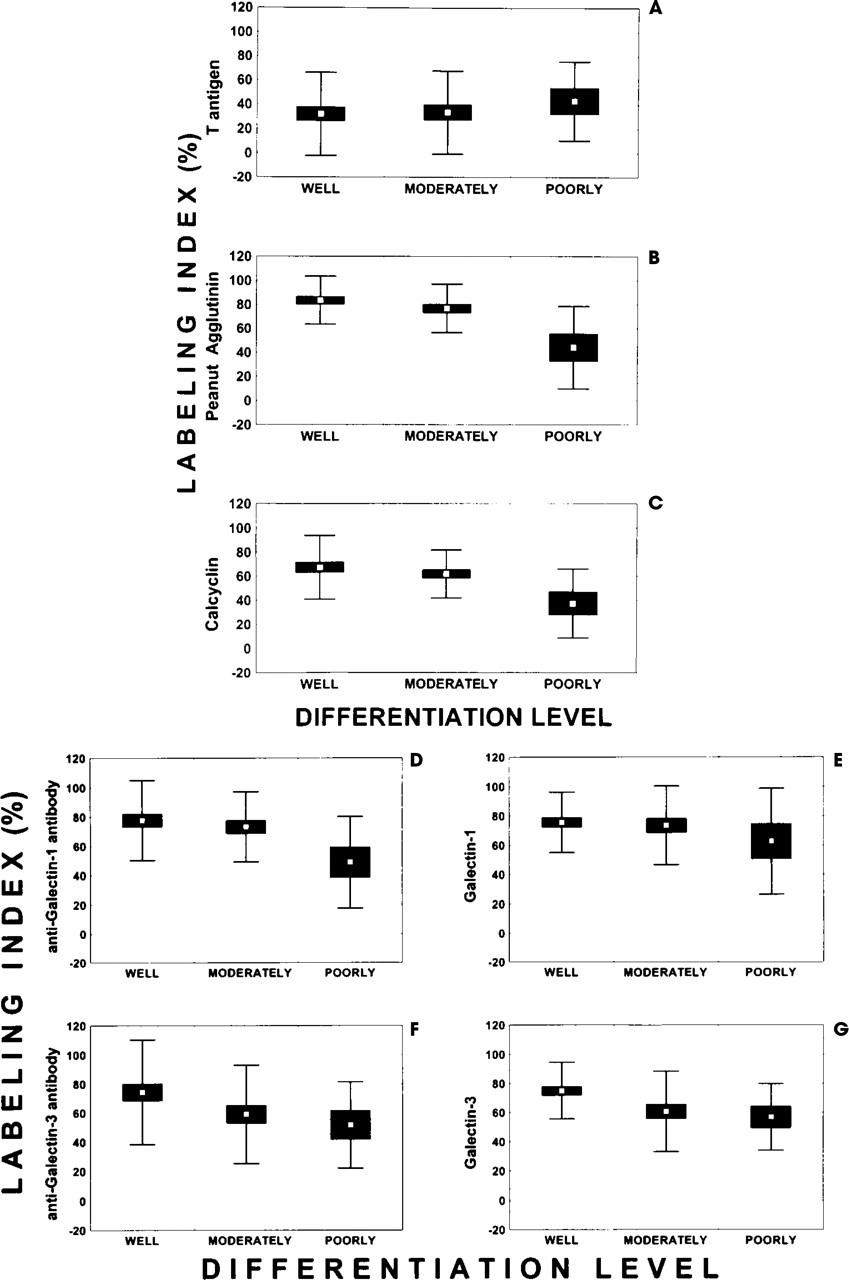
Development of the mean (open squares), SE (solid rectangles), and SD (bars) values quantitatively describing (by means of the LI variable) the percentage of HNSCC tissue area specifically stained by 7 distinct biotinylated probes or target-specific antibodies (carrier-immobilized T antigen (
Coexpression of the various determinants in HNSCCs
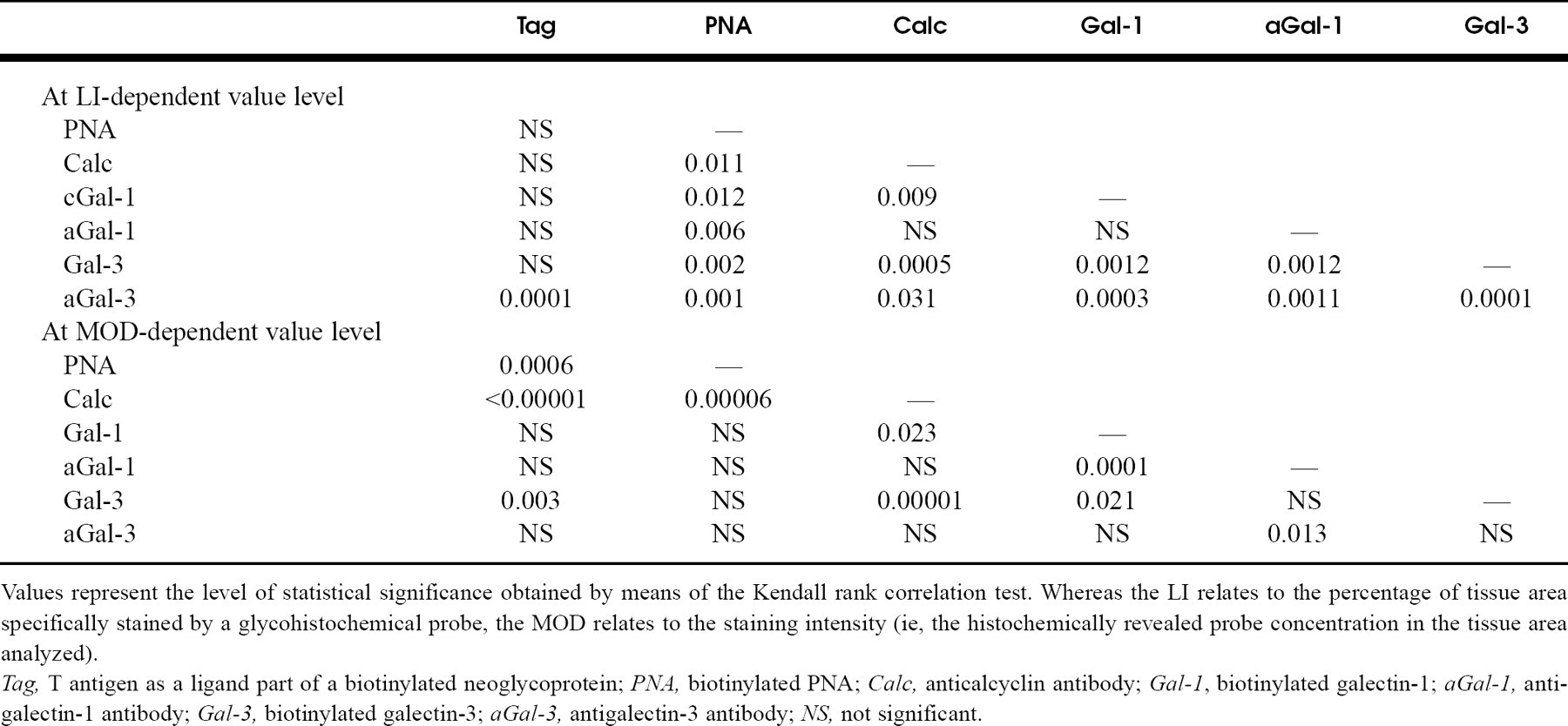
Values represent the level of statistical significance obtained by means of the Kendall rank correlation test. Whereas the LI relates to the percentage of tissue area specifically stained by a glycohistochemical probe, the MOD relates to the staining intensity (ie, the histochemically revealed probe concentration in the tissue area analyzed).
Tag, T antigen as a ligand part of a biotinylated neoglycoprotein; PNA, biotinylated PNA; Calc, anticalcyclin antibody; Gal-1, biotinylated galectin-1; aGal-1, antigalectin-1 antibody; Gal-3, biotinylated galectin-3; aGal-3, antigalectin-3 antibody; NS, not significant.
Computer-assisted Microscopy
The expression of calcyclin, galectin-1, and galectin-3 by the tumor cells of the 61 HNSCCs under study was immunohistochemically shown by the binding of specific anticalcyclin, antigalectin-1, and antigalectin-3 antibodies. The expression of the galectin-1- and galectin-3-binding sites was demonstrated glycohistochemically by means of biotinylated galectin-1 and galectin-3. The expression of the T antigen was glycohistochemically demonstrated by means of biotinylated PNA, whereas the expression of the T-antigen-binding sites (which correspond to endogenous PNA-related lectins) was shown glycohistochemically by means of the biotinylated carrier-immobilized T antigen (ie, with the T antigen used as a ligand part of a biotinylated neoglycoprotein). 39 Seven biologic markers were thus available for each of the 61 HNSCCs. Two quantitative variables were computed for each of the 7 markers by means of a SAMBA 2005 computer-assisted microscope system (Alcatel-TITN, Grenoble, France) with a 20x (aperture 0.50) magnification lens. The way in which we used this system to quantify the histochemical staining followed an optimized protocol, as did the ways in which we dealt with the problems relating to quantitative histochemistry. 32-34 Fifteen fields, each of between 60,000 and 120,000 μm 2 , were scanned on each histologic slide. The 2 quantitative variables computed for each of the 4 histochemical probes included: the labeling index (LI), which corresponds to the percentage of tissue area specifically stained by a glycohisto-chemical probe; and the mean optical density (MOD), which corresponds to the staining intensity (ie, the histochemically revealed probe concentration in the tissue area analyzed).
Statistical Analysis
Some of the results are given as the mean ± SEM. In such cases the statistical comparisons of the data were made by a 1-way analysis of variance after a check on the equality of variance by means of the Levene test and the normal distribution fitting of the data by means of a χ 2 test. The nonparametric Mann-Whitney U test was applied if the variances were not equal or if the value distribution was not normal. Correlations between the expression levels of the various histochemical probes were evidenced by means of the Kendall rank correlation test. All the statistical analyses were performed with Statistica/Windows software (StatSoft, Tulsa, OK).
RESULTS
Development of the Biologic Marker-related LI and Mod Indexes in Relation to the HNSCC Differentiation Levels
Fig 1 shows that for 5 of the 7 markers the mean LI values (describing the percentage of HNSCC tissue area labeled by each histochemical probe) decreased significantly in the monitoring of the well, moderately, and poorly differentiated lesions. These 5 markers were biotinylated PNA, indicating the presence of the T antigen in the tumor cells (Fig 1 B; P = 0.0009), anticalcyclin antibody (Fig 1 C; P = 0.003), antigalectin-1 antibody (Fig 1 D; P = 0.005), antigalectin-3 antibody (Fig 1 F; P = 0.01), and biotinylated galectin-3, which visualized accessible binding sites for galectin-3 (Fig 1 G; P = 0.004). In contrast, the mean LI values obtained after the application of the carrier-immobilized T antigen as a ligand part of a biotinylated neoglycoprotein (Fig 1 A) and biotinylated galectin-1 (Fig 1 E) did not significantly (P > 0.05) follow an unambiguous pattern in line with the differentiation levels.
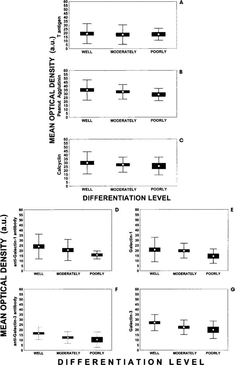
Development of the mean (open squares), SE (solid rectangles), and SD (bars) values quantitatively describing (by means of the MOD variable) the concentration of the target epitopes for each of the 7 probes (carrier-immobilized T antigen (
In comparison with Fig 1, Fig 2 shows that the mean MOD values developed less significantly than the LI ones in relation to the HNSCC differentiation level. Indeed, only the mean MOD values obtained by the immunohistochemical detection of galectin-3 (Fig 2 F; P = 0.006), as well as by the lectin-histochemical detection of galectin-3 ligands (Fig 2G; P = 0.002), significantly decreased in parallel to the loss of extent of differentiation.
Coexpression of Various Carbohydrate-binding Proteins in HNSCCs
The histologic specimens that were used to carry out the histochemical staining came from serial sections of each of the individual tumors analyzed. This means that contiguous areas in each tumor were monitored by each of the markers. This prompted us to investigate whether the pattern of expression of certain target molecules in any given section in fact correlated with the expression of other markers in the contiguous areas. When the distributions of the 61 LI values that characterized the expression pattern of the different determinants were compared, a remarkable correlation surfaced between the T-antigen-dependent (showing PNA-like endolectin) and antigalectin-3 antibody-dependent LI value distributions (Table 1). The PNA-related LI value distribution correlated significantly with each of the other LI value distributions (Table 1). Whereas the calcyclin-related LI value distribution correlated significantly with the galectin-1-, galectin-3-, and antigalectin-3-dependent LI value distributions, it failed to do so with the anti-galectin-1-dependent distribution (Table 1). The same feature was observed in the case of the galectin-1-dependent LI value distribution. The antigalectin-1-related LI value distribution correlated significantly with both the galectin-3- and antigalectin-3-dependent patterns, as did galectin-3 with antigalectin-3 (Table 1).
Table 1 clearly shows that very high levels of statistical significance were obtained from distinct MOD distribution values. This was the case for the Tag/Calc, PNA/Calc, Calc/Gal-3, and Gal-1/aGal-1 paired rank correlations (Table 1).
DISCUSSION
The search for refinements in the set of biologic markers is fueled by the suggested ensuing clinical consequences. Wolf et al 18 stated that the clinical use of biologic staging parameters in the initial assessment of HNSCC patients should enable more aggressive and successful primary treatment strategies to be selected for individual patients. This point of view is motivated, at least in part, our present study, which aims to assess whether the expression of calcyclin, galectin-1, and galectin-3 relates to HNSCC differentiation levels. Two biologic markers were monitored whose intrinsic diagnostic and/or prognostic values in HNSCCs, and particularly in oral types, have already been under-scored. 15-19, 34 These two markers are the T antigen and the T-antigen-binding sites, both of which were visualized by the innovative combination of chemical synthesis and histochemistry. The presence of T-antigen-binding sites (which, in fact, can be considered as an equivalent of a PNA-type endolectin in terms of sugar affinity) was detected in the 61 HNSCCs under study by means of the carrier-immobilized T antigen, whereas biotinylated PNA was used to reveal the presence of the T antigen in the HNSCC tumor cells. Our results on PNA binding dovetail with those previously reported by others. A similar observation was particularly made for the neoglycoprotein, which has so far only been tested in a few cases. Indeed, T-antigen-binding site expression was reduced in parallel with the decrease in differentiation level (ie, from a well to a moderately to a poorly differentiated status). Considering the complete panel of probes selected, this study intimates clearly that, in addition to these two markers, the loss of differentiation in HNSCCs can also affect calcyclin and galectin expression. Furthermore, because the expression level of these various markers was quantified by means of computer-assisted microscopy, we were able to unravel various previously undetected coexpressions of targets for certain probes.
The data in Fig 1 indicate that the loss of HNSCC differentiation from a well to a moderately to a poorly differentiated status is reflected biologically, not only in a decrease in the proportion of cells and/or extracellular matrix tissue presenting the T antigen (but not the PNA-like endolectin; Fig 1 A) but also in a decrease in those exhibiting calcyclin, galectin-1 (but not the galectin-1-binding site; Fig 1 E), and both galectin-3 and galectin-3-binding site positivity. As indicated in Fig 2, in some cases it was not only the proportion of cells harboring binding sites for a given marker that decreased but also the concentration of this marker per cell. This holds true for galectin-3 and its binding sites, for example.
Because no direct measurement of T antigen as a ligand for galectins has been carried out so far, commonly assaying panels of carbohydrates in inhibition assays, a discussion is precluded on the actual potency of the T antigen as a binding partner for galectins. However, the LI parameter correlations suggest an interaction at the galectin-3 level.
Checkerboard correlations enable us to discuss further relationships. Although no T-antigen-binding site-positive cells and only a limited percentage of T-antigen-positive ones were correlated with the percentage of calcyclin-positive cells, both the T-antigen and T-antigen-binding site concentrations correlated markedly (P < 0.00001 and P = 0.00006, respectively) and positively with the calcyclin concentration. Unless this correlation is fortuitous, regulatory mechanisms linking the two marker classes can be tentatively suggested on the basis of this result.
Although initially detected as a growth factor-inducible gene product of quiescent fibroblasts, calcyclin can evidently exert other functions besides playing a part in cell cycle progression. 30,40,41 It has recently been reported that its RNA gene expression can be evidenced in HNSCCs, but not in benign lesions. 42 These data clearly reveal the expression of this protein. However, they show that the loss in HNSCC differentiation level is paralleled by a significant decrease in calcyclin expression.
With respect to galectins, the loss of HNSCC differentiation is linked, at least phenomenologically, to galectin-3 expression. Although malignant thyroid tissue exhibits high levels of both galectin-1 and galectin-3, normal and even benign tissue does not express these markers. 25,43 Schoeppner et al 24 observed that galectin-3 expression in invasive colon cancers varies according to Dukes' stage, thereby indicating a linear relationship with advancing stage. Enhanced galectin-3 expression correlates with a decrease in long-term patient survival, and metastases express a higher level of galectin-3 in comparison with the primary cancers from which they evolve. 24 In sharp contrast, however, Castronovo et al 44 observed that galectin-3 is downregulated in breast cancer and that this decreased expression is associated with the acquisition of an invasive and metastatic phenotype.
Our data show that the loss of phenotypical differentiation in HNSCCs, which is known to be of prognostic value, can be translated at biochemical level not only into modifications in the expression of the T-antigen/T-antigen-binding sites but also into modifications in galectin expression. If the coexpression patterns between certain probes are recalled (Table 1), it can be inferred that galectin-3 may be an acceptor site for the T antigen. Indeed, colon cancer mucins have been evidenced as displaying reactivity to galectin-3. 43 It is thus an intriguing implication to link the detection of the T antigen functionally to that of certain galectins, at least in part. This result has relevance for lectin-histochemical studies on this new class.
It should be emphasized that although our data clearly demonstrate the intergroup correlation between the molecular markers that we used and the tumor differentiation status, there is a substantial overlap between individual patients. Additional markers are therefore needed to allow pathologists to use these markers to provide prognostic information on an individual basis. The characterization of the levels of expression of new galectins (including galectin-9) and S-100 proteins (including S-100A1, S-100A2, S-100A3, S-100A4, S-100A5, and S-100B) is under way in our laboratory.
CONCLUSION
The data from this study reveal that modifications in the level of HNSCC differentiation—a prognosis-related feature—are accompanied by changes in the extent of galectin expression, of their accessible binding sites, and of calcyclin.
We thank Dr J. L. Wang for his kind gift of the expression vector for murine galectin-3.