
Abstract

Two thirds of all desmoid tumors arise from anterior sheath of rectus abdominis muscle. Most extra-abdominal desmoids originate from shoulder and pelvic girdle musculature. Other sites are the extremities, bowel mesentery, and head and neck. 3
Desmoid tumors originating in the neck are rare entities. In a previous report it was stated that only 52 cases were noticed in the literature. 3 Here a patient with extensive desmoid tumor of the neck is presented, and diagnosis and treatment alternatives of the tumor are discussed.
CASE REPORT
A 21-year-old man applied to the Otolaryngology-Head and Neck Surgery Department of Gulhane Military Medical Academy with a report of a neck mass. The patient's initial history revealed an external trauma to the left side of his neck 5 years previously, which soon was followed by a slow-growing mass at the site of trauma. The mass caused no trouble to the patient but was aesthetically unappealing.
Otolaryngologic examination revealed a mass on the left side of the neck occupying the left supraclavicular triangle. The mass was mobile, nontender, firm, and approximately 12 × 15 cm in diameter (Fig 1). There were no other otolaryngologic findings or any neurologic abnormalities. A CT scan of the patient showed a round, lobulated, and well-delineated mass occupying the left supraclavicular fossa, which seemed to be originating from the paraspinal muscles. The mass contained hypodense areas and microcalcifications. Blood tests and other systemic findings were within normal limits.
At the time of surgery a horizontal skin incision was used, and the mass, which seemed to be encapsulated, gray-white, and smooth, was dissected from surrounded structures and removed completely. Pathologic examination revealed that it was an extra-abdominal desmoid tumor. The tumor was composed of fibroblast cells and generally showed hypocellularity, prominent collagenization, and occasional mast cells in routine hematoxylin-eosin staining. In immunohistochemical study, the fibroblastic cells were negative for CD34, which is a myofibroblastic cell marker.
The postoperative period was uneventful, and the patient was discharged on postoperative day 5. Now, 6 months after the operation, there is no sign of local recurrence.
DISCUSSION
Lesions that comprise the so-called fibromatoses occur in a wide range of anatomic locations and age ranges and also vary in their tendency for recurrence. Enzinger and Weiss subdivided the fibromatoses into 2 broad categories as superficial and deep fibromatoses. 2
All of these fibromatoses may recur, but deep ones are more aggressive. 2 There are several theories for the cause of desmoid tumors, such as trauma and endocrinologic disturbances, but none of them have been proved yet. 4 In a report by Enzinger and Shiraki 5 it is stated that more than 60% of desmoid tumors followed a trauma. In the case presented, there was also a history of trauma before the mass occurred. Tissue injury associated with hemorrhage is unlikely given the characteristic absence of hemosiderin on microscopic examination; thus the etiologic significance of trauma remains con-troversial. 3 Also, high levels of estrogens and pituitary gonadotropins have been postulated to be the cause of this tumor, but in most patients there is no evidence of such alteration in hormonal levels. 4
Commonly, these tumors result in an asymptomatic mass, which is firm, solid, and nontender on examination. They can grow as large as 20 cm in diameter. Symptoms may appear as a result of compression to the surrounding structures such as nerves and blood vessels. The tumor can be multicentric in 10% of cases. 1 There may be potential life-threatening manifestations, including airway compromise and mediastinal compression. Mortality from rupture of involved major blood vessels has been reported. 3 Because local extension of tumor is important for planning treatment, CT and MRI are helpful for determination of tumor borders.
Morphologically, desmoid tumors are nonencapsulated, rubbery, gray-white lesions, and they consist of poorly demarcated fibrous tissue that invades surrounding muscle. They have never been shown to invade lymph nodes. Histologically, desmoid tumors consist of a collagenous central core surrounded by large populations of fibroblasts varying in cell and nuclear size and intermixed with giant cells and destroyed muscle cells. The degree of mitotic activity does not correlate with the growth potential and invasiveness of the tumor. The more aggressive lesions are characterized by larger numbers of myofibroblasts, whereas the least aggressive tumors have a greater number of mast cells. The absence of myoblastic cells in desmoid tumors was showed with CD34 for the differential diagnosis of solitary fibrous tumor of soft tissue. 4 There is little evidence to suggest that desmoid tumors undergo malignant transformation. 3
Wide surgical excision of the tumor is the suggested treatment modality in the literature. In cases that are unresectable or multicentric, radiotherapy, chemotherapy, and hormonal treatment are suggested alternatives with limited success. 5 One or more of the treatment modalities can also be used in combination. As mentioned earlier, tumors are locally aggressive and have incidence of local recurrence rate between 10% and 90%. There is also a 10% chance of multicentricity, which may be the cause of some recurrences.
The prognosis of this tumor depends on the size and location. Insinuation of tumor around the great vessels, respiratory passages, and cranial and cervical nerves can make resection a difficult task. Surgical expertise in this area is a requisite for adequate treatment of head and neck desmoids. A conservative approach is unwise and unsatisfactory because recurrence of the tumor is high.
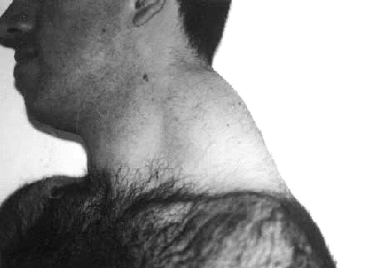
Tumor located on the left side, changing the contour of the neck.
In our case there was a history of trauma, which supports the proposal of trauma being an etiologic factor. There was no endocrinologic abnormality. CT was the most useful diagnostic tool. Tumor was locally aggressive, and wide resection of the tumor was the only treatment used.