
Abstract

CASE REPORT
A 19-year-old healthy man, who underwent FESS in the past, was referred to our department because of recurrent CPS. He did not complain of visual disturbance or impairment in either eye. A computed tomography (CT) scan revealed bilateral pansinusitis (Fig 1A). Revision ESS was undertaken under general anesthesia. His nose was packed with cotton sticks moistened with lidocaine hydrochloride (40 mg/mL) and epinephrine (1:5000) before intranasal injection with lidocaine hydrochloride (10 mg/mL) and epinephrine (1:100,000). Multiple polypectomies, bilateral sphenoethmoidectomies, and frontal sinus and natural ostium window surgery were all performed uneventfully. Hemostasis was achieved by applying Vaseline gauze and Merocel packs into both nasal cavities. In the following period, he was in good condition without specific complaints until 48 hours after surgery, when nasal packing was all removed. Due to nasal oozing, nasal pledgets rinsed with a mixture of 3 mL of lidocaine hydrochloride (40 mg/mL) and 3 mL of epinephrine (1:5000) were packed into both middle meatus to control bleeding. After removing them 10 minutes later, bleeding was stopped and the patient was not uncomfortable. Surprisingly, at 2 hours after removal of the nasal packing, he complained of blurred vision in the left eye. An urgent ophthalmology consultation found the patient's visual acuity to be normal in the right but a total loss in the left. Pupillary light reflexes showed a Marcus Gunn pupil in the left, and funduscopic examination results were normal in both eyes. There was no periorbital ecchymosis, lid edema, chemosis, ophthalmoplegia, or proptosis. A diagnosis of PION was made. An emergent CT scan showed neither injury of optic nerve nor hematoma of orbit (Fig 1B). Simple endoscopic examination under general anesthesia did not show any abnormal bony defect between the sinuses and the orbit. Despite prompt administration of corticosteroid and rheomacrodex suggested by ophthalmologist, the patient's visual acuity remained unchanged. At 1 month after surgery, repeat ophthalmologic examination disclosed that his optic disc has turned into pallor and atrophy. He still did not resume visual acuity at 6 months postoperatively.
DISCUSSION
Blindness secondary to ESS has been attributed to 2 traumatic causes. One is direct injury to optic nerve, and the other is orbital hematoma. The former usually occurs immediately after surgery, and the latter may happen over hours, even days. 1 Urgent CT scanning of paranasal sinuses may identify specific areas of damage or hematoma formation. 2 Exploratory endoscopic examination may reveal abnormal bony dehiscence or optic nerve injury. However, in our case, delayed visual loss after removing nasal packs on the second postoperative day excluded the possibility of direct injury to optic nerve. Meanwhile, the patient had no signs of orbital hematoma such as periorbital ecchymosis, lid edema, chemosis, ophthalmoplegia, or proptosis. 2 Both postoperative CT scan and endoscopic exploration revealed an intact optic nerve and no intraorbital lesion. Therefore, we inferred that the patient's blindness was not due to either of these 2 mechanisms.
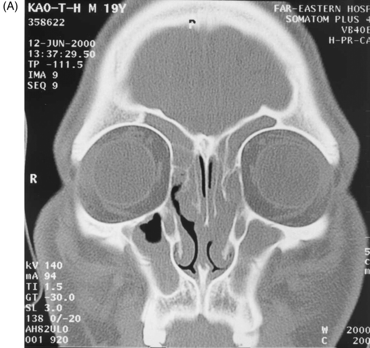
A coronal section of preoperative CT scanning demonstrating opacification over frontal sinuses, anterior ethmoid sinuses, and maxillary sinuses.
Topical use of vasoconstrictive agents on nasal mucosa has been reported to cause acute visual loss due to central retinal artery occlusion 3 or ischemic optic neuropathy. 4 Vasospasm has been postulated as the reason for visual loss due to inadequate blood supply to the optic nerve or retina. Our patient had sudden visual loss associated with a normal-appearing optic disc and retina at the onset, a pale and atrophic disc 1 month later, diagnosed as PION by the ophthalmologist. The posterior segment of optic nerve is nourished by a meningeal-pial vascular network, mainly arising from the branches of ophthalmic artery. 5 ESS predisposing to open vascular channels between sinuses and orbit might be hypothesized even though the orbit was not violated during the operation. Consequently, the pathogenesis of PION in this case is presumed to be vasospasm in the branches of ophthalmic artery, possibly caused by epinephrine diffused via nasal packing.
In addition to being a well-documented case report concerning delayed blindness after ESS, this case is valuable because it might suggest that sudden blindness could occur after epinephrine-rinsed nasal packing, even 2 days after surgery.
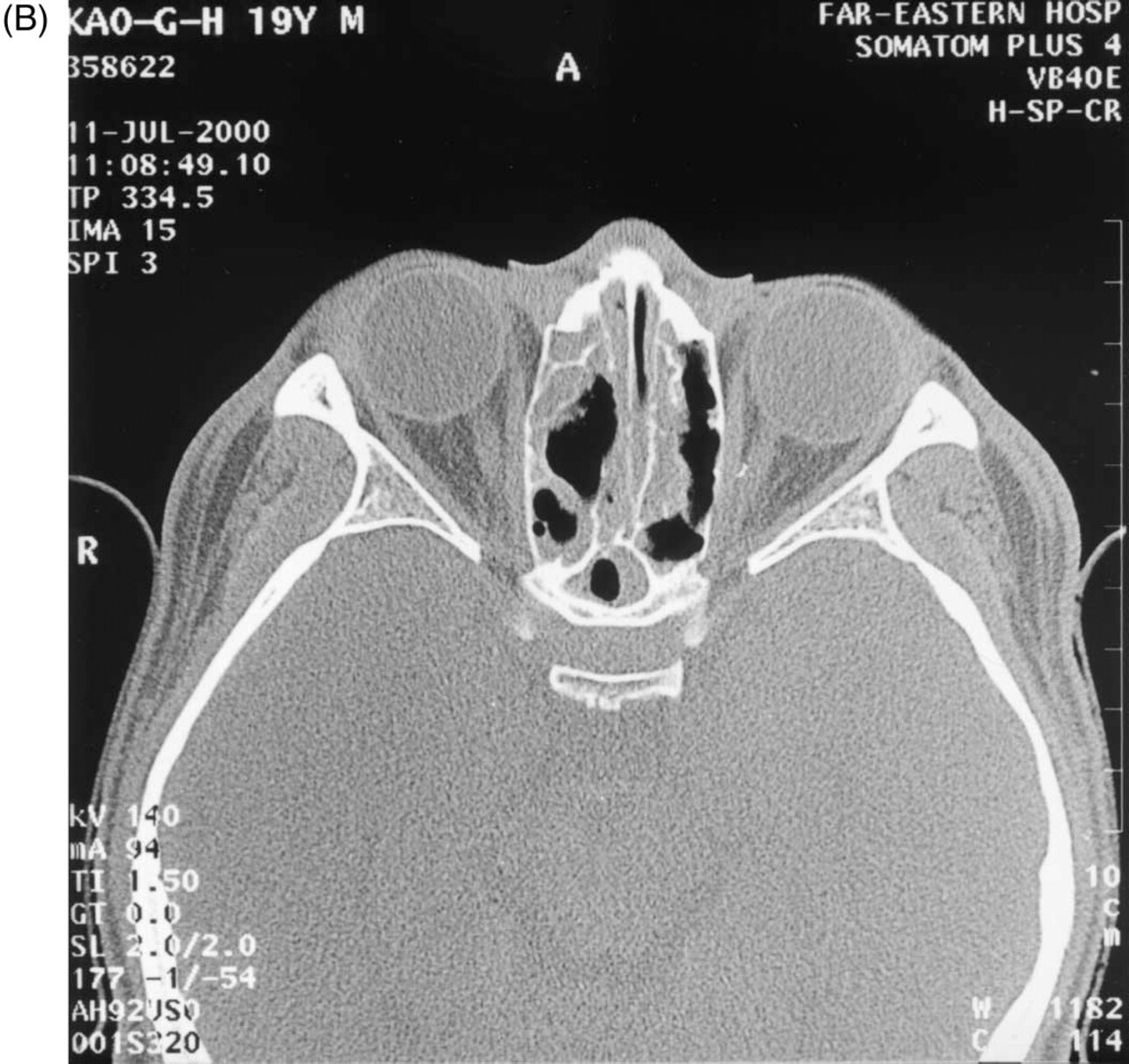
An axial section of emergent postoperative CT scan showing intact lamina papyracea and optic nerve.