
Abstract

Epithelioid HE can arise in soft tissues, viscera, skin, and bone. Common sites include liver, lung, and upper and lower extremities. 2,3 Several cases have been documented in the head and neck region, including oral cavity, thyroid gland, submandibular area, neck, scalp, larynx, and mandible. 4,5 Only one other case, which showed typical histologic features, has been described in the parotid. 3 We describe the first reported case of a parotid epithelioid HE with atypical features.
CASE REPORT
An 81-year-old man was referred to the ENT clinic for evaluation of a left parotid mass. He had noticed the mass approximately 6 months earlier and thought that it was not getting any larger. He was asymptomatic at that time. A CT scan showed a heterogeneous rim enhancing 2-cm left tail of parotid mass (Fig 1). No other masses or lymph nodes were seen. Fine needle aspiration showed a mixed lymphoid population. During the course of his workup, the patient began to have deep boring pain in the area of the mass but otherwise remained asymptomatic. He underwent a superficial parotidectomy with preservation of the facial nerve. Intraoperative frozen section consultation showed an atypical fibroinflammatory process that involved the margin. The deep lobe of the parotid was then removed where the mass had been. No residual tumor was found in the deep lobe specimen.
The superficial parotidectomy specimen revealed a firm nodular mass that measured 2.0 × 1.5 × 1.3 cm. The nodule exhibited a firm gray-white variegated cut surface. The adjacent parotid parenchyma was normal.
The tumor consisted of infiltrative cords, nests, and sheets of large pleomorphic epithelioid cells with irregular round to oval vesicular nuclei, prominent nucleoli, nuclear pseudoinclusions, and abundant glassy cytoplasm. Small intracellular lumen formation was noted focally (Fig 2). Marked cytologic atypia, focal spindling, necrosis, and mitotic figures (>1 per high-power field [HPF]) were present. A solid growth pattern, perineural invasion, and stromal chondroid areas were noted. The tumor appeared to arise from a central thick-walled vascular channel with tumor nests extending centrifugally from the lumen. The tumor cells were positive for CD31, CD34 (focal), and vimentin. The tumor was negative for cytokeratin (AE1/3), desmin, smooth muscle actin, epithelial membrane antigen, S-100, HMB-45, and glial fibrillary acidic protein. A mucicarmine stain showed stromal staining. Controls were appropriate.
The patient has been followed for 7 months postoperatively and is without evidence of disease. The pain he had experienced preoperatively resolved after the surgery. A transient postoperative facial nerve paralysis also resolved. The patient was clinically staged and no evidence of local or regional metastasis was identified.
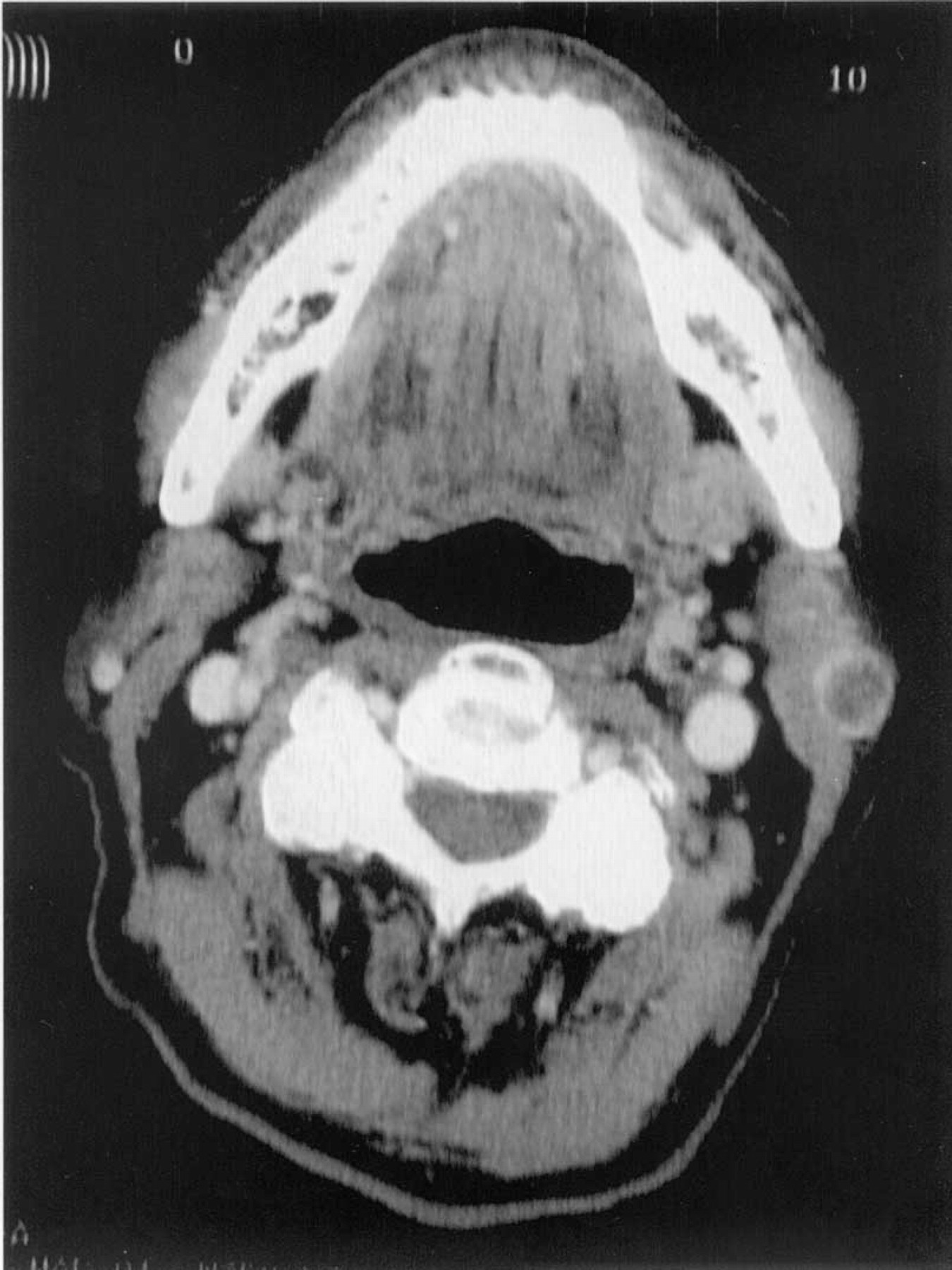
Axial CT scan demonstrating a heterogenous rim enhancing mass in the left parotid.
DISCUSSION
Epithelioid HE is an unique vascular tumor with intermediate/borderline malignant potential. Lesions have been described in a wide variety of anatomic locations, including the head and neck. There is a wide age distribution throughout adulthood. There is no gender predilection. 4,5
Recurrences are seen in 13% of patients and metastases in approximately 31%. 1,3 Sites of metastases include regional lymph nodes, lungs, liver, and bones. 3 There is a 10% to 15% mortality rate. 1 Treatment consists of complete wide local excision. 3 Adjunct radiation and chemotherapy have also been used.
Histologically, most cases appear bland and lack mitotic activity. Atypical features such as significant atypia, mitotic activity (>1 per HPF), focal spindling of cells, or necrosis are correlated with a more aggressive course. 1 Tumors with atypical features appear to have a greater risk of metastases. 1 Mentzel et al 4 studied 30 cases of epithelioid HE and found that only a high mitotic rate (>6 mitoses/10 HPF) correlated with a poor prognosis. They found no clear association between other atypical histologic features, tumor size, and clinical course. In addition, they reported cases with metastases in which the original tumor did not exhibit any atypical features. There remains some controversy regarding the biologic behavior of these tumors.
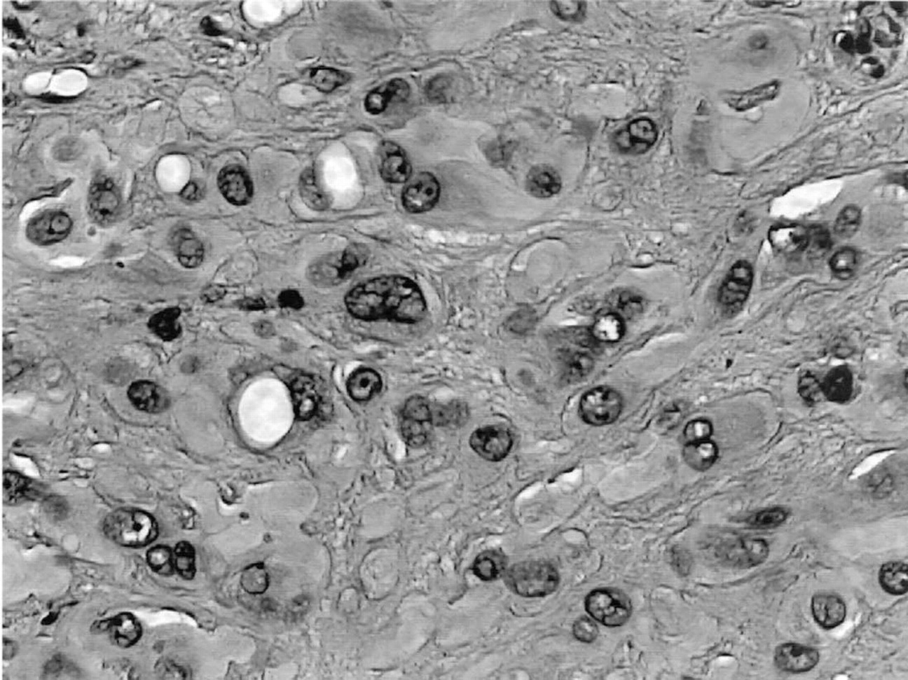
Pleomorphic epithelioid nests with intracellular lumen formation (original magnification, ×20).