





Abstract
Platelets play an important, but often under-recognized role in cardiovascular disease. For example, the normal response of the platelet can be altered, either by increased pro-aggregatory stimuli or by diminished anti-aggregatory substances to produce conditions of increased platelet activation/aggregation and occur in active cardiovascular disease states both on a chronic (e.g. stable angina pectoris) and acute basis (e.g. acute myocardial infarction). In addition, platelet hyperaggregability is also associated with the risk factors for coronary artery disease (e.g. smoking, hypertension, and hypercholesterolaemia). Finally, the utility of an increasing range of anti-platelet therapies in the management of the above disease states further emphasizes the pivotal role platelets play in the pathogenesis of cardiovascular disease. This paper provides a comprehensive overview of the normal physiologic role of platelets in maintain homeostasis, the pathophysiologic processes that contribute to platelet dysfunction in cardiovascular disease and the associated role and benefits of anti-platelet therapies.
Introduction
Under normal physiological conditions when a blood vessel is damaged, the task of platelets within the circulation is to arrest the loss of blood. This process involves the rapid adhesion of platelets to the exposed subendothelium followed by platelet-to-platelet adherence, which ultimately culminates in the formation of a platelet plug temporarily sealing off the damaged vessel wall. In contrast, in pathological conditions, such as atherosclerosis, arterial thrombus formation may limit the blood supply to nearby tissues, thus causing local ischaemia [1] and/or the progression of the atherosclerotic lesion.
In view of the fact that platelets contribute to the etiology and pathogenesis of coronary, peripheral, and cerebrovascular disease, a considerable amount of research into platelet pathophysiology has been undertaken. In this review, we will examine the phenomenon of platelet hyperaggregability associated with cardiovascular disease, and review endogenous and exogenous modulators of platelet function. Commonly used anti-platelet agents will also be reviewed.
Normal platelet function
Platelet physiology
Resting platelets circulate as discoid (Fig. 1a), anuclear cells originating from megakaryocytes in the bone marrow, and have a life span of approximately 8–10 days. Although thrombopoietin is the major hormonal regulator of platelet production [2], Battinelli et al. have recently shown that nitric oxide can also stimulate platelet production from megakaryocytes [3]. Platelets contain a plasma membrane, internal membranes (open canalicular and dense tubular systems), a cytoskeleton (microtubules and microfilaments), mitochondria, glycogen granules, storage granules (α-granules and dense granules), lysosomes, and peroxisomes.
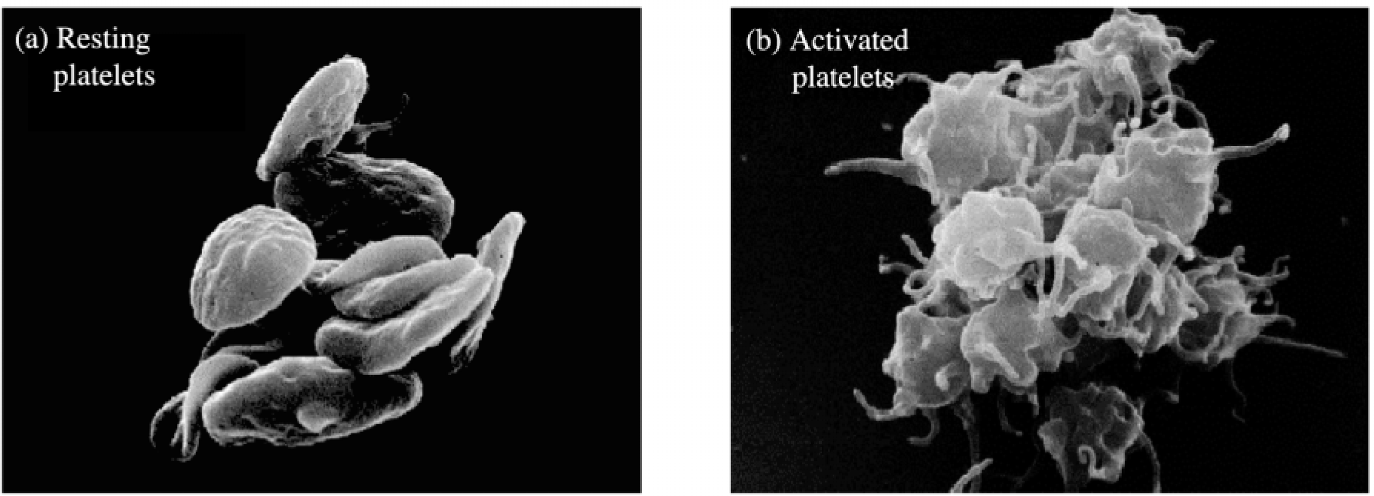
Resting and activated platelets.
The platelet is surrounded by a plasma membrane that extends through the multiple channels of the surface-connected canalicular system, greatly increasing the surface area of the platelet. The plasma membrane contain phospholipids; the negatively charged phosphatidylserine and phosphatidylinositol residues are primarily confined to the cytoplasmic side, where they may serve as substrates for phospholipases. Through this phospholipid bilayer, intrinsic glycoproteins such as glycoprotein (GP) IIb/IIIa extrude, serving as platelet receptors for activating and inhibiting agents.
The dense tubular system is the equivalent of the smooth endoplasmic reticulum in other cells; it is the site where the majority of calcium is sequestered and where enzymes involved in prostaglandin synthesis are localized. This membrane system lies in close contact with the channels of the open canalicular system, forming a membrane complex. There are numerous organelles dispersed in the cytoplasm, including mitochondria, glycogen particles, lysosomes, and peroxisomes. α-Granules and dense granules are platelet-specific storage granules. α-Granules contain mainly proteins, such as platelet factor 4, β-thromboglobulin, platelet-derived growth factor, fibrinogen, fibronectin, thrombospondin, plasminogen activator inhibitor-1, and von Willebrand factor. Dense granules are rich in serotonin, ADP and calcium.
Upon platelet activation, platelets lose their discoid shape, become spherical, and extend long, spiky pseudopods (Fig. 1b). The organelles are contracted towards the platelet center and are enclosed by a tight-fitting ring of reassembled microtubules and microfilaments. Finally, the contents of secretory organelles are expelled. During secretion, granule membranes fuse with those of the surface-connected canalicular system, with the diffusion of internal granular membrane proteins, such as P-selectin, into the plasma membrane. Whereas dense granule contents are easily secreted, α-granule release requires higher agonist concentrations, while lysosomal granule secretion only occurs with potent activating agents. In addition to the contents of the three secretory granules, platelets produce and secrete pharmacologically active substances, such as thromboxane A2 and platelet activating factor, during their activation and aggregation, establishing a positive feedback system for the hemostatic response.
Platelets are activated by several physiological (thrombin, collagen, ADP, epinephrine, vasopressin, serotonin) and non-physiological (divalent cationophores, cyclic endoperoxide analogues) substances. Although activation is produced by substances that vary markedly in chemical structure, platelets respond with the same series of distinguishable responses: (a) shape change; (b) aggregation; (c) three secretory processes; and (d) liberation of arachidonic acid, which is rapidly converted to prostaglandins and lipoxygenase products.
The platelet plasma membrane contains a large number of receptors which specifically bind agonists that stimulate the physiological platelet response, for example, ADP, epinephrine, collagen, thrombin, serotonin, and platelet activating factor. The interaction between a platelet-activating agonist (listed above) and its receptor causes rapid mobilization of signaling molecules within the platelet, notably calcium, diacylglycerol (DAG), and inositol 1,4,5-trisphosphate (IP3), which are sufficient to initiate and complete shape change and aggregation responses (Fig. 2). Low concentrations of these molecules cause some dense granule secretion and arachidonic acid liberation. ADP and prostaglandin endoperoxides released through this dense granule secretion and the prostaglandins and thromboxanes formed through the liberated arachidonic acid are themselves potent platelet agonists, and interact with their specific receptor causing release of more signal molecules. These platelet-derived agonists mobilize signaling molecules by interacting with the primary agonist in a synergistic way. This autocrine stimulation (positive feedback mechanism) increases the overall stimulus to such an extent that dense granule and α-granule secretion is completed. Aggregation causes close cell contact, which also operates as a positive feedback mechanism.
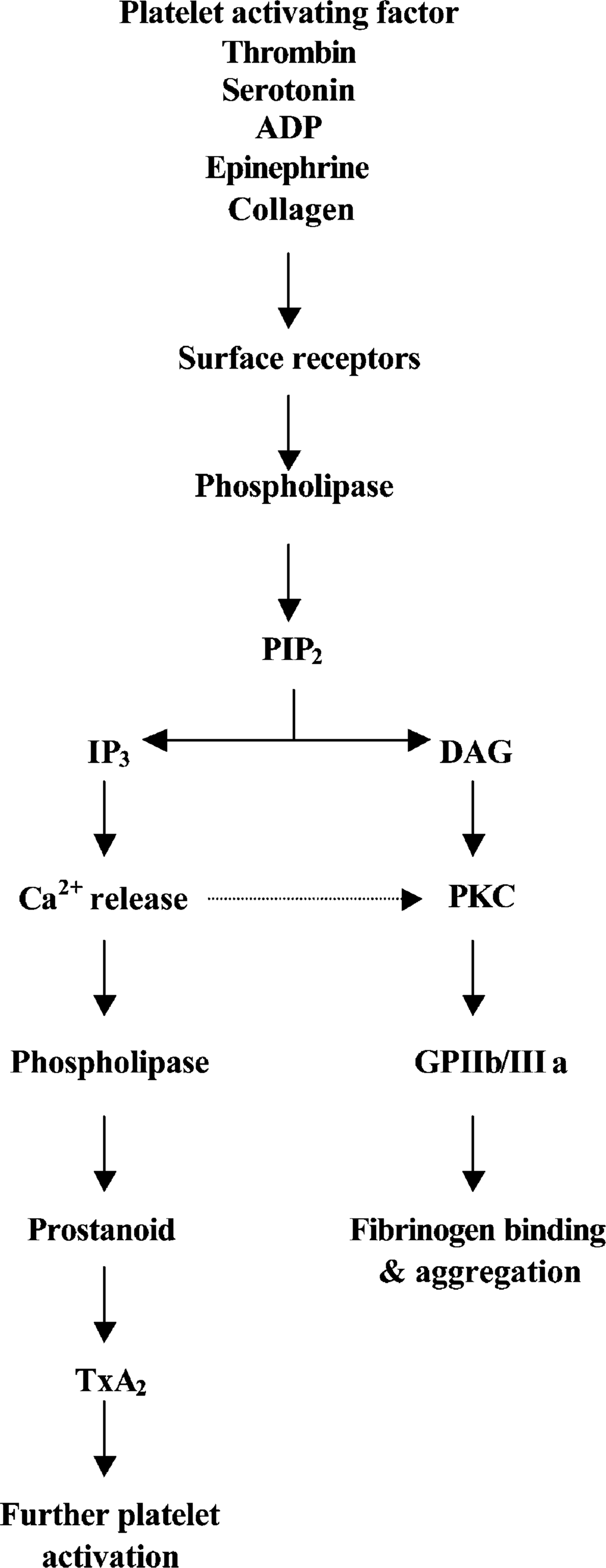
Mechanism(s) of platelet activation.
Some agonists (thrombin and collagen) in high concentrations can mobilize sufficient signal molecules without positive feedback to cause complete activation of all platelet responses; these agonists are ‘strong agonists’. Other physiological agonists at maximal receptor occupancy cause only mobilization of submaximal amounts of platelet signaling molecules; these agonists are ‘weak agonists,’ and rely on positive feedback to elicit all platelet responses. Interestingly, strong agonists in low concentrations behave as weak agonists; that is, they depend on positive feedback to produce full platelet stimulation.
Platelets have an important role in the pathophysiology of cardiovascular disease and its associated risk factors. Many techniques have been developed to detect the extent of platelet activation in these and other disease states. Some of these techniques are described next.
Optical (turbidometric) platelet aggregometry
Optical (turbidometric) platelet aggregometry was one of the first methods developed to assess platelet function and involves quantifying the changes in light transmittance in a platelet sample suspended in plasma following induction of platelet aggregation. Briefly, platelet-rich plasma samples are stirred in a cuvette at 37 °C between a light source and a photomultiplier tube. Upon the addition of a platelet agonist, platelets aggregate and the transmission of light increases. From its inception, this method of assessment of platelet function has been extensively utilized; however, there is evidence suggesting that the method is insensitive at assessing pre-existing or developing platelet micro-aggregates. For this reason, a new aggregometer has been developed that utilizes a combination of laser light scattering and aggregometry to monitor platelet function and the formation of platelet micro-aggregates effectively [4].
Turbidometric platelet aggregation facilitates distinguishing between the primary and secondary phases of aggregation, and is a measure of platelet function. The advantages of the method, however, are outweighed by its many disadvantages: platelet function in vitro does not necessarily reflect platelet function in vivo; sample aging occurs as a result of the time required to prepare platelet-rich plasma; and the presence of substances, such as lipids, in platelet-rich plasma or platelet-poor plasma can alter absorbance at the wavelength of observation. The above disadvantages may be considered as minor; however, the major problem with turbidometric aggregometry is that centrifugation modulates platelet behavior; platelets are heterogeneous in size, density, and metabolic activities, and it is likely that subpopulations of platelets are lost during the preparation of platelet-rich plasma that may be important determinants of hemostatic function in vivo.
Impedance platelet aggregometry
As the optical turbidometric method of assessment of platelet function involves the use platelet-rich plasma or a washed platelet preparation, the contributions from other blood elements that may affect platelet function are absent. Thus, an aggregometer that quantifies platelet function within a whole blood sample was developed utilizing electrical impedance as a means of assessing aggregation. Briefly, a sample of whole blood is stirred at 37 °C between two platinum wire electrodes set at a fixed distance [5]. When exposed to an agonist, platelets aggregate around the electrodes effectively increasing the electrical impedance. Impedance platelet aggregometry has wide-ranging applications, including the in vitro assessment of the disaggregatory capacity of organic nitrates.
The advantages of this technique are that whole blood is a reflection of the in vivo situation, and that no modification of the blood sample is involved (aside from the use of an anti-coagulant). Impedance aggregometry only requires small blood quantities and can be preformed quickly and conveniently. The disadvantages of impedance aggregometry in whole blood are minimal, but those that exist relate to its comparison with platelet-rich plasma. Examples include a more prolonged detection of the effect of aspirin and more variability than platelet-rich plasma.
Platelet activation markers
Platelet activation results in platelet shape change, aggregation, and the release of constituents from α-granule stores, some of which are platelet-specific products. Examples include platelet-activating factor-4 (PAF-4) [6] and β-thromboglobulin [7–9], both of which are easily quantified by ELISA or radioimmunoassay techniques. Other platelet activation assays have quantified the extent of the metabolites of thromboxane B2 in either plasma or urine [10–13].
Utilizing flow cytometry techniques, a number of platelet-specific, cell-surface activation markers have also been utilized to quantify the extent of platelet activation. Such activation markers include P-selectin [14,15], but also CD63 [16,17] and CD40L [18,19]. It is also now possible to assess the activation-dependent changes in the conformation of the GPIIb/IIIa receptor [20–22]. Other methods include binding of secreted proteins (thrombospondin) [23] and exposure of phosphatidyl serine (factor Va and VIIIa binding, annexin V) [24–26].
PFA-100
The PFA-100 system has been extensively used and functions by exposing whole blood samples to high shear within a capillary system. It monitors the drop in flow rate as the platelets within the whole blood sample form a haemostatic plug within an aperture of a membrane that is coated with collagen and other platelet agonists. The test records the closure time, the in-flow rate, and the total time of the test, and has been particularly useful in evaluating patients with von Willebrand's disease [27]. The PFA-100 may also examine the contribution of systemic inflammation on platelet function [28], and the effectiveness of GPII/IIIa pharmacotherapy during coronary intervention [29].
The rapid platelet function assay (RFPA)
The rapid platelet function assay (RPFA) is a simple and rapid means of monitoring the efficacy of GPIIb/IIIa receptor antagonist pharmacotherapy, and is based on the principle that fibrinogen-coated beads agglutinate in whole blood in proportion to the number of GPIIb/IIIa receptors [30]. A whole blood sample is added to a cartridge that contains fibrinogen-coated beads and a platelet agonist. Platelet activation/aggregation commences, resulting in fibrinogen binding to exposed GPIIb/IIIa receptors not already blocked by the receptor antagonist being examined.
The cone-and-plate(let) analyzer
The classical methods for the assessment of platelet function (turbidometric and impedance aggregometry) do not mimic totally all the physiological process that occur in vivo (e.g. platelet adhesion); for which reason a number of other techniques that assess platelet function have been developed. The cone-and-plate(let) analyzer is a system that examines the level of platelet adhesion to an extracellular matrix under shear-induced flow conditions. The analyzer consists of a cone-and-platelet device in which a whole blood sample is subjected to arterial flow conditions for a period of 2 min with platelet adhesion and aggregate formation upon the extracellular matrix monitored using an image analyzer.
Pathological conditions associated with increased platelet aggregation
Risk factors for coronary artery disease
In haemostasis, platelets circulate in close contact with the endothelial cell lining of the vessel wall without adhering to it [31]. Under pathological conditions, however, platelets respond rapidly to alterations of endothelial cells (e.g. fatty streak formation or plaque rupture) and to exposure of subendothelial structures by attaching firmly to the site of the lesion [32,33]. Platelet aggregates (thrombi) subsequently form and are associated with cardiovascular ischaemic events Fig. 3). Furthermore, there is evidence demonstrating a platelet hyperaggregable state in subjects who have various risk factors that are associated with atherosclerosis/coronary artery disease, including diabetes mellitus, hypercholesterolaemia, hypertension, and smoking (Table 1).
Diabetes mellitus
In patients with diabetes mellitus, coronary artery disease tends to be more extensive and severe, with cardiovascular-related deaths being three times more common than in non-diabetic patients [34]. Furthermore, diabetic patients without a documented history of a previous myocardial infarction have the same degree of risk for a myocardial infarction as non-diabetic patients with a previous history of infarction [35]. The mechanisms accounting for this incremental risk remain poorly understood; however, there is some evidence to suggest that a platelet hyperaggregable state may, at least in part, contribute to the observed increased risk.
Platelets from patients with type II diabetes mellitus manifest increased activation compared to platelets from non-diabetic subjects, when comparing the tendency of platelets to adhere to an extracellular matrix [36]. These results are in agreement with the observations made by Iida et al. [37] and others in which platelets from diabetic rats or humans were found to be hyperaggregable or have an increased extent of activation [37–40]. Along with demonstrating platelet hyperaggregability, a significant degree of spontaneous platelet aggregation has been observed in platelet samples obtained from type II diabetics compared to age-matched non-diabetic subjects [41].
Hypercholesterolaemia
Atherosclerosis is an inflammatory disease that is mediated, at least in part, by the progressive accumulation of cholesterol, more specifically low-density lipoprotein (LDL) cholesterol, within the walls of the vasculature. Owing to the influence of the integrity of the vasculature on platelet function, it is not surprising to observe that those subjects with hypercholesterolaemia also have dysfunctional platelets.
Early studies demonstrated platelet hyperaggregability to a range of platelet agonists in subjects with familial hypercholesterolaemia compared to platelets obtained from healthy controls [42]. Furthermore, patients with hypercholesterolaemia have elevated levels of β-thromboglobulin and other markers of platelet activation compared to age-matched control subjects. Aoki et al. demonstrated that platelet-dependent thrombin generation was increased in patients with hypercholesterolemia and in patients with hypercholesterolemia plus hypertriglyceridemia compared with patients with hypertriglyceridemia and control subjects [43]. Following a 5-min pre-incubation of platelets with LDL, a dose-dependent increase in bound molecules of fibrinogen per platelet was found after optimal stimulation with α-thrombin [44], further implicating hypercholesterolaemia in the phenomenon of platelet hyperaggregability.
Cardiovascular disease states associated with platelet hyperaggregability
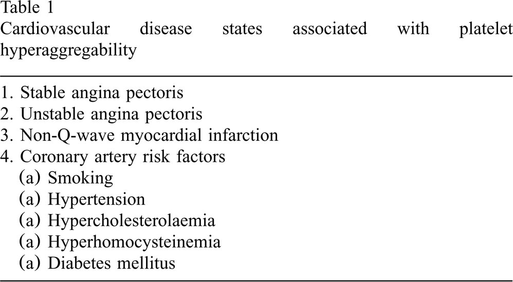
Cardiovascular disease states associated with platelet hyperaggregability
Initial observations of platelet dysfunction in subjects with hypercholesterolaemia led to a series of experiments that demonstrated that oxidised LDL (ox-LDL) serves as a pro-aggregant [45,46]. Platelets exposed to ox-LDL alone undergo immediate platelet shape change and pseudopodia formation [47] that enhance adhesion of platelets to endothelial cells under flow condition [48].
In a series of recent studies addressing the exact mechanism by which ox-LDL serves as a pro-aggregant, Siess et al. suggested that the lysophosphatidic acid (LDA) formed during oxidation of LDL serves as the active moiety in both mildly and minimally modified LDL [49]. Antagonists against LDA were demonstrated to prevent both mildly and minimally modified LDL-induced platelet activation. Interestingly lovastatin has been demonstrated to prevent this ox-LDL activation of platelets via a mechanism that involves a lysophosphatidic acid-dependent pathway [50].
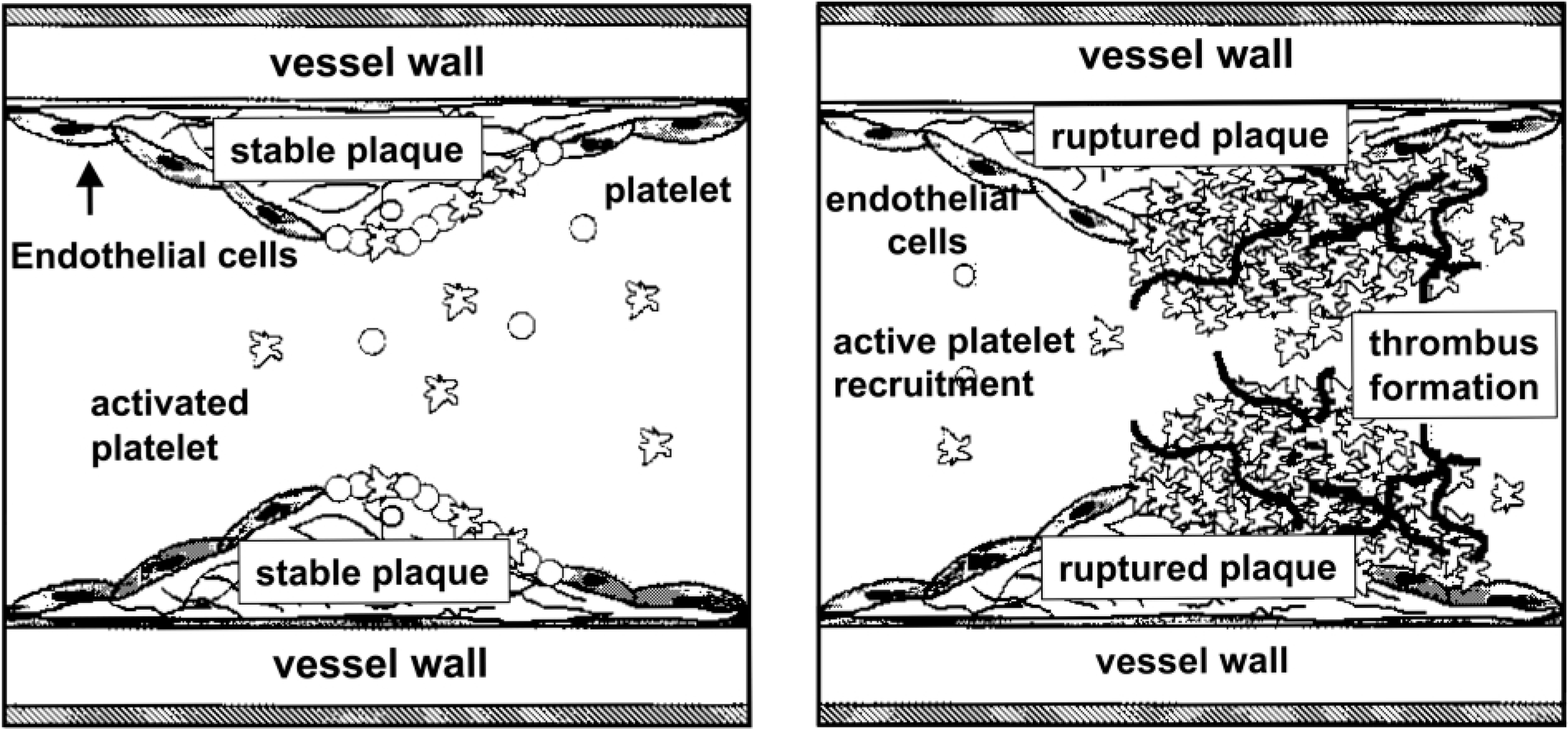
Platelets and vessel closure. (a) Platelets circulate in both the resting (unstimulated) and activated state. (b) Upon stimulation (for example plaque rupture) large numbers of activated platelets are rapidly recruited to the growing thrombus. This may ultimately lead to vessel closure.
Kjeldsen et al. [8] measured plasma concentrations of β-thromboglobulin (a marker of platelet activation), in untreated essential hypertensive men, and found them to be significantly higher than levels in healthy normotensive control men. In agreement with these observations, a number of other investigations have also observed increased platelet activity in subjects with essential hypertension [51,52].
A number of studies have indicated that the observed platelet dysfunction in subjects with essential hypertension can be normalized following anti-hypertensive drug therapy. Islim et al. determined that the degree of platelet activation in subjects with essential hypertension being treated with either β-adrenergic blockers or diuretics were not significantly different from levels of activation observed in untreated hypertensive subjects. However, angiotensin converting enzyme (ACE) inhibition was associated with a significantly lower plasma β-thromboglobulin concentration compared to control subjects [53]. Moser et al., whilst comparing the anti-platelet properties of captopril, enalapril, and fosinopril, determined that there was no significant effect on the degree of platelet aggregation induced by a range of agonists, but did observe a significantly decreased level of TXB2 generated by platelets from hypertensive subjects who had been treated with fosinopril [54].
Smoking
Cigarette smoking is strongly associated with coronary artery disease and has also been shown by some, but not all, investigations to increase the extent of platelet aggregability [55,56]. Smokers also have more small, spontaneous platelet aggregates and more medium and large platelet aggregates induced by epinephrine than non-smokers [57], reconfirming results from a previous investigation in which similar observations were reported [58].
Hyperhomocysteinemia
In epidemiological studies, hyperhomocysteinemia has been associated with premature peripheral artery disease, cerebrovascular disease, and coronary artery disease, independent of the effects of factors such as hyperlipidemia, cigarette smoking, and hypertension [59,60]. Addition of homocysteine to isolated endothelial cells induces functional abnormalities in the release of endothelium-derived NO [61] and detachment of cells [62]. In addition, methionine-induced moderate hyperhomocysteinemia in non-human primates leads to abnormal vasomotor activity [63].
In humans, homozygous homocystinurina is associated with markedly accelerated atherosclerosis and thrombosis, and with endothelial dysfunction in children as young as 4 years old [64]. Furthermore, subjects with no known vascular risk factors other than hyperhomocysteinemia demonstrate impaired arterial endothelium-dependent flow-mediated vasodilation [65]. In this group, hyperhomocysteinemia was not associated with renal failure or deficiencies in folate or vitamin B12. Treatment with folate has been shown to restore endothelial dysfunction during a methionine challenge in healthy volunteers without affecting plasma homocysteine levels [66]. Hyperhomocysteinemia also effects platelet activation. Homocystinuric patients have abnormally high TXA2 biosynthesis [67], which is associated with increased platelet activation. In addition, homocysteine rapidly auto-oxidizes in plasma, and, as a consequence, reactive oxygen species, such as superoxide anion, hydroxyl radical, and hydrogen peroxide, are formed [68]. The hydroxyl radical subsequently initiates lipid peroxidation. Di Minno et al. demonstrated that short-term administration of probucol, which prevents the oxidative modification of low-density lipoprotein, to homocystinuric patients results in a 40–60% decrease in TXA2 biosynthesis, while having no effects on TXA2 biosynthesis in normal volunteers [69]. The decrease in TXA2 biosynthesis was not correlated with either plasma homocysteine levels or plasma cholesterol levels in this study.
Coronary artery disease states
Stable angina pectoris
In a study by Meade et al., patients with angina, a history of myocardial infarction, or electrocardiographic evidence of ischaemia demonstrated increased (although not significant) responses towards ADP compared to those without clinical evidence of disease [70]. Further to this observation, Elwood et al. demonstrated a significant relationship between ischaemic heart disease and platelet aggregation in patients with past myocardial infarction and electrocardiographic evidence of ischaemia; however, no correlation was detected for ADP-induced platelet aggregation and angina [71]. Previously, Chirkov et al. demonstrated that patients with stable angina exhibited significantly greater extents of aggregation than normal subjects, when assessed in platelet-rich plasma using ADP to induce aggregation [72]. More recently, utilizing whole blood aggregometry, Chirkov et al. demonstrated that platelets from stable angina patients are more aggregable than those from normal subjects [73].
In contrast to the above data, Thaulow et al. in a large prospective study demonstrated that platelet concentration and ADP-induced aggregation were significantly correlated only with long-term, fatal coronary heart the development of angina pectoris or a positive exercise electrocardiographic response [74].
Moderate and strenuous exercise is known to enhance platelet aggregability in both healthy subjects [75,76] and patients with stable angina pectoris [77,78]. Within those subjects with stable angina pectoris, there is a debate as to what extent exercise-induced ischaemia or the presence of coronary atherosclerosis generating an arterial obstruction contributes to the enhanced platelet aggregability. Some investigations have reported increases in platelet aggregability markers (such as PAF-4 or β-thromboglobulin) following exercise testing [79,80], but results are not uniform in this regard [81,82].
Diodati et al. demonstrated that platelets became activated across an atherosclerotic bed following rapid atrial pacing and the consequent increase in coronary blood flow [83]. This increase in platelet reactivity, however, was only demonstrated in those subjects who had a haemodynamically significant narrowing of a coronary artery, implying that the mechanism behind platelet hyperaggregability following exercise may be partially shear stress-related [83]. Recently, Andreotti et al. demonstrated that low grades of exertion enhance whole blood platelet aggregability in patients with stable angina pectoris, an effect that was not observed in subjects without coronary artery disease [84].
Unstable angina pectoris/acute myocardial infarction
Patients with acute coronary syndromes, demonstrate hypersensitive platelets, circulating platelet aggregates, or elevated levels of circulating platelet secretory products [85,86]. For example, Langford et al. demonstrated that patients with unstable angina and acute myocardial infarction exhibit systemic platelet activation when blood samples were assessed using flow cytometry surface markers for platelet activation [86]. Available data are consistent with not only localized intracoronary [87] but also generalized [88,89] activation of platelets in unstable angina pectoris. Furthermore, a recent study by Chirkov et al. demonstrated that patients with acute coronary syndromes had an increased extent of aggregation when compared to normal volunteers. In addition, the authors also showed that platelet responsiveness to the anti-aggregatory effects of the nitric oxide donor sodium nitroprusside were diminished in patients with stable angina and acute coronary syndromes when compared to control subjects [90].
Another large body of evidence for the importance of platelets in acute coronary syndromes derives from the clinical efficacy of anti-platelet therapies. For example; aspirin is effective for the secondary prevention of myocardial following unstable angina [91–93], while glycoprotein IIb/IIIa antagonists are effective in reducing acute ischaemic complications of percutaneous coronary intervention [94–97], and in improving clinical outcomes among patients with acute coronary syndromes without persistent ST-segment elevation [98].
Modulators of platelet function
Activation of platelets is negatively controlled by biochemical processes that attenuate or prevent agonist-induced responses. Mechanisms of inhibition intrinsic to the platelet, activated by normal agonist-induced signal pathways include inositol 1,4,5-trisphosphate, lipocortin, lipoxygenase metabolites, and protein kinase-C. Physiologically two molecules are of fundamental importance for inhibition of platelet responses, cyclic guanosine-39′,59′- monophosphate (cGMP) and cyclic adenosine 39′,59′-monophosphate (cAMP).
Endogenous inhibitors of platelet function
Prostaglandin I2 (PGI2)
Endothelial cells and platelets can both generate eicosanoids from arachidonic acid via cyclooxygenase; however, the products formed in each cell type have opposing effects. Endothelial cells contain prostacyclin synthetase, which can produce PGI2 from endoperoxides (PGH2) [99], while platelets contain thromboxane synthetase, which produces thromboxane A2 (TXA2) [100]. Thromboxane A2 formation in the platelet induces platelet aggregation, which is accompanied by the platelet release reaction whereby serotonin and other granule components are expelled from platelet stores. Thromboxane A2 also induces a rise in the concentration of ionized calcium in the platelet cytosol and a decrease in platelet cyclic AMP formation by inhibiting adenylyl cyclase. In addition, TXA2 acts on vascular smooth muscle cells to produce vasoconstriction. As platelets are unable to resynthesize cyclooxygenase, in the presence of the irreversible cyclooxygenase inhibitor aspirin, TXA2 production is completely blocked. In contrast, endothelial cells are able to resynthesize cyclooxygenase; thus, aspirin only leads to a transient decrease in PGI2 production.
Endoperoxides (PGG2, PGH2) released from the platelet by the conversion of arachidonic acid can also be used by the endothelium to produce PGI2 [99]. In addition, to its potent vasodilatory effects, PGI2 inhibits platelet function. Prostaglandin I2 binds to a specific receptor on the surface of the platelet and stimulates adenylyl cyclase [101]. The resulting increase in platelet cyclic AMP leads to calcium reuptake by the dense tubular system and, thereby, inhibits platelet activation, platelet granule secretion, platelet aggregation, and the development of platelet procoagulant activities [102].
Nitric oxide
Nitric oxide is a potent endothelial product that is principally involved in the regulation of vascular tone and in maintaining the anti-thrombotic properties of the endothelium. Nitric oxide achieves these ends by decreasing vascular smooth muscle tone and by inhibiting platelet responses, such as platelet adhesion, platelet granule secretion, platelet aggregation, and arachidonic acid liberation [103–105].
Nitric oxide released by the endothelium diffuses readily across the platelet membrane and within the platelet binds to the heme moiety of soluble guanylyl cyclase. Heme binding leads to guanylyl cyclase activation and an increase in cyclic GMP production, which, in turn, activates a cyclic GMP-dependent protein kinase that phosphorylates myosin light chain kinase and regulates actomyosin ATPase activity [106,107]. Activation of cyclic GMP-dependent protein kinase also leads to phosphorylation of calcium transporters, which decrease intracellular calcium concentration [108]. Nitric oxide also inhibits the normal activation-dependent increase in platelet surface glycoprotein expression, such as P-selectin and the active conformation of glycoprotein (GP) IIb-IIIa [109]. Endothelial NO production is stimulated by shear stress and by substances released during platelet activation (such as ADP and 5-HT), as well as by determinants of the coagulation cascade, such as thrombin [110].
In addition to the effect of endothelial NO, platelets themselves posses both constitutive and, possibly, inducible NOS (cNOS and iNOS), whereby NO is produced during platelet aggregation [111–113]. Platelet aggregation can be enhanced by incubation with inhibitors of NOS and decreased by the NOS substrate
Exogenous inhibitors of platelet function (anti-platelet pharmacotherapies)
Aspirin
It has long been recognized that platelets exposed to aspirin have a diminished extent of aggregation in response to a range of platelet agonists [115,116], via a mechanism that was originally thought to involve the acetylation of prostaglandin synthase (see below) [117,118]. Platelets are extremely sensitive to aspirin. Patrono et al. reported a time- and dose-dependent inhibition of platelet production of thromboxane B2 (the stable product of the hydrolysis of TXA2) following 100 mg aspirin administration in 45 healthy subjects [119]. These results were later confirmed by others with even lower doses of aspirin producing a similar effect [120,121].
In a prospective, randomized, double-blind, placebo-controlled trial, the RISC study group examined low dose aspirin administration in 796 men with either unstable angina pectoris (UAP) or a non-Q-wave MI, and showed a reduction in the risk of both an MI and death after 5 days of therapy [93]. These benefits of aspirin utilization were then reconfirmed for the medium- (3 months) [93] and long-term (1 year) periods of observation [122]. Similar reductions in event rates for UAP and non-Q-wave MI patients were observed in other trials utilizing higher doses of aspirin [123,124]. The Second International Study of Infarct Survival (ISIS-2) examined the efficacy of aspirin (160 mg/day) utilization post myocardial infarction. In ~17000 infarction patients, aspirin utilization during the first 24 h of an infarct and for 1 month subsequently significantly reduced the incidence of 5-week cardiovascular mortality, non-fatal re-infarction, and non-fatal stroke [92]. This early benefit was maintained long-term with neither a loss nor gain in benefit of aspirin randomization during 10 years follow-up [125].
Aspirin utilization also prevents further vascular events in patients who have had a transient ischaemic attack or ischaemic stroke. In an overview analysis of 31 randomized trails, daily doses of aspirin ranging from 300 to 1500 mg/day reduced the risk of subsequent stroke, myocardial infarction, or vascular death by ~22% [126]. These benefits were then reconfirmed with a lower dose of aspirin. In the Swedish Aspirin Low-dose Trial (SALT) randomization to aspirin (75 mg/day) resulted in an 18% reduction in the risk of further stroke or death in patients with a history of an cerebrovascular event [127].
Mechanism
Cyclic prostanoids such as prostaglandins, prostacyclin, and thromboxane A2 are the products of the metabolism of arachidonate by prostaglandin endoperoxide H synthase, also known as cyclo-oxygenase (COX-1/2) [128–130]. Cyclo-oxygenase converts arachidonate to prostaglandin G2 by a cyclo-oxygenase reaction, then further catalyzes a peroxidation reaction to form prostaglandin H2 (PGH2). After the biosynthesis of PGH2, this endoperoxide is then converted to one of a series of potential prostanoid products, depending on the location of the enzyme. Within platelets PGH2 may be converted into the potent platelet agonist and vasoconstrictor thromboxane A2 (TXA2) by thromboxane synthetase. Within the vascular endothelium, PGH2 is converted into the potent anti-aggregatory and vasodilating agent prostacyclin (PGI2) by prostacyclin synthetase.
The two cyclo-oxygenase enzymes (COX-1, COX-2), also referred to as prostaglandin G/H synthase isozymes (PGH synathase 1 & 2), both exhibit cyclo-oxygenase and peroxidase activities. Both COX-1 and COX-2 enzymes are inhibited by aspirin through irreversible acetylation of an hydroxyl group of a single serine residue at position 529 within COX-1 and position 516 in COX-2 [129,131]. When blocked by the actions of aspirin, both COX-1/-2 are incapable of producing PGH2, the necessary precursor of a number of prostaglandins and thromboxane.
Primary prevention
As aspirin was shown to demonstrate a significant protective effect in the secondary prevention of an ischaemic event, the possible benefit of its use in primary prevention has also been examined in a series of randomized trials. In the Physicians' Health Study [132], a randomized, double-blind, placebo-controlled trial of alternate days of aspirin (325 mg) ~22000 men were treated and followed for 5 years. Randomization to receive aspirin resulted in a 44% reduction in the risk of myocardial infarction. This included significant benefits for aspirin on both non-fatal and fatal events [132]; however, this benefit was apparent only amongst those subjects 50 years and older [133] with the protective effect becoming apparent soon after initiation of therapy [134]. A slightly increased risk of stroke among those taking aspirin was also observed, although not significant. No reduction in mortality from all cardiovascular causes was associated with aspirin utilization [133]. Interpretation of this study must be made with caution, as the subject population was not truly representative. Only 88 cardiac deaths among ~22000 subjects studied were recorded, representing a cardiovascular mortality 88% less than expected [135]. There is some suggestion that this selective study group may have made lifestyle changes that lowered the likelihood of their having serious coronary artery disease.
This may very well be one of many contributing factors in the failure of the British Doctor's Trial to show a significant difference between subject groups in the combined endpoint (important cardiovascular events, MI, stroke, and total cardiovascular mortality). Two-thirds of more than 5000 British male physicians were randomized to either receive aspirin (500 mg/day) or were instructed to avoid aspirin-containing products totally for the study period [136]. Like the Physicians' Health Study there was a slight, but non-significant increase in disabling strokes among those subjects receiving aspirin. Despite the limitations of both investigations, an overview of these two trials of primary prevention demonstrated an overall reduction in nonfatal MI by 33% that was highly statistically significant [137,138].
In the Swedish Angina Pectoris Aspirin Trial (SAPAT), ~2000 stable angina patients were randomized to receive aspirin (75 mg) or placebo on a background of sotalol. Following a mean follow up of 4 years, aspirin randomization was associated with a 34% reduction in the primary outcome events (myocardial infarction and sudden death). A significant reduction in the incidence of first myocardial infarction was associated with aspirin randomization [139]. These results confirmed the observations made by Ridker et al. [140] in a small cohort of chronic stable angina patients from the Physicians' Health study.
Some patients with other cardiovascular conditions may also benefit from the anti-platelet effect of aspirin as suggested in a comprehensive overview [141]. In this overview of 46 randomized trials, the authors demonstrated that anti-platelet therapy (chiefly aspirin alone or aspirin plus dipyridamole) greatly reduced the risk of vascular occlusion in a wide range of patients.
Organic nitrates
Organic nitrates are in widespread use for the treatment/management of patients with stable angina pectoris, acute coronary syndromes, and heart failure. In addition to their vasodilator properties, the beneficial effects of organic nitrate use are related, in part, to their potent anti-platelet properties.
It is now widely accepted that nitroglycerine along with other organic nitrate preparations possess potent anti-platelet properties, initially demonstrated in vitro with high concentrations [142–144] but now shown at an ex vivo level with therapeutically achievable dosing regimens [145–147].
In unstable angina pectoris and acute myocardial infarction patients, therapeutic concentrations of nitrates were demonstrated to inhibit the extent of platelet aggregation significantly assessed ex vivo [148]. Moreover, platelet hyperaggregability induced during the passage of blood across the coronary circulation following rapid atrial pacing in patients with stable angina pectoris was inhibited by therapeutic doses of nitroglycerine or sodium nitroprusside [149].
Glycoprotein IIb/IIIa receptor antagonists
The final step in the process of platelet activation is the calcium-dependent transformation of the GPIIb/IIIa receptor complex to reveal previously cryptic binding sites for fibrinogen, fibronectin, and vitronectin. Much progress on development of antagonists that interfere with the binding of fibrinogen to the GPIIb/IIIa receptor has been made over the last 15 years, with three therapeutic classes of GPIIb/IIIa receptor antagonists being developed. Monoclonal antibodies, synthetic pep-tide and non-peptide receptor antagonists, and disintegrins have undergone various levels of basic experimental and clinical investigation, with the monoclonal antibody abciximab being the most extensively studied.
Abciximab
Utilizing chimeric 7E3Fab, Anderson et al. showed that inhibition of GPIIb/IIIa was a valid treatment for prevention of abrupt closure of a coronary artery during percutaneous transluminal coronary angioplasty (PTCA) [150,151]. Tcheng et al., studying patients with a moderate to high risk of sustaining an ischaemic complication, demonstrated a dose-dependent inhibition of platelet aggregation and GPIIb/IIIa receptor blockade [152]. A bolus dose of 0.25 mg/kg was found to result in blocking >80% of the GPIIb/IIIa receptors and a reduction to <20% extent of aggregation compared to samples prior to treatment. In a randomized, placebo-controlled study of 60 patients with ST-T changes, Simoons et al. demonstrated that a 0.25 mg/kg bolus followed by 10 mg/min for 18–24 h of c7E3Fab reduced the recurrence of ischaemic episodes significantly [153].
Following these initial investigations, a number of large clinical trials have been performed demonstrating the effectiveness of c7E3Fab in a number of different clinical settings. The Evaluation of c7E3 for the Prevention of Ischemic Complications (EPIC) trial first demonstrated the importance of GPIIb/IIIa blockade in high-risk patients with unstable angina or evolving myocardial infarction, with pronounced and sustained benefit [94,154–156]. Subsequent large-scale trials, such as EPILOG and EPISTENT, extended the findings of EPIC to lower-risk patients undergoing PTCA/stent implantation [157–160].
Tirofiban
Tirofiban is a non-peptide GPIIb/IIIa receptor antagonist that also serves as an effective anti-platelet agent by inhibiting the binding of fibrinogen to the activated GPIIb/IIIa receptor complex. In vitro administration of tirofiban dose-dependently inhibited platelet aggregation induced by ADP, collagen, arachidonic acid, thrombin, and the thromboxane analogue U46619 [161]. These in vitro anti-aggregatory effects were then reconfirmed ex vivo in dogs [162], healthy volunteers [161,163], and high-risk patients with coronary artery disease undergoing coronary angioplasty [164]. On the basis of these initial investigations, a series of multicenter trials, such as PRISM, PRISM-PLUS, and RESTORE, were performed in patients undergoing coronary intervention, including subjects with unstable angina/non-Q wave MI [97,165,166], and the benefits of the agent were confirmed in all groups studied.
Eptifibatide
The synthetic peptide eptifibatide, developed from a peptide found in the venom of the south-eastern pigmy rattlesnake Sistrurus barbouri that inhibits and disaggregates platelets, is another GPIIb/IIIa receptor antagonist and the only disintegrin to undergo extensive clinical investigation for its anti-platelet/anti-thrombotic properties [167]. Its anti-platelet properties have been extensively examined in a number of healthy subjects [168], patients undergoing elective PTCA [169,170], and patients with unstable angina pectoris or myocardial infarction [171,172]. Following from these initial investigations, eptifibatide has also undergone extensive clinical evaluation in such trials as IMPACT-11 and PURSUIT [96,173].
Oral preparations
As described above, intravenous GPIIb/IIIa antagonist administration at the point of care has demonstrated a clear and significant clinical benefit. It was initially thought that an extension of the platelet inhibitory effect beyond the initial hospitalization should enhance the long-term clinical outcome of these patients. Thus, a series of potent, orally active GPIIb/IIIa inhibitors have been developed, including xemilofiban, orbofiban, roxifiban, and sibrafiban. Unlike their intravenously administered counterparts, however, these agents have all demonstrated disappointing results regarding clinical benefit [174–178]. A pooled analysis of four trials of oral GPIIb/IIIa inhibitors showed a 37% relative increase in mortality with the use of oral GPIIb/IIIa inhibitors (from 1.3% to 1.7%) [179], probably owing to increased affinity of platelet GPIIb/IIIa for fibrinogen immediately after dissociation of the inhibitor from the receptor.
ADP receptor antagonists
Ticlopidine (Ticlid) and its structural analogue clopidogrel (Plavix) are thienopyridine derivatives that comprise a class of anti-platelet agents which function as ADP receptor antagonists [180].
Di Minno et al., in a comprehensive study examining platelet from healthy subjects, demonstrated that ticlopidine (500 mg) inhibited ex vivo platelet aggregation to a variety of platelet agonists including ADP, epinephrine, A23187, collagen, arachidonic acid, and thrombin via a mechanism independent of the GPIIb/IIIa complex [181]. Addition of the ADP scavengers apyrase or CP/CPK, added in vitro to a washed platelet preparation obtained prior to ticlopidine or clopidogrel administration, demonstrated a similar anti-platelet effect to ticlopidine/clopidogrel treatment alone, suggesting that they both selectively inhibit human platelet responses to ADP [182,183].
Using flow cytometry analysis of intracellular vasodilator-stimulated phosphoprotein (VASP) phosphorylation, Schwarz et al. were able to confirm that the anti-platelet effect of ticlopidine and clopidogrel occurred via the P2YAC ADP receptor [184]. Phosphorylation of VASP, a cytoskeleton and integrin-associated platelet protein, correlates with inhibition of binding of soluble fibrinogen binding to the platelet GPIIb/IIIa receptor and inhibition of platelet aggregation [185]. Reconfirming the hypothesis that VASP phosphorylation was a useful marker for inhibition of platelet aggregation, Schwarz et al. demonstrated a time-dependent increase in VASP phosphorylation when platelets were treated with sodium nitroprusside (SNP 100 μM) [184]. Ticlopidine and clopidogrel pretreatment of platelets showed an attenuation of the inhibitory effect of ADP on prostaglandin E1-stimulated, cAMP-mediated VASP serine 239 phosphorylation.
The clinical utility of ticlopidine and clopidogrel has been extensively examined, especially in the prevention of coronary stent thrombosis [186–188], with ticlopidine utilization being shown to be beneficial in the prevention of adverse outcomes in stroke patients [189]. Several studies, however, have demonstrated that these agents are capable of inducing thrombotic thrombocytopenic purura, although very infrequently [190–194].
Conclusion
Platelets play an important role in the maintenance of hemostasis. However, the normal response of the platelet can be altered, either by increased pro-aggregatory stimuli or by diminished anti-aggregatory substances. These factors contribute to conditions of increased platelet activation/aggregation and occur in the cardiovascular disease states of stable angina pectoris and acute coronary syndromes. In addition, they are also associated with the risk factors for coronary artery disease, such as smoking, hypertension, hypercholesterolaemia, diabetes mellitus and hyperhomocysteinemia. Finally, the utility of current anti-platelet therapies in the management of the above disease states further emphasizes the pivotal role platelets play in the pathogenesis of cardiovascular disease.