
Abstract

Introduction
Primary penile lymphoma is very rare. Approximately 50% of non-Hodgkin's lymphoma (NHL) presents at extranodal sites, most commonly the gastrointestinal tract [1]. Diffuse large B cell is more likely than other types of intermediate and high-grade lymphoma to develop in extranodal sites [1]. In view of the rarity of primary penile lymphoma, there is no consensus as to treatment, and patients sometimes have surgery which could have been avoided if the diagnosis had been considered.
Case report
A 91-year-old man presented with a 1-month history of a red indurated area on his glans penis. He had dysuria but no systemic symptoms. He had a past medical history of haemorrhoidectomy, basal cell carcinoma of temple, measles, polypectomy, total hip replacement and cataract removal.
He was taking aspirin, nifedipine, dosulepin, celecoxib and salbutamol. Examination revealed an area of erythema with an elevated lesion measuring approximately 2cm diameter associated with central crusting on the glans penis (Fig. 1). He had no inguinal lymphadenopathy. Biopsy revealed a diffuse large B cell lymphoma. CT scan of chest, abdomen and pelvis showed no significant lymphadenopathy.
Taking into account the presence of stage 1 disease, the patient's age and relative frailty it was felt that toxicity of combination chemotherapy was not justified. Prior to radiotherapy the lesion had grown and was causing partial obstructive symptoms and blood stained discharge. He was treated with external beam radiotherapy, 40Gray (Gy) in 20 fractions to the penis with immobilisation with a wax block using 6MV photons and two fields. Three months later the lymphoma on his penis had resolved leaving only a small scab (Fig. 2)—the patient was symptom free; however he had developed an irregular firm swelling at the inferior of the base of the penis, just outside the previous treatment field.
Further biopsy confirmed recurrence, and CT showed no disease elsewhere. He therefore had 36 Gy in 18 fractions to the base of penis with 6MV photons and a single field with 5 mm bolus and the slight overlap with the previous field was accepted. This also responded well to radiotherapy. A CT scan at 17 months showed no lymphadenopathy.
When last seen in clinic, 2 years after initial presentation he was well with no signs of recurrent local disease.
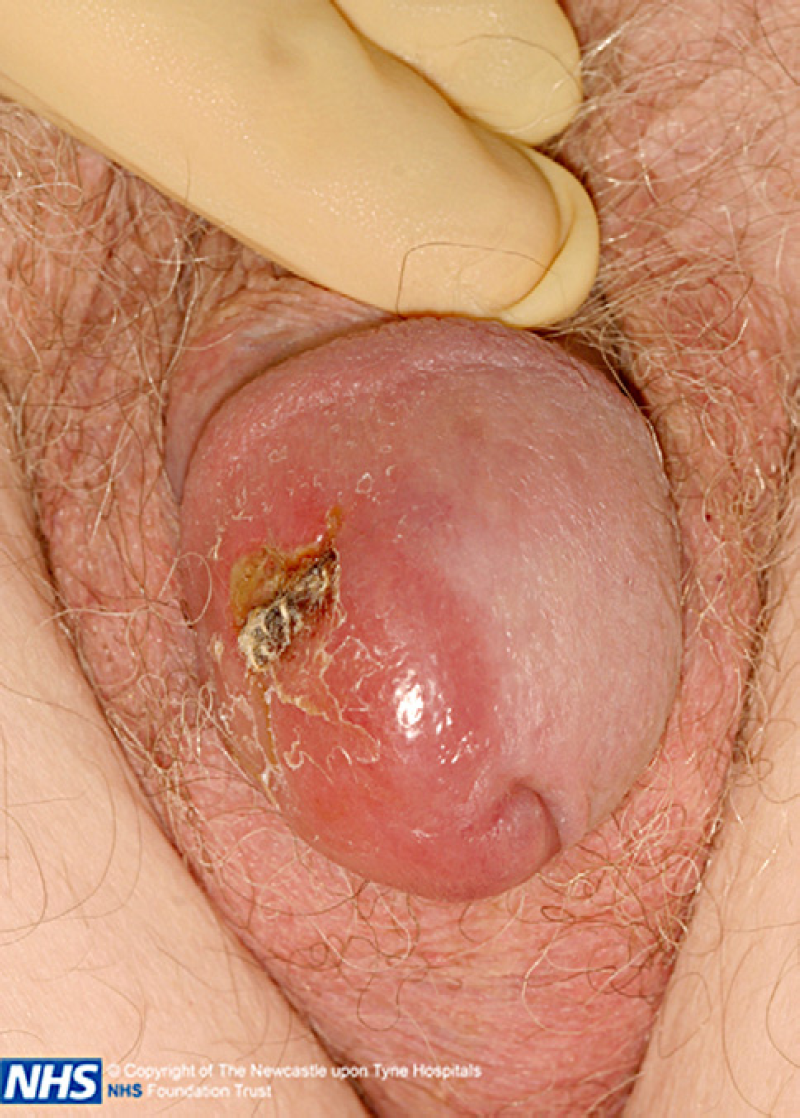
Pre-radiotherapy.
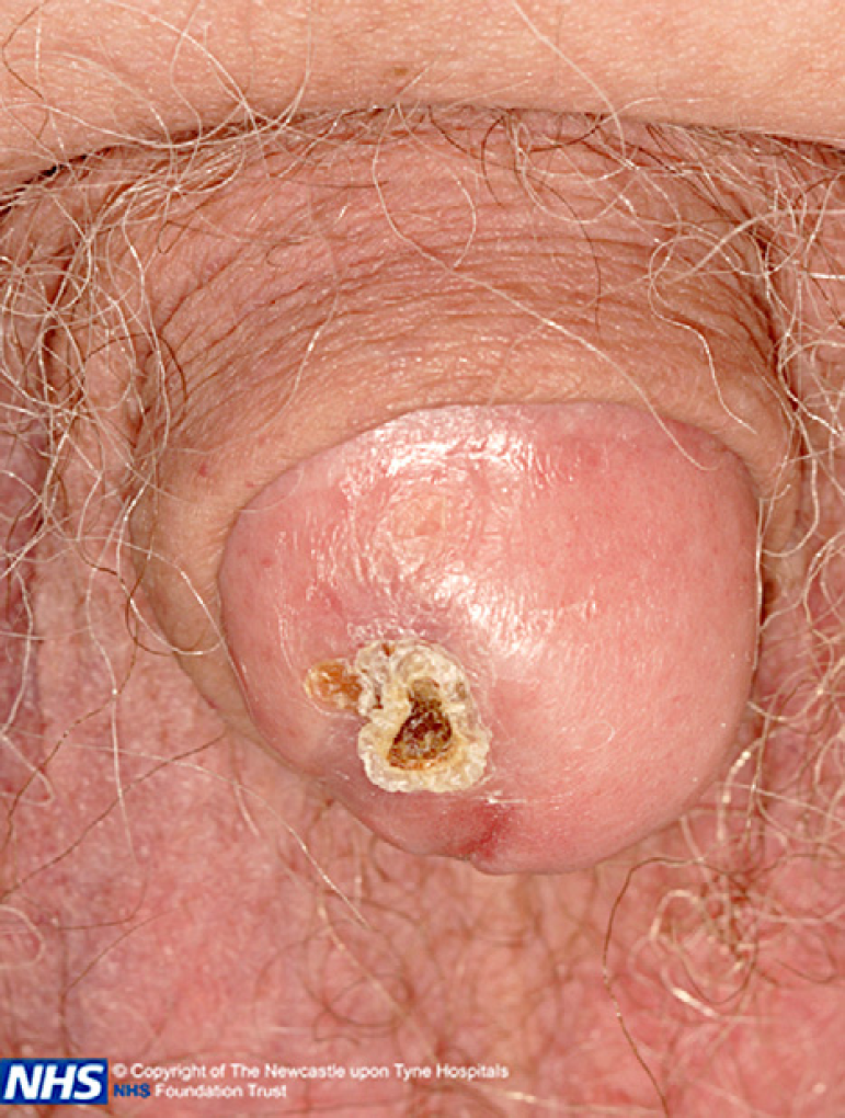
Post-radiotherapy.
Discussion
A review of the literature reveals 22 reported cases over 40 years. Eight of them had surgery, 11 chemotherapy, 7 radiotherapy, and 1 laser surgery. Sometimes combined modality treatment was used, depending partly on the stage of disease, age and performance status of patient, and partly on whether it had been diagnosed pre-operatively [2 –6]. There is no consensus as to radiotherapy dose, but 40 Gy seems to be most popular. It is known that stage IE lymphoma has a good prognosis, with a potential cure rate of 65% when treated with radiotherapy alone [5], making it a good treatment option in a patient such as this for whom chemotherapy would not be appropriate, and avoiding the need for surgery.
Footnotes
None declared.