
Abstract

Clinical Problem
A 45-year-old man with diabetes presented to the emergency department with acute pain, swelling, and erythema on the lateral aspect of his right foot.
Research Question
Is magnetic resonance imaging (MRI) the best imaging modality for the diagnosis of suspected osteomyelitis of the foot or ankle?
Literature Search
A computer-based literature search by using Ovid, PubMed, and Google was performed based on a PICO question (patient, intervention, comparison, and outcome).
The Evidence
A systematic meta-analysis [1] found 16 studies from which 2×2 contingency tables could be constructed and used to extract information about foot and ankle cases suspected of osteomyelitis. The studies selected for review used extraction instruments derived from the Cochrane Methods Group checklist of Systematic Review of Screening and Diagnostic Tests [2]. These studies compared MRI with either plain radiography, technetium Tc99m bone scan, or white blood cell (WBC) scan. Of the studies included for review, at least 80% of the patients were 16 years of age and older. The diagnostic accuracy in each study was determined by using bone biopsy. Sensitivity and specificity were calculated for all articles, and summary receiver operator curves (ROC) were calculated for each imaging modality. The diagnostic odds ratio (DOR) describes the ratio of the odds of a positive test result in patients with disease relative to the odds of the same test result in patients free of disease. Mathematically, it is defined by the formula; (true positive/false negative)/(false positive/true negative), or more simply, LR(+)/LR(–). The DOR has no dependency on prevalence, thus it is an invalid measure of a test's error rate given specific disease prevalence. According to Glas et al [3], the DOR is ideal for meta-analysis, because it enables the combination of results from different studies to be more precisely used in the comparison of various diagnostic tests. In this study, the DOR was used for a head-to-head comparison of MRI with plain radiography, technetium Tc99m bone scan, and WBC studies.
From the results of this meta-analysis, MRI consistently demonstrated higher diagnostic accuracy in comparison with technetium Tc99m bone scan, plain radiography, and WBC studies (Table 1). The summary DOR for MRI was 42.1 (95% confidence interval [CI], 14.8–119.9). The sensitivity and specificity of MRI ranged from 77%–100% and 40%–100%, respectively. At a clinically relevant cut-point of 90% sensitivity, the specificity of MRI was 82.5%.
Comparison of MRI to selected studies based on diagnostic odds ratio
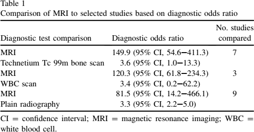
CI = confidence interval; MRI = magnetic resonance imaging; WBC = white blood cell.
Clinical Applicability
Compared with technetium Tc99m bone scan, WBC studies and plain radiography, current evidence suggests that MRI is a superior modality in the diagnosis of suspected osteomyelitis of the foot or ankle and is helpful in ruling out other diagnoses.
Comments
Morrison et al [4] found in their study that the sensitivity and specificity for nonenhanced MRI of osteomyelitis was 79% and 53%, respectively, and could be increased with fat-suppressed contrast-enhanced MRI to a sensitivity of 88% and specificity of 93%. The investigators of the meta-analyses addressed the increased use of gadolinium in present practice and performed a subset analysis that did not reveal a significant variation in the use of MRI with gadolinium. However, a study performed by Johnson et al [5] showed a sensitivity of 95% and a specificity of 91% when using T1-weighted MRI in the diagnosis of suspected foot osteomyelitis, and they suggest that more emphasis should be placed on the unenhanced images to get an accurate diagnosis, given the concerns surrounding gadolinium administration and its potential relation to nephrogenic systemic fibrosis.
Currently available literature on imaging of osteomyelitis has many flaws. For instance, no randomized control trial has been conducted that compares MRI with other imaging modalities in the workup of osteomyelitis. Furthermore, the majority of studies do not address issues of practicality (ie, price, availability, etc) and lack suitable information to create a 2×2 contingency table.
The current meta-analysis does account for the above factors, which consequently limits the review to 16 studies. Unfortunately, the majority of studies used were not blinded and did not document comorbid foot or ankle disease (ie, Charcot foot). In addition, MRI was not compared with positron-emission tomography, combination-imaging strategies, or directly to bone biopsy. Given these limitations, the current evidence available suggests that MRI is the preferred imaging modality in the diagnosis of osteomyelitis of the foot or ankle.
Given the significant difference in diagnostic utility between MRI and a technetium Tc99m bone scan, the small difference in cost after interpretation by a radiologist, and the overall cost of missing a diagnosis of osteomyelitis (OM), MRI is more cost effective than a bone scan, provided there is a sufficiently high pretest probability [1].
Unfortunately, the absence of a formal decision model means that an actual value for such a probability has yet to be established [1].