
Abstract

Case Report
This is a case of a middle-aged man living in a remote area of Bolivia who was recently hospitalized due to progressive difficulty breathing. On admission, multiple lesions involving the mucosa of the nares, palate, and pharynx were apparent (Figs 1 and 2). There was obvious nasal obstruction and narrowing of the nasopharynx and oropharynx. Leishmaniasis was suspected given his history, and the diagnosis was confirmed with a skin smear. The patient is being treated with amphotericin B.
Discussion
Leishmaniasis is a protozoal disease transmitted by sandflies in both the New and Old World. Some species cause only cutaneous disease, whereas others may cause mucocutaneous or visceral involvement.1 Mucocutaneous leishmaniasis is frequently caused by Leishmania braziliensis and characterized by a primary cutaneous lesion that may be followed months to years later by destructive nasopharyngeal lesions (espundia).2 The time between the primary lesions and mucosal involvement may be anywhere from 1 month to several decades and occurs in a relatively small percentage of patients originally infected with L. braziliensis. 3
Initial signs and symptoms of mucocutaneous leishmaniasis are erythema and edema of the involved mucosa and often nasal stuffiness, serous and crusted rhinorrhea, discomfort, or epistaxis. This is followed by ulcerations, often covered with a mucopurulent exudate, and granulomatous-like lesions. These usually lead to destruction and perforation of the nasal septum, palate, lips, pharynx and larynx and occasionally the trachea. The lesions are chronic and progressive and eventually lead to an inability to eat and a high risk for fatal aspiration.
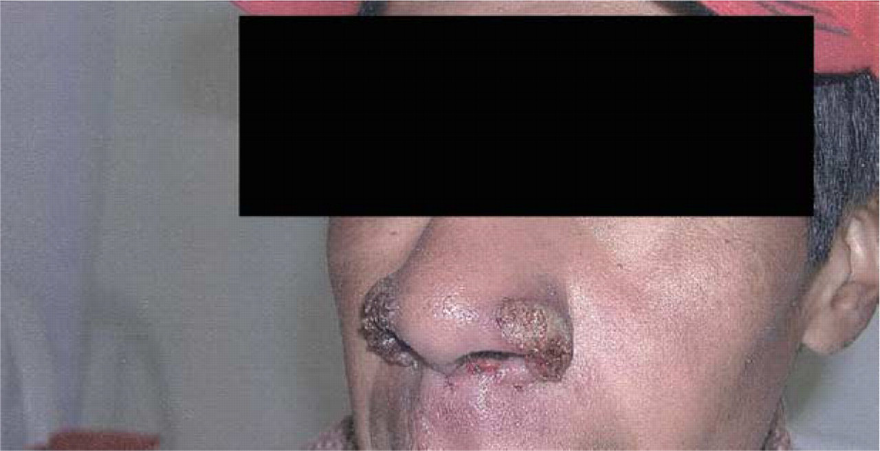
Mucosal leishmaniasis affecting the nares.
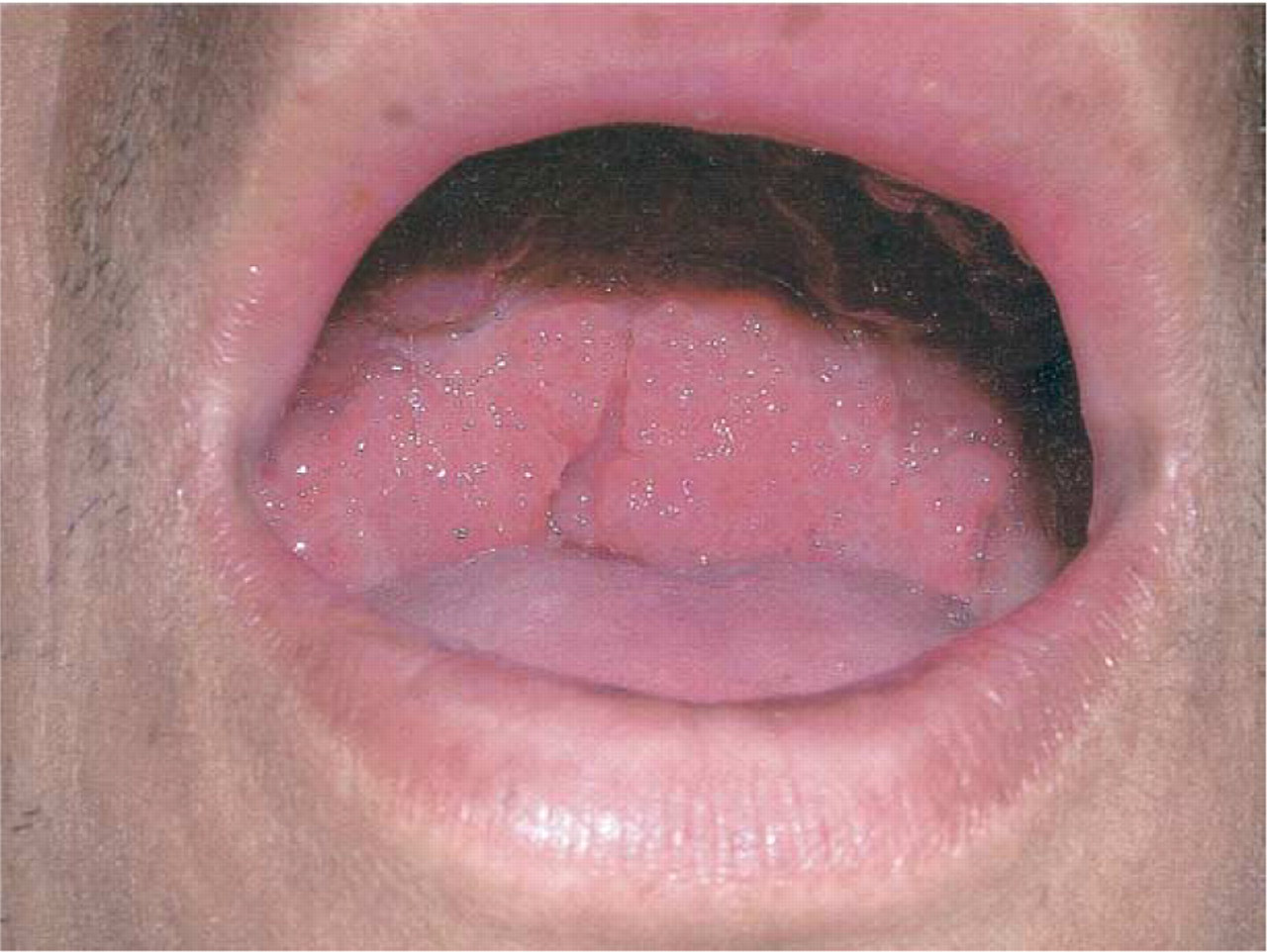
Mucosal leishmaniasis affecting the palate.
The diagnosis of mucosal leishmaniasis is confirmed when amastigotes are identified in touch preparations or biopsy specimens or when promastigotes are isolated from a culture of a tissue aspirate. Diagnosis is often made on the basis of clinical findings. The differential diagnosis includes syphilis, leprosy, basal cell carcinoma or other malignant tumors, midline granuloma, paracoccidiomycosis, tertiary yaws, histoplasmosis, sarcoidosis, tuberculosis, or rhinosporidiosis.3 Traditionally, therapy has involved the use of a pentavalent antimonial. Alternate therapies include amphotericin B, oral ketoconazole, and interferon gamma. In addition to drug therapy, adequate nutrition must be provided for the patient and the need for possible ventilatory support assessed. Post treatment plastic surgery may benefit patients with symptoms secondary to extensive tissue destruction.