
Abstract
OBJECTIVE: To investigate the effect of 5-fluorouracil (5-FU) ointment on the inner ear of guinea pigs.
STUDY DESIGN AND SETTING: In group A (n = 7), 5-FU ointment was applied into the left external auditory canal. In group B (n = 10), 5-FU ointment was applied to the left middle ear through myringotomy. In both groups, the right ear served as a control. One week later the endocochlear DC potential (EP) was measured and morphology of the cochleae was examined using scanning electron microscopy (SEM) and light microscopy.
RESULTS: In group A, there was no significant difference between the EP values of the experimental side and the control side. In group B, there was a statistically significant difference between them (P < 0.05). Morphologic findings showed no damage.
CONCLUSION: 5-FU ointment application to the external ear seems to be safe but its application to the middle ear may pose some risk of ototoxicity.
METHODS
Animals
Seventeen healthy male guinea pigs (weight, 295 to 365 g), free of external or middle ear disease, were used in this study. All animals had free access to commercial food and water, and were maintained in an environment with controlled temperature and 12 hr light—dark cycles. Animal care and experimental procedures were carried out in accordance with the Guidelines for Animal Experimentation of Nagasaki University with approval of the Institutional Animal Care and Use Committee.
Anesthesia and Drug Administration
All animals were anesthetized with Nembutal (sodium pentobarbital 35 mg/kg intraperitoneal [i.p.], Abbott Laboratories, North Chicago, IL), and immobilized with Relaxin (suxamethonium chloride 15 mg/kg, i.p., Kyorin Pharmaceutical Co., Ltd, Tokyo, Japan). Depth of anesthesia was maintained during the experiment using half doses of Nembutal each hour or as required. Breathing was assisted with a respirator (New England Medical Instruments, Meadway, MA).
Guinea pigs were divided into 2 groups. In group A, approximately 100 mg of 5-FU (5%) ointment (Kyowa Hakko Kogyo Co., Ltd, Tokyo, Japan) was applied into the left external auditory canal with a syringe, and the right ear served as a control. One week later, endocochlear DC potential (EP) was measured in both ears, and then both cochleae were removed. Fourteen cochleae from 7 animals were processed for the scanning electron microscopy (SEM) study. In group B, approximately 200 mg of 5-FU ointment was needed to fill the left middle ear and external auditory canal. The right ear served as a control. Myringotomy was carried out carefully, and 5-FU ointment was infused very slowly to prevent damage to the middle and inner ears by mechanical insertion. One week later, EP was measured in both ears and cochleae were removed. Fourteen cochleae from 7 animals were processed for SEM, 6 cochleae from 3 animals were processed for light microscopy. The total number of group B was 10.
Electrophysiologic Recordings
The EP was recorded from the basal turn of the cochlea. Guinea pigs were sedated and placed in an electrically shielded, double-walled, sound-attenuating booth (Industrial Acoustics Co., Bronx, NY). Each animal was fixed on a head holder with ear bars designed for guinea pig. Body temperature was maintained at 37°C using an animal warming blanket. The bulla was opened to expose the middle ear cavity. The EP was recorded using a glass microelectrode filled with 150 mM KCL, which was mounted on a micromanipulator and connected to a DC amplifier (FD223, WP Instruments, Sarasota, FL), and data were collected on a 16-bit data recording system (Power Lab 400 AD Instruments, Castle Hill, Australia). Ag-AgCL wire was placed over the neck muscles as the ground electrode. The glass microelectrode was advanced into the scala media through the round window membrane (RWM) until a positive EP was obtained.
Differences in the EP between the control and the experimental ears were analyzed statistically using paired Student's two-tailed t test. Difference was considered to be significant when the P value was <0.05.
Scanning Electron Microscopy
Immediately after the completion of the measurement of the EP, sedated guinea pigs were euthanized with Nembutal (sodium pentobarbital 175 mg/kg, i.p.). After decapitation of the guinea pigs, cochleae were removed and immediately immersed in 2.5% glutaraldehyde in 0.1 M phosphate buffer. After small holes were made in the apex, the round and oval windows, the cochleae were fixed by perilymphatic perfusion of 2.5% glutaraldehyde in 0.1 M phosphate buffer and refrigerated overnight. The following day, the cochleae were rinsed in and perfused with buffer and postfixed with a slow perfusion of 2.0% osmium tetroxide in 0.1 M phosphate buffer. They were then rotated for 15 min and rinsed with buffer again. The bony capsule of the cochlea was picked away using a fine needle, and the remaining bone and lateral wall were dissected away with the use of a single-edged razor blade and forceps to expose the organ of Corti. During this process the stria vascularis was badly damaged or destroyed and was discarded. Dissected cochleae were dehydrated in serially increasing concentrations of ethanol from 50% to 100% and were critical point dried. Cochleae were mounted on scanning electron microscopy (SEM) stubs and were sputter coated with silver. Fully processed specimens were examined and photographed with a JEOL JSM-6700F scanning electron microscope.
Hair Cell Count
Semiquantitative hair cell counts were carried out with a modified version of the method used by Korver et al. 6 Fourteen cochleae from 7 animals in group A and 14 cochleae from 7 animals in group B were used. Representative areas of the basal turn, middle turn, and apical turn were photographed. In each area, inner or outer hair cells were counted in a section that was 10-pillar cell heads in length. The results were shown as the average survival percentage rates compared to control. Statistical analysis was carried out using non-paired Student's two-tailed t test. Difference was considered to be significant when the P value was <0.05.
Light Microscopy
For histologic study of stria vascularis, 6 cochleae from 3 animals in group B were used. The temporal bones were fixed in 10% formalin, decalcified in EDTA, and embedded in paraffin. The specimens were sectioned along the mid-modiolar plane at a thickness of 5 μm and every twentieth section was stained with hematoxylin-eosin.
RESULTS
Electrophysiologic Findings
In group A, the EP values of experimental ears and control ears were 90.0 ± 7.3 mV (mean ± SE) and 87.9 ± 6.9 mV, respectively (Fig 1). There was no significant difference between them (P > 0.05). In group B, the EP values of experimental ears and control ears were 57.5 ± 36.7 mV and 86.2 ± 9.9 mV, respectively (Fig 2). This difference was statistically significant (P < 0.05). As shown in Figure 2, the EP values of the experimental ears had a tendency to fall into 2 groups, almost normal and extremely low.
Electron Microscopic Findings
Because of extensive damage to the stria vascularis that was caused as the lateral wall was removed to expose the organ of Corti, the morphology of the stria vascularis was not examined. Figure 3 shows SEM micrographs of a guinea pig cochlea from an experimental ear in group B, which showed an extremely low EP. Almost normal stereociliary arrangements and surface structure on the inner and outer hair cells and an almost normal number of microvilli on the surface of the inner pillar cells can be seen in the basal (Fig 3a), middle (Fig 3b), and apical (Fig 3c) turns. No notable changes can be seen in any turns. Because the results from group A were almost the same as in group B, the SEM data was not described here.
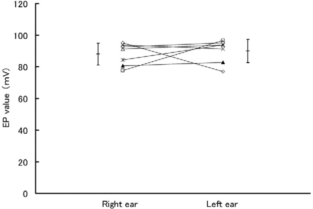
The EP values of animals in group A (5-FU ointment in the left external auditory canal and no drug in the right) are shown (n = 7 for each ear). There was no significant difference between left and right ears (P > 0.05). AV, average. Error bar = SD.
Hair Cell Count
Group A. The outer hair cell survival rates for this group on the left side (5-FU side) were 97.7 ± 2.9, 97.3 ± 4.8, and 93.3 ± 5.4% in basal, middle, and apical turn, respectively. The inner hair cell survival rates were 98.8 ± 2.9, 98.8 ± 3.1, and 98.6 ± 3.4% in basal, middle, and apical turn, respectively.
The outer hair cell survival rates for this group on the control side were 99.4 ± 1.6, 95.2 ± 7.9, and 86.4 ± 15.6% in basal, middle, and apical turn, respectively. The inner hair cell survival rates were 98.6 ± 3.8, 98.6 ± 3.8, and 98.6 ± 3.8% in basal, middle, and apical turn, respectively.
There were no significant differences in either outer or inner hair cell counts between the 5-FU side and the control side for any turn (P > 0.05).
Group B. The outer hair cell survival rates for this group on the 5-FU side were 98.0 ± 3.2, 99.0 ± 1.3, and 93.5 ± 6.4% in basal, middle, and apical turn, respectively. The inner hair cell survival rates were 100 ± 0, 100 ± 0, and 100 ± 0% in basal, middle, and apical turn, respectively.
The outer hair cell survival rates for this group on the control side were 98.8 ± 3.1, 98.6 ± 2.7, and 94.6 ± 6.4% in basal, middle, and apical turn, respectively. The inner hair cell survival rates were 98.8 ± 3.1, 100 ± 0, and 98.6 ± 3.8% in basal, middle, and apical turn, respectively.
There were no significant differences in either outer or inner hair cell counts between the 5-FU side and the control side for any turn (P > 0.05).
Light Microscopic Findings
Three cochleae from the 3 animals on the 5-FU side and 3 cochleae from the 3 animals on the control side were compared histologically. No notable differences between 5-FU and control ears were seen in the stria vascularis (Fig 4).
DISCUSSION
Topical 5-FU ointment caused a reduction in EP when applied to the middle ear (group B, 4 ears), but not when 5-FU ointment was applied into the external auditory canal. It is unclear why only 4 ears in group B had a low EP, whereas the remaining 6 ears did not. 5-FU ointment was observed around the RWM in the 4 ears with low EP, but not in the other 6 ears. We speculated that when 5-FU ointment came in contact with the RWM, it diffused through the round window or oval window, causing a reduction in EP. However, no structural damage was observed in either the organ of Corti SEM or in the stria vascularis light microscopic observations. These findings suggest that when 5-FU penetrates into the inner ear, it is not likely to cause degenerative changes to the inner ear structures. The EP can be affected without seeing structural changes. For example, Kikuchi et al 7 reported that one of the functions of the gap junctions in the cochlea is recirculation of K+ ions from hair cells to strial marginal cells, and interruption of this recirculation would deprive the stria vascularis of K+ resulting in hearing loss. Thus, the EP reduction that we observed in group B of the present study may be due to the impairment of the functional rather than the morphologic aspect of the inner ear.
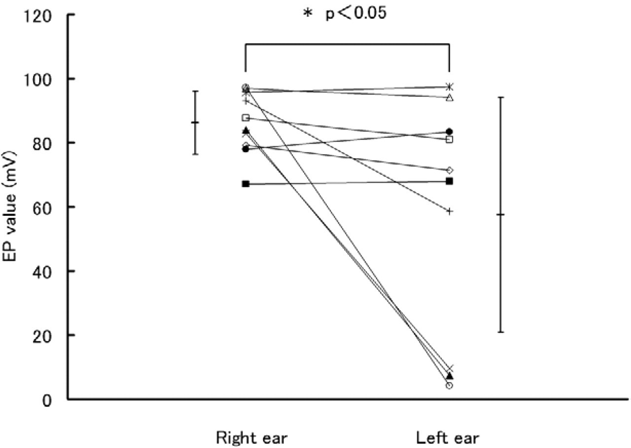
The EP values of animals in group B (5-FU ointment in the left middle ear and no drug in the right) are shown (n = 10 for each ear). There was a significant difference between the 2 ears (P < 0.05). The EP values in the experimental side fell into 2 groups: almost normal and very low. AV, average. Error bar = SD.
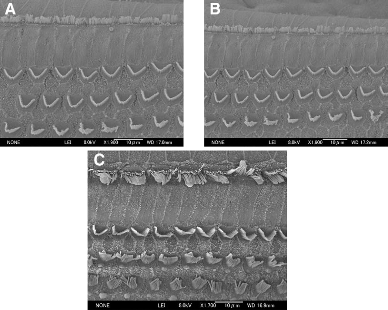
Representative SEM photomicrographs of the organ of Corti from a guinea pig in group B at 1 week after treatment (5-FU ointment in the left middle ear) that had a very low EP. Intact inner and outer hair cells and supporting cells can be seen in the basal
Sudoh et al 8 reported the incidence of apoptotic endothelial cells of the aorta was higher near the tip of the catheter associated with 5-FU indicating that 5-FU can cause vascuritis. We speculated that 5-FU may have caused vascuritis in the stria vascularis, resulting in mild to severe reduction in EP according to the extent of damage in the stria vascularis. No structural damage was observed in the stria vascularis on light microscopic observation. Further investigations are required to determine how 5-FU affects the stria vascularis using transmission electron microscopy (TEM) or other methods.
In this study, not only 5-FU but also ointment vehicle might be a potential source of toxicity. 5-FU is a low molecular weight (MW) compound (130.8) and is expected to readily cross the RWM. The ointment vehicle contains low MW materials (ie, propylene glycol [MW, 76.09], stearyl alcohol [MW, 270.40], and petrolatum). It was reported previously that the passage across RWM of higher MW compounds is more selective, but low MW compounds can cross the RWM freely. 9,10 Ointment vehicle is a possible source of ototoxicity, but Federspil 11 reported that there was no ototoxicity when he applied low concentration gentamicin, sisomicin, and tobramycin by ointment into the middle ear. The ointment vehicle of Federspil 11 might be different from ours, however, and his results may support the idea that 5-FU rather than the ointment vehicle caused a reduction in EP.
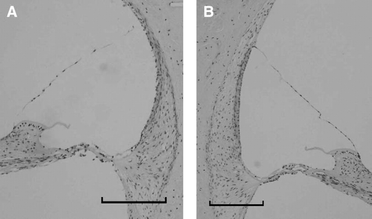
The stria vascularis in group B (5-FU ointment in the left middle ear and no drug in the right).
In this study, doses of 5-FU are approximately 500 or 1000 times as much as are used usually in humans, but they were applied topically. We suggest concentration and contact time of 5-FU are more important than topical dose. When we opened the bulla to expose the middle ear cavity, we could not find any ossicular chain damages or perilymph leakage. We suggest that mechanical insertion was the least possible cause of hearing changes.
If we had examined the inner ear at a later time point, we might have observed structural changes to the stria vascularis or the hair cells. However, the shortest interval for clinical 5-FU use is 1 week, so we investigated cochlear structure and function 1 week after 5-FU ointment application.
These results suggest some limitations to the local application of 5-FU treatment for cholesteatoma. This treatment may only be indicated for cholesteatoma without eardrum perforation or cochlea or semicircular canal fistula, or we should not apply it close to RWM. Further investigation is required to clarify the mechanism of ototoxicity of 5-FU, using TEM for the stria vascularis or longer period experiments.
The authors wish to thank Takashi Suematsu for his technical assistance with electrophysiologic recordings and scanning electron microscopy.