
Abstract

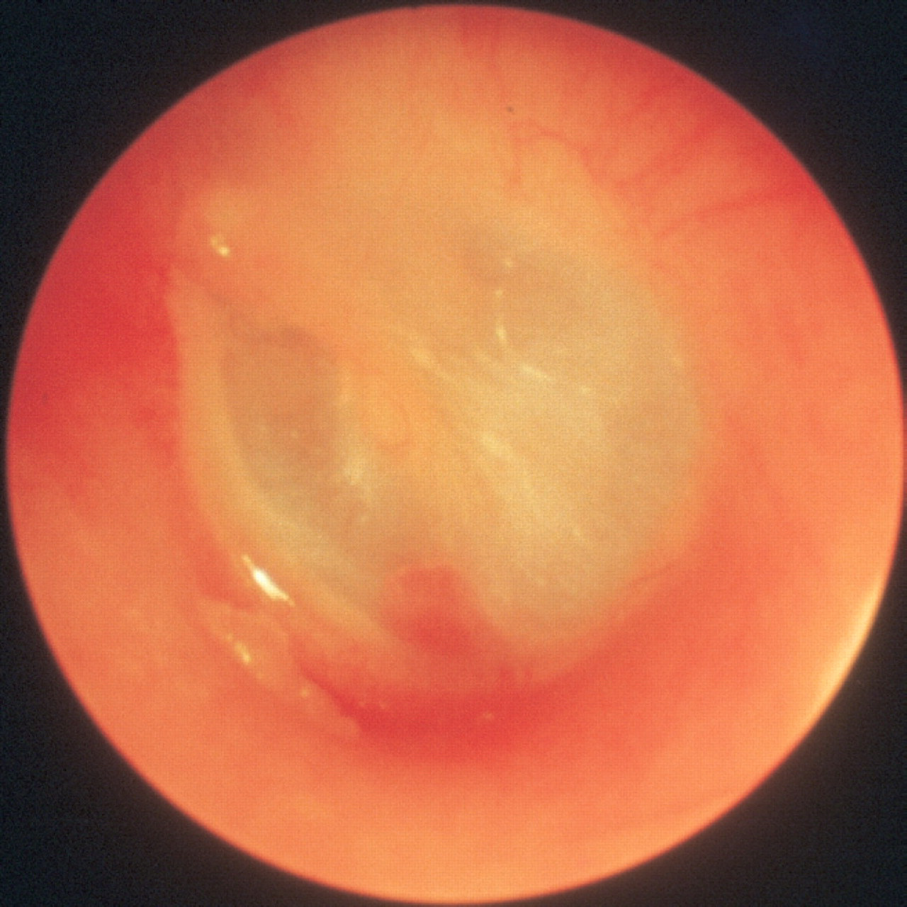
Otoscopic examination revealed a small bright red mass lying in the inferior portion of the left middle ear in contact with an intact tympanic membrane.
The patient was given a general anesthesia and underwent exploratory tympanotomy through a postauricular approach. After elevation of the tympanomeatal flap, the posterior and inferior bony annulus was drilled and a small red vascular tumor, which was not in contact with the ossicles, was found at the cochlear promotory. The mass measuring 5 × 3 × 2 mm in size was completely excised with a surrounding cuff of muscosa. The ossicular chain and chorda tympani nerve were preserved. The pathologic examinations showed a capillary hemangioma (Fig 2). The immunohistochemical stain studies revealed that there was no proliferation of the paraganglial cells in this lesion. The postoperative course was uneventful. The patient's pulsatile tinnitus disappeared completely, and there was no deterioration of hearing after surgery. A repeat high-resolution CT of temporal bone 5 years later revealed no recurrence.
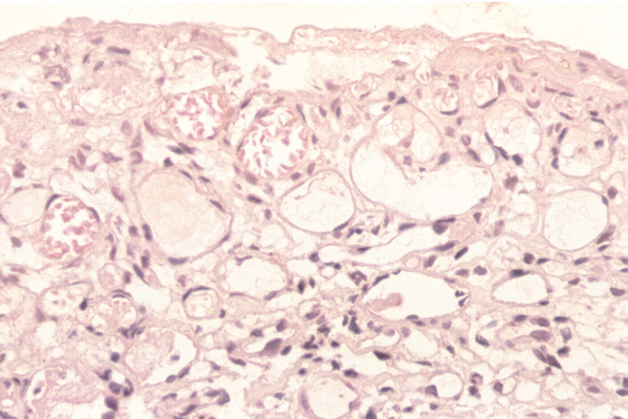
Capillary hemangioma composed of tightly packed capillaries. (Hematoxylin and eosin stain; original magnification × 200.)
Discussion
The incidence of hemangioma in the temporal bone is uncommon, and its occurrence in the middle ear is very rare. Mangham et al 1 reported a series of 1430 intratemporal tumors, of which 10 (0.7%) were vascular tumors (vascular malformations and hemangiomas); the most frequent sites of occurrence were the geniculate ganglion and the internal auditory canal with only 1 tumor that involved the middle ear. 1 A survey of English-language literature reveals only 11 cases of hemangioma of the middle ear were reported. 1 – 3 When the present case is included, there would be total of 12 reported cases (5 males and 7 females); 7 are capillary hemangiomas, 3 cavernous hemangiomas, and 1 cellular juvenile hemangioma. No detailed histologic classification was available for 1 of these patients. The patients' ages ranged from 4 months to 80 years; 4 patients were less than 4 years of age and 5 patients aged over 51 years. In 7 cases, the right ear was involved, in 4 cases the left ear was involved, and in 1 case, both ears were involved.
A variety of signs and symptoms has been reported for hemangiomas of the middle ear. They may be asymptomatic and may only be recognized incidentally with removal of the incus, 3 but the most common clinical manifestations are retrotympanic vascular mass and pulsatile tinnitus. The hemangioma may also present as a polypoid mass in the external ear canal, bleeding from the ear after ventilation tube insertion, vertigo, and hearing loss. 2 , 3
The differential diagnosis of an otoscopically visible capillary hemangioma of the middle ear includes glomus tympanicus tumor, glomus jugulare tumor, aberrent carotid artery, high jugular bulb, cholesterol granuloma and meningioma, hemangiopericytoma and Masson's intravascular hemangioendothelioma. 2 – 5 Preoperative high-resolution CT scan is indicated to demonstrate the extent of the tumor because a vascular tumor may be the harbinger of a skull base vascular tumor. 4 Angiography, retrograde venography, MRI, and magnetic resonance angiography allow more definitive identification of the nature of a vascular mass of the middle ear. 4 , 5 However, definitive diagnosis can only be obtained by exploration surgery with biopsy or excision of the tumor with histologic examination. Although spontaneous resolution has been reported, 2 the treatment of choice for a sympatomatic middle ear capillary hemangioma is total excision with meticulous dissection, to preserve hearing and the normal structure of the middle ear. Methods of surgical intervention depend on the size and the extent of hemangioma. A small lesion confined to the promontory, as in the present case, may be treated with exploratory tympanotomy via transcanal approach 2 , 3 ; however, transmastoid, translabyrinthine, middle fossa approach, or combined approach is necessary for a large lesion with intratemporal extension. 1 – 5 Inadequate excision always renders postoperative recurrence. There were 2 postoperative recurrence in the 11 cases of hemangioma of the middle ear reported in the literature. 2