
Abstract

On microscopy, the section of the specimen showed epitheloid and spindle cells arranged in lobules and small bundles. The nuclei were pleomorphic, hyperchromatic, and contained prominent nucleoli. Cytoplasm showed abundant melanin pigment.
Immunohistochemical stainings for S100, Vimentin, Melan-A, and HMB45 antigens were strongly positive. A diagnosis of nasal mucosal melanoma (NMM) was made.
The patient underwent a paralateronasal surgical “box resection” of the mass with the septum, the lateral nasal wall, and a partial maxillectomy. A reconstruction of the nasal pyramid was achieved through a nasal flap rotation. Postoperative course was uneventful. She has been followed for 12 months and has presented no evidence of recurrence or epistaxis.
Discussion
NMM represents less than 1 percent of all melanomas and from 2 percent to 9 percent of head and neck melanomas. More than 75 percent of the lesions originate in the nasal cavity. Both genders are affected equally, with the peak age incidence between fifth and eighth decade.1
NMM arises from the melanocytes that have migrated during embryologic development from neural crest to the nasosinusal mucosa. One third of the NMMs are amelanotic lesions. Epistaxis, nasal obstruction, and headache are the most common presenting symptoms. NMM is liable to be mistaken for other nasosinusal malignancies, particularly when melanin is scanty or absent. The differential diagnosis of NMM includes several neoplasms as the nasopharyngeal carcinoma, the large B-cell lymphoma, the extramedullary plasmocytoma, the sarcomatoid squamous carcinoma, the rhabdomyosarcoma, the neuroendocrine carcinoma, and the olfactory neuroblastoma. Immunohistochemistry is essential for the correct diagnosis. NMM is positive for S100 protein, vimentin, and specific melanocytic markers such as melan-A and HMB45 antigens.2
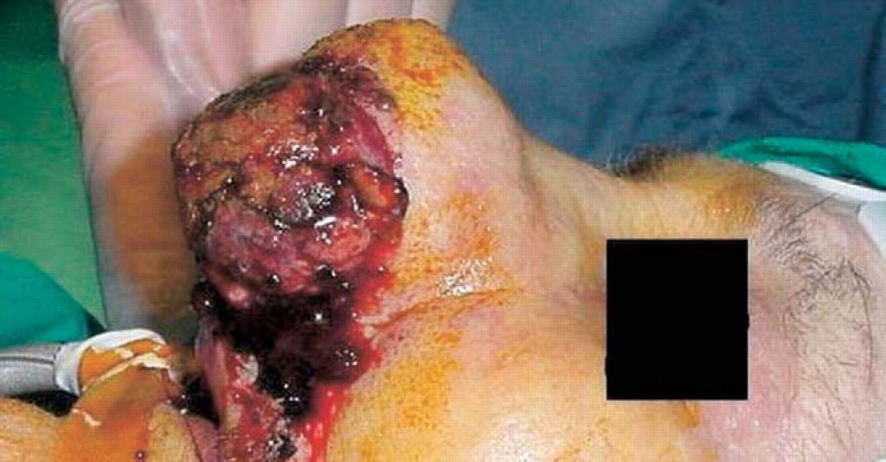
Preoperative image of nasal mucosal melanoma of the left cavity.
Prognosis of NMM is uniformly poor. Tumor size, site of origin, bone invasion, histologic aggressiveness, and metastasis at the time of diagnosis are the main prognostic factors.2,3 Wide surgical excision is the treatment of choice. The local recurrences are common because of the difficulty in achieving complete tumor removal with wider negative margins at this anatomic site.2 Chemotherapy should be reserved for patients with systemic disease. Therapeutic regimens with dacarbazine, cisplatin, ranimustine, and tamoxifen have been used with varying results.4,5
The elderly patients present treatment dilemmas that include prognosis quoad vitam, quality of life, life expectancy, surgical and anesthesiological risks, and prolonged postoperative care. In our case, the leading problem of the patient was epistaxis, which was potentially life-threatening. Decisions with respect to radical surgery in an elderly population must encompass quality-of-life issues in addition to that of disease-free survival. This is a classic demonstration of how the surgery may improve the quality of life rather than extend it.
Author Information
Author Contributions
Financial Disclosure
None.