
Abstract

We describe a new tool, “Sleep MRI,” a real-time device that detects and characterizes the anatomic site, magnitude, and duration of airway obstruction simultaneously with MRI-compatible physiologic measures of peripheral arterial tone, hemoglobin oxygen saturation, and pulse rate in patients with obstructive sleep apnea syndrome (OSAS). The goal of the study is to present the safety and feasibility of Sleep MRI. Sleep MRI is a novel technique of airway imaging during sleep that assesses respiratory events measured by ambulatory sleep study technology with simultaneous real-time MR imaging (RT-MRI).
Methods
Seventeen adult OSAS subjects were prospectively enrolled after Institutional Review Board approval at Stanford University. An open MRI system (0.5T Signa SP, General Electric, Waukesha WI) with a twin magnet oriented vertically and separated by 60 cm allowed the subjects to sleep in their preferred positions. A small, oval receive coil was placed encircling the face of the subject to optimize images of the upper airway. Images were acquired with the RTHawk, 1 a real time 2D spiral sequence with 6 interleaves, that acquires a midline sagittal view with a resolution of 2.6 × 2.6 × 5 mm. Constrained by the gradient hardware (12 mT/m gradient amplitude, 16 T/m/s slew rate), the system was able to acquire images at an actual frame rate of 5.5 fps. This rate was fast enough to accurately capture the respiratory events that occurred at much slower rates. A sliding window algorithm was used to reconstruct the images at 33 fps providing a smooth transition between frames.
A small, battery-operated physiology monitoring system (Watch-PAT WP100, Itamar Medical Ltd, Caesarea, Israel) continuously recorded wrist actigraphy, pulse arterial tone (PAT), pulse rate, and pulse oximetry. The WP100 internal clock was synchronized with the MRI clock. Both RT-MRI and physiologic data were recorded simultaneously and continuously for 90 minutes. Sleep state, arousals, and respiratory events were automatically scored based on an algorithm (WatchPat, zzzPAT algorithm). Sleep versus wake was based on actigraphy changes. Arousals were defined as a 40 percent drop in the PAT signal amplitude associated with a pulse rate increase. 2 Respiratory events (RE) were defined as a PAT attenuation associated with pulse rate elevation and/or drop in blood oxygen saturation greater than 4%. We then correlated the REs during sleep with the imaging data to identify obstructive events coincident with acquired RT images.
OSA Subject Demographics
The study sample included 13 males and 4 females with a mean (standard deviation) age of 39.5 (10.1) years. Physiologic measures from the prior polysomnogram (PSG) data revealed a mean (standard deviation) Apnea-Hypopnea Index (AHI) of 36.8 (26.9) events per hour with a range between 9.2 and 108.3 and a mean polysomnogram lowest oxyhemoglobin saturation (PSG LSAT) of 86.6 percent (6.5) with a range of 76 percent to 97 percent.
Results
Sleep MRI determined AHI, LSAT, total sleep time (TST), RE, and percentage of desaturations and are included in Table 1. Mean TST during the 90 minute measurement period was 56.9 (12.0) minutes with a range between 34 and 74 minutes. There were no airway or MRI-related complications associated with Sleep MRI. The mean WP AHI was 35.1 (26.0) events per hour with a range between 1.6 to 81.3 events per hour. The mean WP LSAT was 90.9 percent (3.25%) with a range between 85 percent and 95 percent. The mean REs during sleep were 31.9 (27.5) events per hour and the mean rate of desaturation was 19.4 (22.7) events per hour with a mean duration of 25.6 (6.4) seconds.
Measurement of Watch-PAT results during RT-MRI
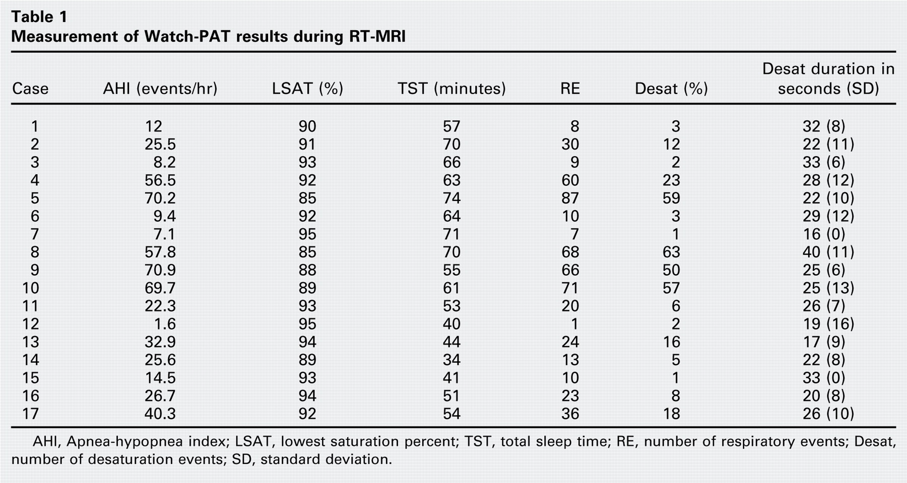
AHI, Apnea-hypopnea index; LSAT, lowest saturation percent; TST, total sleep time; RE, number of respiratory events; Desat, number of desaturation events; SD, standard deviation.
WP AHI correlated with REs (Pearson correlation coefficient = 0.98). WP LSAT correlated with PSG LSAT (Pearson correlation coefficient = 0.73). A lack of correlation was found between PSG AHI and WP AHI (Pearson correlation coefficient = 0.14). Airway obstruction on RT-MRI was defined as an observed isolated palate or base of tongue obstruction or combined complete obstruction (as opposed to a long duration narrowing at these levels), or as an isolated epiglottis obstruction. PAT signal attenuation, PAT amplitude reduction, pulse rate acceleration, and desaturation events were coincident with airway obstructions during nonsedated sleep (Fig 1). Every RE event was associated with a narrowing of the upper airway visualized on Sleep MRI.
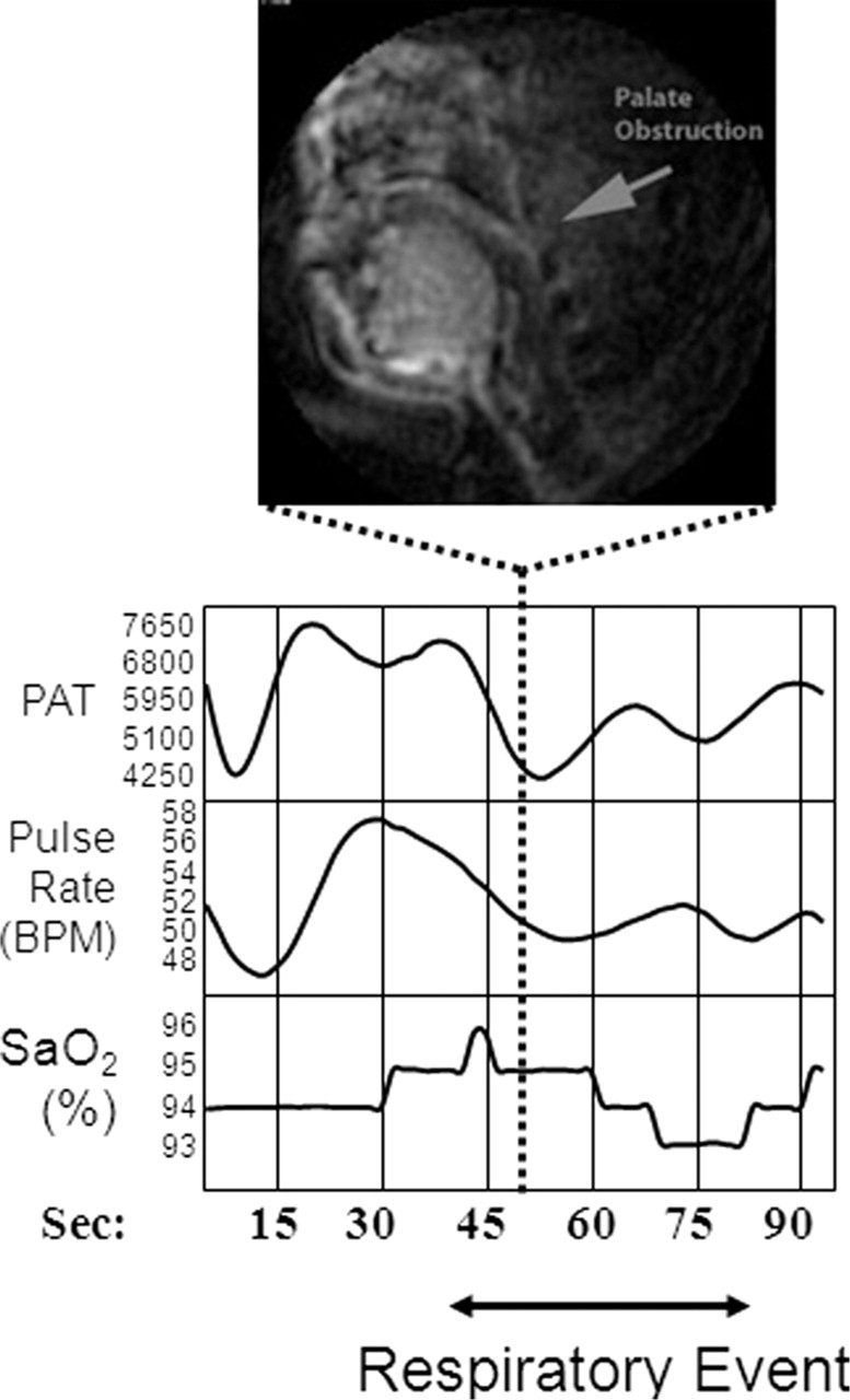
RT-MRI Palate obstruction coincides with respiratory event. PAT signal attenuation and amplitude reduction, pulse rate acceleration, and a desaturation event are seen.
Discussion
Our study shows a novel technology, “Sleep MRI,” that can characterize airway obstruction in association with REs in OSAS patients. Although the midsaggital plane was used for evaluation, axial and coronal views are feasible. Sleep MRI offers the ability to see the site of obstruction, whether palate, tongue, epiglottis, or combined. It also allows one to evaluate the cause of a respiratory event due to simultaneous recording of the RT-MRI. This approach may lay the foundation to improve medical and surgical management of OSAS patients.
The Watch-PAT AHI was correlated with respiratory events. Therefore, respiratory events may be used as an indicator for obstructive events. Further agreement was found between the WP LSAT and PSG LSAT. A lack of correlation was found between the WP AHI and PSG AHI, probably because this was a nap study with a TST of 57 minutes. The authors are investigating the use of overnight Sleep MRI to improve the accuracy of diagnosing site of obstruction and documenting physiologic events during both non rapid eye movement (NREM) and rapid eye movement (REM) sleep. In addition, we are evaluating procedures for automatic characterization of imaging with physiologic measures and analyzing the temporal relationships associated with these events.
Conclusion
Our study shows a novel technique to simultaneously evaluate airway obstructions and respiratory and desaturation events in real time during sleep without sedation. By simultaneously observing site of obstruction dynamically, as detailed by RT-MRI, and quantified respiratory events, this approach can characterize the actual site of dynamic obstruction.
Author Contributions
Financial Disclosure
None.