
Introduction
Recent studies suggest that cerebrovascular dysfunctions occur early in Alzheimer's disease (AD), as reflected primarily by regional decreases in cerebral perfusion that, often, precede the neurological deficits (Iadecola 2004, Nat Rev Neurosci 5: 347). Oxidative stress induced by amyloid beta (Aβ) and cerebrovascular fibrosis associated with increased levels of the transforming growth factor-beta 1 (TGF-β1) have been proposed to underlie these dysfunctions. In the present study, we attempted to reverse cerebrovascular alterations in vivo in order to better understand their underlying mechanisms.
Methods
Transgenic mice overexpressing mutated forms of the amyloid precursor protein (APP mice) or TGF-β1 (TGF mice) were treated when cerebrovascular dysfunctions were fully developed, together with their respective wild-type littermates. Mice (n=3-6/group) received the antioxidant Tempol (1 mM in drinking water, 6 weeks) or N-acetyl-L-cysteine (NAC; 100 mg/kg/day i.p., 4 weeks), or pioglitazone (Actos, 20 mg/kg/day in chow, 6 weeks) which exhibits anti-inflammatory, -oxidant and -angiogenic properties. The ability of isolated and pressurized middle cerebral artery (MCA) segments to dilate to calcitonin gene-related peptide (CGRP) and/or constrict in response to nitric oxide synthase (NOS) inhibition (10 μM L-NNA) was determined by in vitro videomicroscopy. Changes in cortical and hippocampal microvessels were evaluated by immunocytochemical staining of the oxidative stress marker manganese superoxide dismutase (MnSOD) or structural components of the blood vessel wall.
Results
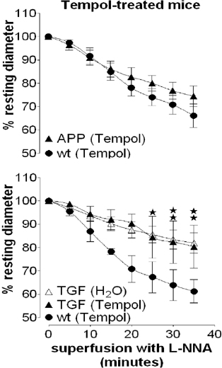
APP and TGF mice exhibited impaired dilatations to CGRP (↓56% and 69%, respectively, p<0. 05) and constrictions to NOS inhibition (↓54% and 53%, respectively, p<0.05). Tempol and NAC fully restored cerebrovascular responsiveness in APP mice. In contrast, Tempol had no beneficial effects in TGF mice and NAC only normalized the NO-mediated response, but not the CGRP-mediated dilatation, indicating that increased TGF-β1 levels differentially affect these two pathways. Interestingly, pioglitazone fully restored cerebrovascular functions in both mouse models. Preliminary immunocytochemical data suggest that Tempol and NAC normalize the perivascular MnSOD upregulation selectively observed in APP mice.
Conclusion
These results show that the apparently similar cerebrovascular dysfunctions in APP and TGF mice are mediated by different pathogenic mechanisms. They also show that perivascular oxidative stress-mediated deficits in APP mice can be fully reversed even at an advanced stage of the pathology. In contrast, other pathogenic factors are involved in TGF mice as antioxidants could not fully restore cerebrovascular functions. The efficacy of pioglitazone warrants additional work so that we may elucidate the molecular and cellular mechanisms leading to recovery from Aβ and TGF-β1 pathologies, both present in AD. This may offer a means to re-establish brain hemodynamics in AD patients with cerebrovascular deficits (See Figure 1).
Footnotes
Acknowledgements
Supported by CIHR (MOP-64194 (EH), studentship (NN)) and Alzheimer Society of Canada.