Background and objective
Diffusion weighted imaging (DWI) has become a prominent method in the early detection of ischemic stroke, yet typical images are plagued by artifact, low spatial resolution, and low signal to noise ratio (SNR). We hypothesized that improving image quality by reducing voxel size and increasing SNR using a diffusion tensor (DTI) sequence would improve lesion conspicuity.
Methods
42 patients imaged between September 2003 and March 2004, diagnosed with ischemic acute stroke and imaged both with conventional DWI (3 direction, 7 mm thick, 28 sec) and DTI (19 direction, 3.5 mm thick, 256 sec) within 24 h of stroke onset were included in this study. Hyper-intense regions were segmented independently on co-registered trace-weighted images from both techniques by two readers blinded to patient identifiers then averaged between both readers. Discrete lesions were defined by a 3D morphometric analysis as contiguous hyper-intense voxels surrounded by non ischemic tissue. On a per patient basis, the volume and number of lesions were compared between both techniques using a Student T test and a Wilcoxon signed ranked test respectively.
Results
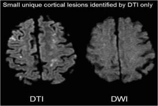
The total lesion volume (sum across all lesions) per patient did not differ between techniques (7042 and 7052 mm3 in DWI and DTI, respectively). Readers identified 253 lesions on DTI as compared to 129 lesions on DWI, for an average of 6.04 and 3.07 discrete lesions per patient respectively (p<0.001) and an average volume per lesion of 2866 mm3 and 3643 mm3 for DTI and DWI respectively (p<0.001). A total of 134 discrete lesions (53%) identified on DTI, having an average volume of 77 mm3 per lesion, were not identified on DWI, while 7 lesions (0.05%) identified on DWI, having an average volume of 147 mm3 per lesion, were not identified on DTI. Lesions found only on DTI tended to be located in cortical gray matter. Lesions identified only on DWI tended to be near regions of artifact. Moreover, single DWI lesions tended to appear as a geometrically complex, but contiguous lesion on DTI.
Conclusions
Higher resolution DTI showed a higher number of discrete small cortical gray matter lesions than DWI. The clinical implications of those lesions is currently under investigation (See Figure 1).