Introduction
There is a desire to develop PET probes for in vivo imaging of second-messenger systems. Cyclic-AMP (cAMP) is a continuously produced nucleotide which is catabolised by phosphodiesterase-4 (PDE4) enzymes. PDE4 expression and conformation is altered by cAMP concentrations, and therefore PDE4 measures are an indirect measure of cAMP function. Using the selective PDE4 inhibitor, [11C]rolipram 1 , it is possible to image the distribution of PDE4 enzymes with PET 2 . Rolipram exists as two enantiomers, with S(+)-rolipram having approximately 10-fold lower affinity for PDE4 than R(−)-rolipram in vitro and in vivo 3 . cAMP levels may be altered acutely or chronically. The aim of this study was to investigate whether acute agonism of cAMP-coupled receptors induces regional changes in the [11C]rolipram PET signal consistent with the known receptor pharmacology.
Methods
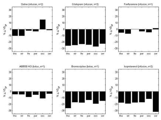
In all studies, Yorkshire/Landrace porcines (40Kg, n=10) were anaesthetised by induction with ketamine and midazolam, and maintained with isofluorane. Baseline [11C]R(−)- and [11C]S(+)-rolipram scans were performed followed by a pharmacological challenge and further [11C]R(−)- and [11C]S(+)-rolipram scans in each subject. The pharmacological agents were administered at least 30 minutes prior to the subsequent [11C]rolipram scans; citalopram (SSRI, 20 mg/hr), fenfluramine (serotonin-transporter blocker, 50 mg/hr), A68930 HCl (D1-dopamine receptor agonist, 0.075 mg/Kg), bromocriptine (D2-dopamine receptor agonist, 1.25 mg/Kg) and isoproterenol (β2-adrenoceptor agonist, 7.5 μg/Kg) and saline as a control (n=2). Dynamic data were collected over the brain for 90 minutes (EXACT HR scanner-3D) and frequent arterial samples were taken to assay concentrations of parent radioactivity. Six regions of interest (ROI; thalamus(tha), striatum(str), frontal cortex(fro), parietal cortex(par), occipital cortex(occ) and cerebellum(cer)) were defined on each baseline [11C]R(−)-rolipram scan and applied to all scans in that subject to generate time activity curves (TACs). Logan analyses (plasma input) were applied to all TACs to estimate total tissue volumes of distribution (V D ) and two outcome measures were calculated; (i) V D R− and (ii) V D Sp =V D R− −V D S+ .
Results and Discussion
The changes between baseline and challenge conditions were similar for both outcome measures (V D R− and V D Sp ). The percentage changes for V D Sp post challenge are given (figure 1). For challenges with multiple studies the mean is presented for each region. A68930 and (+)fenfluramine did not produce changes in the rolipram signal above that observed with saline. Challenges with citalopram, bromocriptine and isoproterenol exhibited, at best, small global changes in rolipram binding with no apparent regional differentiation. cAMP turnover is generally rapid in response to an acute challenge, leading to a transient signal that may not be detectable in the time frame of a PET study. To conclude, these data suggest acute agonism of cAMP-coupled receptors may be difficult to image using PET. Further studies are planned to assess the effect of chronic modulation of cAMP on PDE4 expression, as measured by PET.