
Introduction
The combination of transgenes encoding for prodrug-activating enzymes is a promising approach in current gene therapy protocols. The enzymes E. coli cytosine deaminase and herpes simplex virus type 1 thymidine kinase have been shown to act synergistically. Using positron emission tomography (PET) we determined the in vivo transduction efficiency mediated by HSV-1 amplicons using the HSV-1-tk as PET marker gene to monitor the effects of prodrug therapy.
Methods
Human Gli36dEGFR glioma cells were grown as s. c. tumors in 22 nude mice and transduced in vivo by HSV-1 amplicons carrying the genes for cytosine deaminase (cd), viral thymidine kinase (tk39) and gfp (HSV-cdIREStk39gfp). Non-transduced Gli36dEGFR tumors served as negative controls, retrovirally transduced Gli36dEGFR cells stably expressing cdIREStkgfp as positive control. Following transduction, therapeutic i.p. prodrug application was performed with 5-fluorocytosine and ganciclovir. Tumor sizes were measured using calipers and growth slopes were calculated. PET-imaging was performed in 11 mice. [18F]FHBG-PET was performed after in vivo transduction prior to therapy to evaluate transduction efficiency [%ID/g]. [18F]FLT-PET was performed as baseline evaluation prior to in vivo transduction and as therapy monitoring after 1 week of therapy. Background-corrected accumulation of [18F]FLT [%ID/g] was determined. Therapeutic efficiency was quantified by the difference of [18F]FLT-accumulation before and after therapy (ΔFLT).
Results
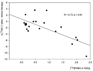
Positive control tumors were successfully treated with prodrugs leading to disappearance of tumors within 10 days. 15/22 in vivo transduced tumors responded to prodrug therapy; 4 tumors disappeared completely (complete responders) and 11 tumors showed decelerated growth compared to respective negative controls (partial responders). Growth slopes of tumors responding to gene therapy were significantly less steep compared to negative controls (p<0.05). Transduction efficiency as measured by [18F]FHBG-accumulation was 1.22 ± 0.83%ID/g for stably transfected tumors and 0.37 ± 0.30%ID/g for in vivo transduced tumors. In stably transduced tumors, therapeutic effects could be monitored by PET with significant differences in [18F]FLT-accumulation before (3.38 ±3.65%ID/g) and after therapy (0.06 ±0.19%ID/g; p=0.01). 8/11 in vivo transduced tumors showed a significantly lower [18F]FLT-accumulation after therapy (1.91 ±1.12 vs. 0.42 ±1.31%ID/g; p<0.01), while in 3 tumors [18F]FLT-accumulation was increased. For stably and in vivo transduced tumors, the level of exogenous gene expression or transduction efficiency as measured by [18F]FHBG-PET correlated well to the resulting therapeutic efficiency as measured by [18F]FLT-PET (r=0.73; p<0.01, Fig. 1.). Volumetric data did not correspond to [18F]FLT-PET data in assessment of therapeutic response. Consecutively, transduction efficiency in [18F]FHBG-PET did not correlate to therapeutic efficiency as measured by volumetry.
Conclusion
Therapeutic efficiency can be non-invasively followed and quantified by microPET using [18F]FLT. Transduction with HSV-1 amplicon vectors in vivo causes distinct levels of gene induction, which correlate well to the effect of gene therapy as measured by [18F]FLT-PET imaging. Volumetry gives complementary information in assessing response to gene therapy.
Footnotes
Acknowledgements
Grant support: Supported in part by MSWF 516–400 002 99, ZMMK-TV46, DFG-Ja 981/1-2, EU-6thFW-EMIL