
Abstract
We report our experience with the repair of the orbital floor fractures and present new technical findings. We evaluated 30 subjects with pure blowout fractures treated at the Department of Maxillofacial Surgery of the Federico II University of Naples, Italy, between 2005 and 2007. A preoperative examination by computed tomography scans provided classification of the orbital floor fractures into small and large fractures by measurement of the bone defect to choose the appropriate reconstructive implant materials, resorbable or nonresorbable. The clinical follow-up has been performed at 1 week, 1 month, 3 months, and 6 months. We observed a resolution of preoperative symptoms. The scar was not evident, and there was an absence of postoperative complications. We concluded that the use of resorbable materials for small orbital floor fractures and nonresorbable materials for large orbital floor fractures offers satisfactory results in both functional and aesthetic considerations. Furthermore, the new technical findings allow standardization of the surgical technique to be more accurate, also reducing the economic costs.
Orbital fractures account for 40% of craniofacial injuries; of the four walls of the orbit, the floor, which is extremely thin, is the most frequently injured. According to the pertinent literature, such fractures represent 67 to 84% of cases of orbital fractures.12
Orbital floor fractures can be broadly classified as pure or impure blowout fractures; the first are isolated orbital floor fractures, and the second are also associated with an orbital rim fracture, involving other skeletal elements: zygomatic, frontal, nasoethmoidal, or maxillary bones.3 A blowout fracture mechanism is not very clear; experimental and clinical studies have generally proposed two main theories: the hydraulic and the buckling mechanisms.
According to the buckling theory, the fracture is produced as a result of transmission of the trauma forces directly to the orbital floor through the orbital rim; the hydraulic mechanism considers that the effects of the kinetic energy of the blow are transferred via the incompressible orbital soft tissue to the floor of the orbit.45
The most common causes of injury are motor vehicle accident, physical assault, and sport-related injuries. More rarely, it can be the result of a fall, gunshot, or industrial accident.3
Management of orbital floor fractures is still debated and controversial. In the literature, there are many conflicting reports about classifications, type of implant materials, and ideal time to perform surgery.
The aim of this study was to report our experience about the repair of the orbital floor fractures and new technical findings.
The patient series was divided into: (1) pure blowout fractures with large defect and (2) pure blowout fractures with small defect.
Materials and Methods
In the Department of Maxillofacial Surgery of the Federico II University of Naples, Italy, between 2005 and 2007, we observed 44 unilateral fractures of the orbital floor, pure and impure.3 Of these patients, only 30 with pure blowout fractures were included in our study. They fulfilled the following inclusion criteria: (1) clinical diagnosis of an orbital floor defect; (2) imaging showing an orbital floor defect. The other 14 patients were excluded because they were affected by impure orbital floor fractures (exclusion criteria) with associated facial fractures. Of these, the zygomatic-malar fracture was the most common. The patients were 22 males (73.33%) and 8 females (26.67%) with a mean age of 48.9 years (range 16 to 86 years).
The most common causes of fracture were sports, vehicle accidents, assaults, fireworks and falls. The clinical signs and symptoms were periorbital ecchymosis (75% of cases), subconjunctival hemorrhage (92%), periorbital swelling and/or edema (33%), diplopia (60%), altered ocular motility (25%), infraorbital nerve anesthesia (75%), and dystopia (83%) (enophthalmos in 76% and hypoglobus in 24%; Tables 1 and 2). Ocular injury was observed in two patients (6%): one had corneal abrasion and the other had subluxation of the crystalline. Only one patient had neurological symptoms represented by epileptic seizure due to brain edema. Three patients had associated extremity fractures, the arm in two cases and shoulder in the third.
Preoperative Ophthalmic Signs and Symptoms
Inclusion criteria.
Preoperative Ophthalmic Signs and Symptoms

All are inclusion criteria.
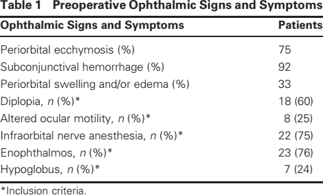
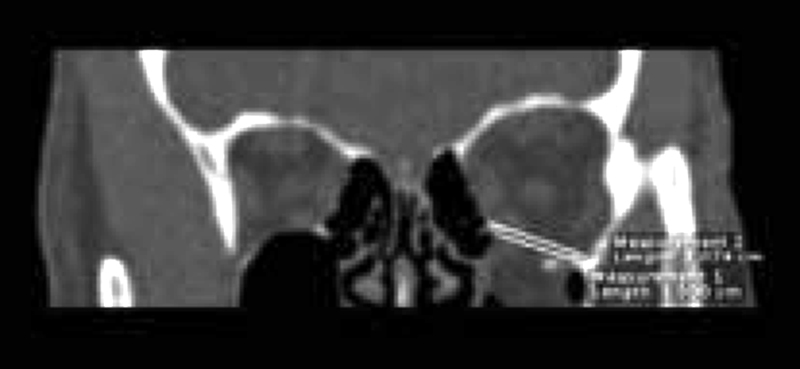
All patients were examined by high resolution multislice computed tomography (CT), evaluated with coronal and sagittal images (Figs. 1, 2) of the floor displacement preoperatively. CT scans were performed with 1-mm thickness contiguous slice section under bone window settings. An open source image-processing software (OsiriX, CA) was used in each patient to reconstruct and manipulate CT scan data.
Computed tomography scan, coronal view with superior and inferior measurements (C) of the bone defect.
Computed tomography scan, sagittal view with superior and inferior measurements (S) of the bone defect.
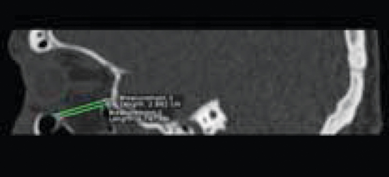
The CT soft tissue window allowed us to identify the protrusion of the orbital fat and the entrapment of the extraocular muscles with special regard to the inferior rectus and presence of foreign bodies. We classified orbital floor fractures in two types: small and large. Our classification is based upon the measurements of the bony defect area performed on the coronal and sagittal scans. The method applied consists of depicting two lines corresponding to the fractured floor, calculating the mean value obtained from the higher and the lower line on each view (Figs. 1, 2). The elliptical area was calculated by multiplying the width (C on coronal view) and the length (S on sagittal view), then multiplying the result by π (Fig. 3).
Schematic drawing of the orbital floor defect measurements that we evaluate using the radiological data based on coronal (C) and sagittal (S) views, used to obtain the area of the fracture, according to the mathematical formula (width×lengthπ×).
We have classified as “small” the fractures with an area of bony defect less than 3 cm2, and “large” with an area more than 3 cm2. Among the patients, 23 had large fractures and seven had small, according to our classification. The average time interval between accident and surgery was 7±4 days.
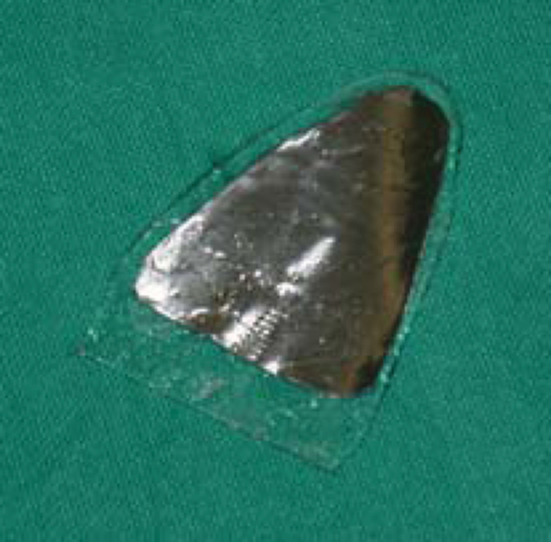
In all the 30 patients, the orbital floor was explored via subciliary approach. The skin incision was placed 2 or 3 mm below lash line, from the medial cantus laterally up to the lateral cantus to reach the orbital floor, avoiding the orbicular muscle and preserving the orbital contain. The lower maxilla and orbital floor were always dissected free, preserving the periosteum.
The reconstruction of the floor in the cases with small fractures was performed by using resorbable implants (poly-l-lactide, poly-d, l-lactide, trimethylene carbonate, and polyglycolide). In the large fractures, we used nonresorbable material (high-density porous polyethylene). The resorbable implants we used were 30×40 mm in size, and the nonresorbable were 38×50 mm in size (Table 3).
Summary of Cases Treated from 2005 to 2007
Intraoperatively, all patients received a prophylactic dose of intravenous antibiotics (ceftriaxone 2 g) and, postoperatively, for 3 days intramuscular therapy (ceftriaxone 1 g).
The clinical follow-up was performed at 1 week, 1 month, 3 months, and 6 months after treatment. At the 6-month follow-up, the patient had a CT scan according to our method described above.
Among the 18 patients who had diplopia before surgery, resolution of symptoms postoperatively was observed in 15 (83.3%) and in the other 3 (16.7%) patients, diplopia disappeared within 3 months. The altered ocular motility was resolved in all eight patients who had this before surgery.
In 18 (81.8%) of the 22 patients, the infraorbital nerve anesthesia was resolved after surgery, three patients (16.6%) had residual hypesthesia, and 1 (1.6%) had residual paresthesia. Enophtalmos resolved postoperatively in 20 (89%) of the 23 patients and hypoglobus in 5 (66%) of the seven patients who had these symptoms before surgery (Table 4).
Postoperative Ophthalmic Signs and Symptoms

Postoperative complications were observed in three patients (10%). Two of them had dehiscence of wounds and one had facial infection; all resolved with local and systemic antibiotics and corticosteroid therapy. No technical complications such as improper position of implants, hematomas, or infraorbital or optic nerve injuries were observed. In all 30 patients, the scar was not evident.
Discussion
The orbit protects the visual apparatus through the bone structures, and it acts as a receptacle. Nevertheless, the slightest trauma can provide serious damage. This is why the integrity of the eye and associated tissues must be evaluated accurately and quickly to avoid irreversible damage. The repair of the orbital floor fractures is not without risks, which must be taken into consideration when surgery is decided as a treatment of choice.
A significant facial asymmetry, imaging evidence of the fracture, the age of the patient, and clinical signs and symptoms are extremely important in determining the surgical indications. In most patients, time allows for disappearance of initial edema and hemorrhage that in some cases are the causes of diplopia and enophthalmos. Fractures of the orbit that do not have functional or aesthetic injuries do not need surgical treatment.
In our opinion, the indications for surgery are increased orbital pressure, persisting diplopia, enophthalmos, visual impairment, and hypoanesthesia of the infraorbital nerve. We perform early surgery if there is CT evidence of entrapped muscle or periorbital tissues associated with oculocardiac reflex and also in the presence of symptoms of defects in the orbital structures. In other circumstances, we prefer observing patients for ∼7 days because some of deficits can resolve spontaneously.67 In all patients, we use the subciliary surgical approach. We prefer this approach when there is no need to expose the medial or the lateral orbital wall. Furthermore, the scar is not evident.89
The choice of resorbable material is based on its biological features: it is degraded into α-hydroxy acid (natural and no toxic components) and it guarantees a solid buttress for more than 8 weeks. Furthermore, it is easy to shape because of its plasticity, and its three-dimensional structure offers high stability. Nonresorbable materials, which we use in the large fractures, are implants with an open-pore structure. This structure allows rapid vascular soft tissue and bone ingrowth that serves to stabilize the implant in relation to the orbital content.10
Management of orbital fractures is controversial because of the difficulty in evaluating the anatomy of the defect area, and amount of soft tissue herniation. All patients were imaged preoperatively by high-resolution multislice CT, with coronal and sagittal scans (Fig. 1, 2) showing the displacement of the floor.
Using a mathematical formula to calculate the oval area based on sagittal and coronal data obtained by CT scans, we estimate the area of the fracture to be repaired. This method has enabled us to standardize the surgical technique to be more accurate, also reducing the economic costs.
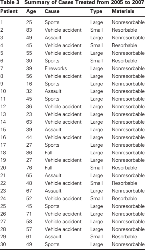
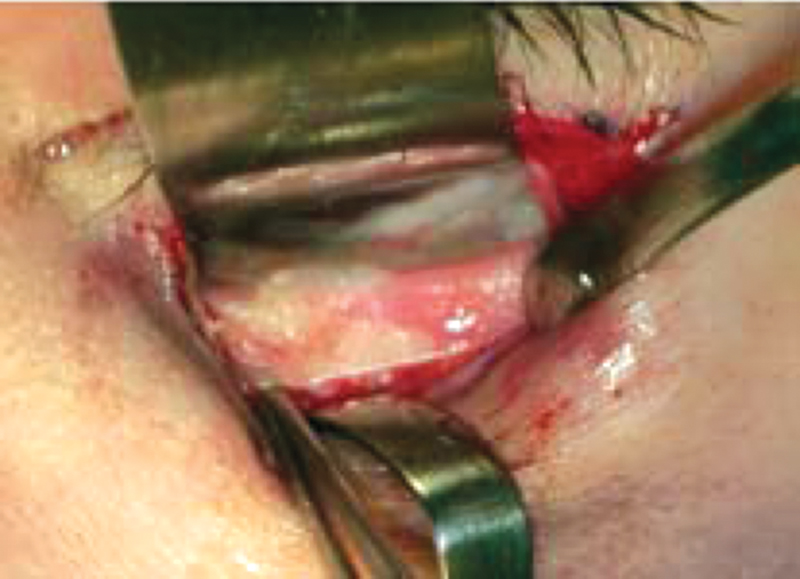
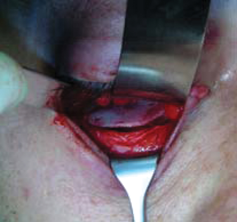
The choice of the material to repair the orbital floor is due to the type of fracture; in fact, in small ones, we use a resorbable sheet. This form of controlled regeneration allows complete healing of the orbital floor and the defect; the involved bone is regenerated with the bioresorbable plate acting as a template during the healing process.1112 The resorbable implant simply acts as a scaffold by establishing a supportive structure on which the periorbita will heal. The implant is gradually resorbed and replaced completely by fibrous collagenous tissue13 (Figs. 4, 5). In large fractures, we need to use nonresorbable sheets because of the large loss of substance and the herniation of the content of orbit into the maxillary sinus.14 The porous structure of nonresorbable material allows the soft tissue to incorporate the sheet, so we prevent the formation of a fibrous capsule and a foreign body reaction, with migration or extrusion, and resistance to infection (Figs. 6, 7).
The resorbable implant adapted to the small bony defect using a template based on computed tomography data and intraoperative view.
The resorbable implant placed to repair the fractured floor.
The nonresorbable implant adapted to the large bony defect using a template based on computed tomography data and intraoperative view.
The nonresorbable implant placed to repair the large fractured floor.
Long-term sequelae may be associated with incomplete healing of the fractures and of deformities associated with malaligned fractures, lid tears, persistent pain.
Conclusion
We concluded that the use of resorbable materials for small orbital floor fractures and nonresorbable materials for large orbital floor fractures, in accordance with our classification, provides reliable and reproducible stabilization of orbital wall defects, offering satisfactory results, both functional and aesthetic. Furthermore, this study suggests that our new technical findings allow us to standardize the surgical technique to be more accurate and also reduce the economic costs.