
Abstract
Dermoid cysts are unusual neoplasms and can occur in every part of the human body. They represent only 6.9% of all dermoid cysts in the head and neck region; in the oral cavity, the incidence is low, approximately 1.6% of all dermoid cysts. Our aim is to present an unusual case of a large sublingual dermoid cyst with mandibular prognathism caused by cyst growth. We reported a case of a large sublingual dermoid cyst in an 8-year-old female patient. A bibliographic research from 1937 to 2013 is reviewed and we found only three cases of mandibular deformity, of which only one was a dermoid cyst of the floor of the mouth. Removal of dermoid cysts of the floor of the mouth should be completed as early as detected, especially in newborns and infants when osseous growth abnormalities could result if removal is delayed.
Dermoid cysts are unusual neoplasms that often present in childhood and can occur in every part of the human body,12 mostly found in the periorbital lateral eyebrow area.345 In the oral cavity, the incidence is low, approximately 1.6% of all dermoid cysts,1678 and usually found in the anterior portion of the floor of the mouth.
Typically, dermoid cysts present either as slow-growing, painless, nontender midline floor of the mouth or as submental swellings that can develop to significant dimensions before producing symptoms.3 Once the diagnosis is established, surgical removal of the lesion is the treatment of choice.14891011
Case Report
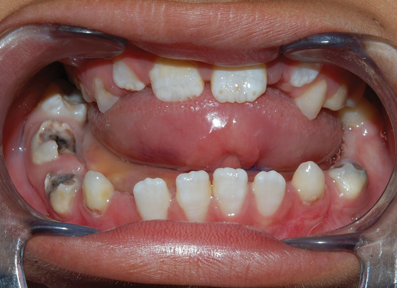
An 8-year-old female patient was referred to our department with mandibular prognathism (Fig. 1), lip incompetence, and salivary incontinence. Intraoral examination revealed a large midline sublingual swelling elevating the tongue into the palate, diastemas, and dental caries (Figs. 2 and 3). Her parents reported that this swelling appeared since the age of 3 years and this was associated with difficulties in mastication as well as speech and oral health care difficulties caused by the size of the cyst. CT scans (axial, coronal, and sagittal sections) revealed a large radiolucent space occupying lesion in the sublingual space.
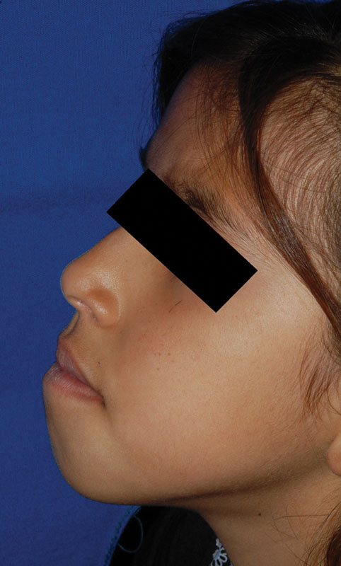
Lateral view. Mandibular prognathism.
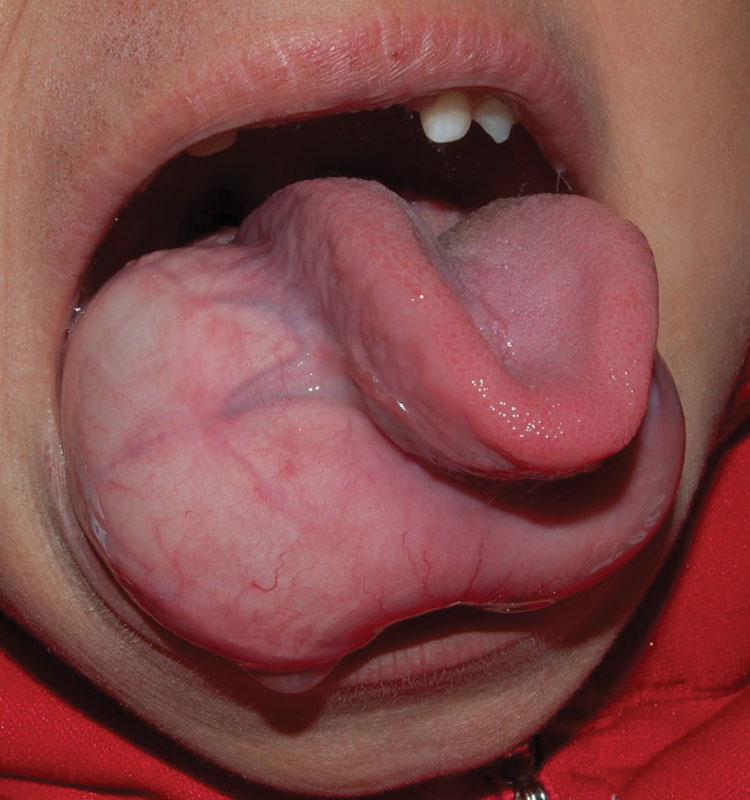
Large sublingual swelling raised the tongue upward.
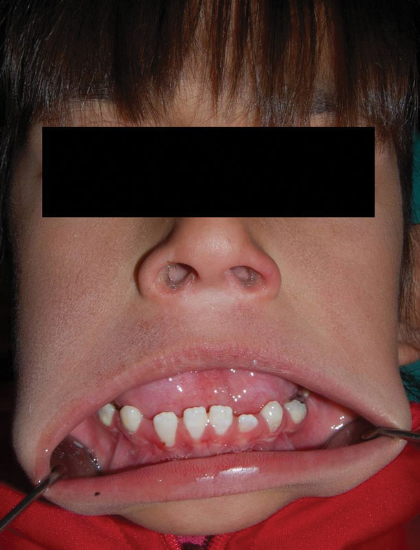
Diastemas and dental caries.
The surgery was performed under general anesthesia with nasal intubation, and sublingual transverse incision was made, taking care to avoid any damage to the Wharton duct on both sides. Careful submucosal dissection revealed the large cyst, which was then enucleated (Fig. 4).
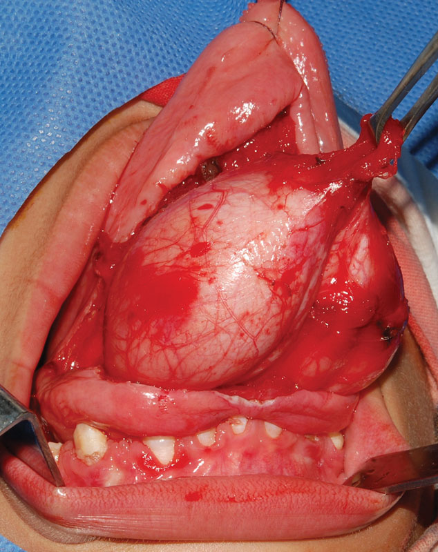
Intraoperative view of the cyst being delivered from the floor of the mouth.
The surgical access was closed with long-term 3–0 absorbable sutures and these sutures were removed 1 week later. The postoperative course was uneventful, and the patient was discharged home 72 hours after surgery. After enucleation, the mandibular expansion caused by the growing cyst could be evaluated (Fig. 5). The patient was followed up for 12 months and no signs of recurrence could be found.
7 days after surgery. Note the resulting mandibular prognathism and dental caries caused by the impossibility of dental care before cyst enucleation.
The specimen, measuring 6 × 5 cm, was sent for histopathological examination and the diagnosis of dermoid cyst was made (Figs. 6 and 7).
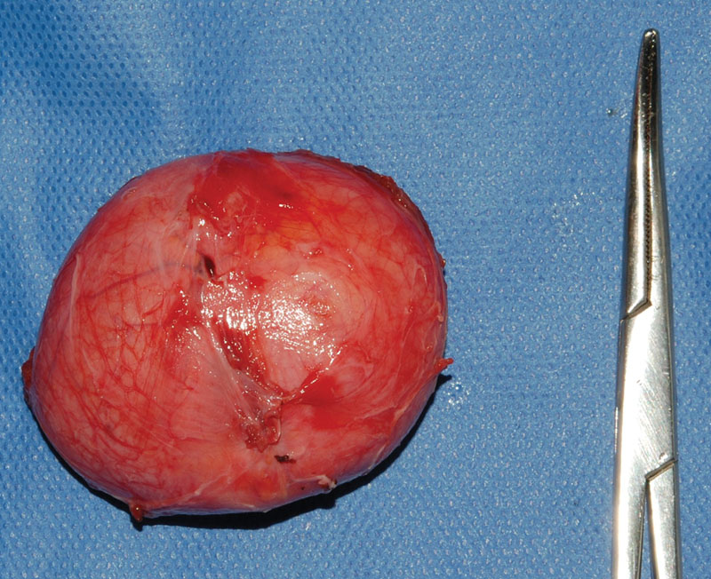
Surgical specimen after total enucleation.
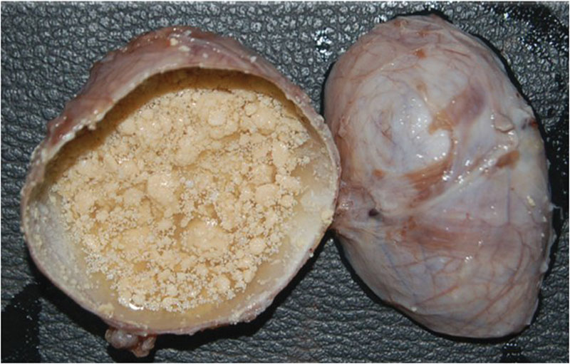
Cross section of the cyst revealing its contents.
Discussion
Dermoid cysts are unusual neoplasms that often present in childhood and can occur in every part of the human body.12 Cases have been described in the region of the nose, sinuses, orbits, scalp, rectum, ovaries, abdomen, testicles, salivary glands, spinal cord brain, etc.12 In the oral cavity, they represent only 1.6% of all dermoid cysts.1678
From 1910 to 1935, New and Erich6 examined 1,495 patients with dermoid cysts, and in the oral cavity they accounted for approximately 25% of the cases occurring in the head and neck region.
In the 195 cases reviewed by King et al,8 using 5-year age intervals, they found the highest incidence in the newborns to 5-year-old group (27.6%). Identifying the highest incidence of cysts in the newborn–infant age group challenges the commonly presented statement that these cysts rarely appear at birth. The average age of patient at the time of first identification of the cyst was 15.8 years.8
As the sublingual cyst grows, a swelling of the floor of the mouth appears, and the tongue is normally pushed up and backward causing difficulty in mastication and speech.158913 Surgical enucleation is the only effective treatment for dermoid cysts.123458
According to the anatomic relationship between the cyst and the muscles of the floor of the mouth, dermoid cysts can be designated: submental (between geniohyoid and mylohyoid muscles), sublingual (above mylohyoid and genioglossal muscles), and submandibular (lateral to the musculature).41314 Although some authors have suggested that the cyst always appears superior to the mylohyoid muscle, the geniohyoid muscle determines the space in which the swelling appears.81516
The location of the cyst appears to be a factor in determining the surgical approach. Most authors recommend the intraoral approach for the sublingual cyst, whereas the extraoral approach is usually chosen for submandibular and submental space involvement.89101317 In the case of an intraoral approach, a midline vertical mucosal incision is performed along the ventral surface of the tongue; however, only small cysts can be enucleated using this kind of incision.17 Other incisions were proposed in the ventral surface of the tongue like an inverted “T”-shaped incision.5 The cystic fluid can be aspirated to reduce the mass of the lesion. Although it cannot be the definitive treatment,18 it is useful to facilitate control of the deeper pole of the cyst.813
Brusati et al19 proposed a midline glossotomy and Di Francesco et al13 described a modification of this surgical technique consisting of an extension along the ventral surface of the tongue.
The extraoral approach is usually chosen for submental and submandibular dermoid cysts.814 Large cysts that perforate the mylohyoid may require both intraoral and extraoral incisions to provide direct visualization of important adjacent structures.18102021
Recurrences are unusual after absolute surgical excision.1258 The malignant transformation, when longstanding, has been reported but is exceedingly rare.322 The most common is squamous cell carcinoma,323 and has been documented only in the teratoid variant.22
Conclusion
Mandibular growth can be altered at different stages of development as a result of functional manifestations of growth of surrounding tissues.24 Dermoid cysts of the floor of the mouth require immediate surgical intervention, especially in newborns and infants when osseous growth abnormalities could result if removal is delayed,4819 and may interfere with deglutition and speech, or can pose a risk to the airway.216 Although some authors manifest the delayed treatment may cause growth abnormalities, we did an extensive bibliographic research and found only three cases of mandibular deformity—one of them was a congenital teratoid cyst of the floor of the mouth,21 other was a dermoid cyst of the tongue,11 and only one was a dermoid cyst of the floor of the mouth.4 For this reason, the case we reported is extremely rare.