
Abstract

PCNs of the head and neck have been classified by Batsakis as extramedullary plasmacytoma, solitary plasmacytoma of bone, and manifestation of multiple myeloma (MM). They represent distinct manifestations of a disease continuum in which, due to the significant implications for treatment and for survival, the clinical and pathologic findings are critical for diagnosis and for distinguishing one from the other.
MM is the most common PCN, with clonal proliferation of abnormal cells in bone marrow. The clinical manifestations of the disease, such as localized areas of bone pain or swelling, fatigue, or anemia, result from an expanding plasma cell mass in the bone marrow. In the head and neck region, symptoms also rarely include soft tissue masses in the upper respiratory tract or in the oral cavity. The most common sites of these tissue masses are the nasal cavity, nasopharynx, paranasal sinuses, tonsils, gingiva, larynx, thyroid, lymph nodes, and orbital cavity. We describe the course of a patient with a rare case of MM originating in the thyroid cartilage and presenting primarily as a neck mass.
CASE REPORT
A 50-year-old man sought medical attention due to an anterior neck mass that had progressively enlarged during the preceding 6 months and was associated with a sensation of suffocation. On admission, hoarseness and mild stridor were noted. Medical history was unremarkable except for mild anemia due to β-thalassemia trait.
On physical examination, the patient had mild respiratory distress and stridor. A 6- × 6-cm non-tender, firm, and fixed anterior was noted neck mass above the thyroid gland, with no lymphadenopathy. Flexible fiberoptic laryngoscopy revealed supraglottic stenosis with supraglottic and glottic edema. Both vocal cords were swollen with mild limitation of movement.
A computed tomography (CT) scan (Fig 1) revealed an expansile mass originating within the thyroid cartilage, with small punctate calcifications within the 2 laminae. The supraglottic region was narrowed. No lymphadenopathy was seen. Fine-needle aspiration from the mass revealed atypical lymphocytes with plasmacytoid characteristics. Binucleated plasma cells were seen that were suspected to be malignant. The patient underwent an incisional biopsy of the neck mass. Histologic examination of the specimen revealed fibrotic tissue infiltrated by plasma cells, which stained positively by kappa light-chain monoclonal antibodies.
Whole body bone scan and systemic skeletal radiographic survey revealed multiple “punched-out” osteolytic lesions involving the skull and the left femur. Bone marrow biopsy revealed a normocellular bone marrow with normal representation of all three hematopoietic lines. Small groups of plasma cells, 5% to 20% per field, stained positively by immunohistochemistry for κ light-chain immunoglobulin. Urinalysis was negative for albumin and Bence Jones protein. A monoclonal peak was present on serum protein electrophoresis that was identified as immunoglobulin G with κ light chain (IgG, 2260 mg/dl). Serum β2-microglobulin was elevated (2597 mg/dl; normal, <2000 mg/dL). Rhodamine staining for Multidrug Resistance-1 in bone marrow was negative. The patient was diagnosed as having MM.
The patient was treated with 1500-cGy radiotherapy to the neck mass, with improvement in his respiratory complaints. Chemotherapy with vincristine, Adriamycin (doxorubicin), and dexamethasone (VAD) was started, for a total of 3 courses. Because there was no change in the size of the mass, the patient received additional 3000-cGy irradiation. Chemotherapy with high-dose melphalan was also initiated. The patient died several months later due to sepsis and cerebral hemorrhage.
DISCUSSION
PCNs, in the neck region, are rare and may be clinically confused with more common tumors of the neck. They may appear or originate in different structures such as the larynx, trachea, thyroid gland, parotid gland, aerodigestive tract, hyoid bone, or cervical lymph nodes. In MM, disseminated extraosseous disease is common, even though it may be asymptomatic. Neck mass as primary manifestation of MM is uncommon, especially a mass that originates from the thyroid cartilage. There have been only 2 prior reports of MM originating in the thyroid cartilage. Debain et al 2 and Van-Dyke et al 3 described 2 cases of MM involving the thyroid cartilage. Cricoid cartilage involvement in MM is also rarely reported in the literature. When there is involvement of thyroid or cricoid cartilaginous tissues by PCN, the mucosa of the larynx and subglottic regions usually remain intact, without evidence of tumor erosion into the airway. Symptoms such as dyspnea, hoarseness, and stridor may be seen when a neck mass is present.
There are 2 theories for the cartilaginous involvement in MM: (1) invasion of the cartilaginous tissue by adjacent plasmacytoma 4,5 and (2) osseous metaplasia within the cartilaginous tissue with formation of true marrow space. 6 Osseous metaplasia has been described in the cricoid and thyroid cartilages, mainly in adults. In such cases, a plasmacytoma may originate directly within the new formed marrow.
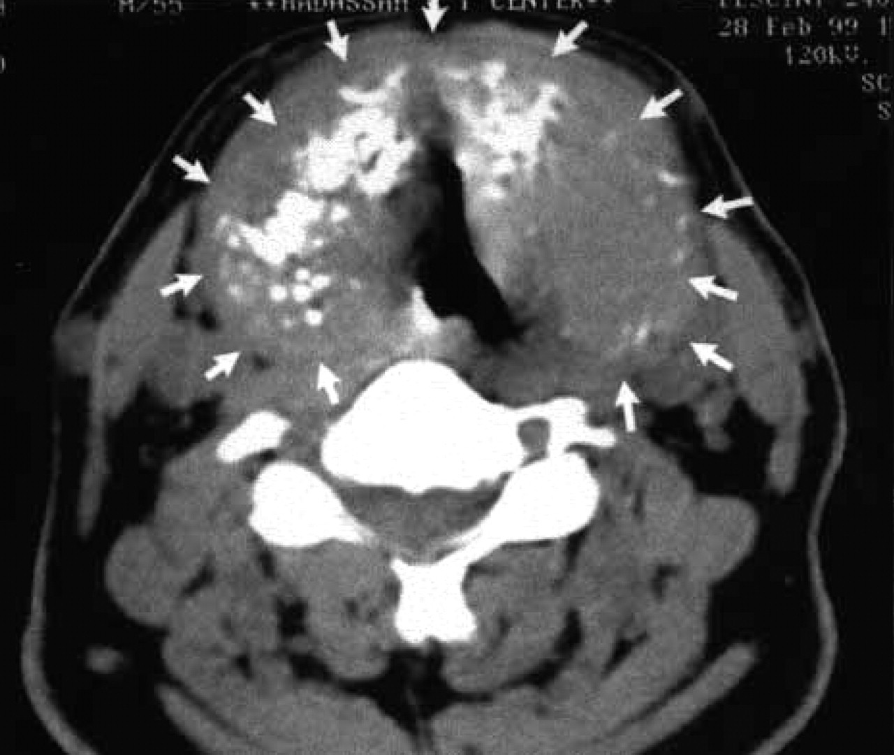
An axial computed tomography scan of the patient with a neck mass, demonstrating an expansile mass with small punctate calcifications within the thyroid cartilage. The airway passage is narrowed.
The term “extraosseous manifestation of multiple myeloma” is accepted when cartilaginous involvement occurs, because ossified cartilage is not developmentally true bone and because it is a very rare presentation compared with the ubiquitous intramedullary lesion seen in MM. The pattern of involvement in our patient, the thinning and expansion of the thyroid cartilage laminae, the “exploded” appearance of the thyroid cartilage, and the lack of a soft tissue mass adjacent to the thyroid cartilage, strongly suggest that the plasmacytoma originated within the thyroid ala rather than via erosion by an adjacent tissue plasmacytoma.
When untreated, patients with MM rarely survive beyond 1 year. The standard treatment of MM is based on cytotoxic drugs. The most effective regimens include VAD and MP (melphalan and prednisolone). Radiation therapy is added to achieve local control of advanced disease, to relieve bony pain, or to reduce mass effects leading to severe complications such as airway obstruction. A recent development in the treatment of MM is high-dose chemotherapy followed by autologous bone marrow transplantation using peripheral blood stem cells. Chemotherapy leads to remission in 75% of patients; median survival is only 3 years.
We presented the case of a patient with a neck mass originating from the thyroid cartilage that was found to be MM. When a PCN is detected in a neck mass, complete evaluation is necessary for to distinguish localized plasmacytoma from MM due to the differences in prognosis and treatment. The otolaryngologist may play a definitive role in diagnosis and care of these patients, some of whom present with upper airway narrowing or with other head and neck symptoms.