






Abstract
In ultra-supercritical power plants, Ni-base alloys are candidate materials for long-term, high-temperature applications, operating at temperatures and pressures as high as 750°C and 35 MPa. Alloy IN740 and its modification, alloy IN740H, are considered for such applications. Their microstructural evolution, at 750°C for times ranging between 3000 and 5000 hours, has been investigated by means of scanning electron microscopy, electron back-scattered diffraction, energy dispersive X-ray analysis and phase quantification. All phases were identified and quantified allowing comparison between the two microstructures, their evolution and stability. Particular attention was paid to γ′, η and G phases. The results are used within a broader investigation aimed at improving and further developing a predictive creep model based on continuous damage mechanics.
Keywords
Introduction
In the field of coal power generation, the drive for environmental protection has led to the engineering of more efficient power stations in conjunction with carbon capture technology. New advanced ultra-supercritical (USC) power plants are, therefore, being developed with the aim of operating with a steam temperature as high as 750°C and pressures of 35 MPa.1,2 Ni-base alloys are among the candidate materials for applications such as boilers, able to withstand such service conditions. Such high-temperature applications led the research focus on high creep resistance. Forecasted requirements in the power generation sector have set the targets to minimum creep strength of 100 MPa at 100 000 hours of service.1–7 Two possible candidate materials for USC boiler applications are the high chromium Ni-base precipitation-hardenable superalloy IN740 and its modification, IN740H, both developed by Special Metals.5,7,8–15 The alloy IN740H has been produced with the desire of improving the IN740 high-temperature properties (e.g. creep resistance). Compared with IN740, the Ti/Al ratio in IN740H is lowered in order to stabilise the microstructure at long aging times. In addition, the Nb content is lowered to improve the weldability. The present investigation concentrates on characterising and comparing the high-temperature microstructural evolution of the two alloys. Alloy IN740 has been aged at 750°C for 3000 hours. Alloy IN740H has been aged at 750°C for 5000 hours. Although the annealing conditions and aging time for the aged IN740H specimens are different than that of its parent alloy, in view of the unchanged precipitation sequence within the annealing and aging conditions, 16 the comparison of the two microstructures does nonetheless provide a clear microstructural indication of several improvements. The analysis provides characterisation of the microstructural constituents and the quantification of phase fractions, mean particle size and inter-particle spacing (IPS). The quantitative data obtained through the microstructural analyses also serve as input and validation for modelling predictions. Although the aging times are shorter than the aimed service life, in the absence of experimental data for such times, the data gathered are nonetheless very useful as they represent a way to assess the key parameters needed to develop the predictive model. The model uses a continuum damage mechanics (CDM) approach which has been successfully used for the prediction of creep rupture properties of 9 wt-%-Cr ferritic steel17,18 and which is now being adapted to the case of Ni-base superalloys systems. The modelling results are presented conjunctively in another publication. 16
Experimental procedure
Compositions of IN740 amd IN740H alloy investigated

The IN740 samples were investigated in the as-received (AR) solubilised state (1170°C for 20 minutes – this specimen will be referred to as sample ref-AR) and in the aged condition. The aging treatment was isothermal holding at 750°C for 3000 hours (this specimen will be referred to as ref-aged). The IN740H samples were investigated in the AR state (1150°C for 1 hour – this sample will be referred to as sample H-AR) and in the aged condition. The aging treatment was isothermal holding at 750°C for 5000 hours (this specimen will be referred to as sample H-aged). Alloy IN740H also underwent a pre-service aging conditions of 4 hours at 750°C allowing γ’ precipitation.
Cutting of the samples was carried out using a Struers Accutom-5 cutting machine. Bakelite mounting for all samples was carried out in a Struers ProntoPress-10 machine. A Struers TegraPol-25 automatic polishing machine was used for both grinding and polishing. The latter was carried out with a 6 μm and finally 1 μm particle size diamond solution. Colloidal silica polishing was carried out for electron back-scatter diffraction (EBSD) analysis. Glyceregia (a mixture of glycerine, HCl and HNO3) was used as etching reagent for preparing the samples to reveal the microstructural features. A LEO 1530VP field emission gun scanning electron microscope (FEG-SEM) was used to image the sample surfaces. The most useful detection mode for this analysis was high resolution secondary electron referred to as In-lens. Energy dispersive X-ray analysis (EDX) was performed in the FEG-SEM system using an Oxford Instruments X-Max 80 mm2 detector. Elemental mapping and point spectra were obtained for all the samples. EBSD analysis provided grain size and morphology. The system uses HKLNordlys F high speed Camera and Oxford Instrument Aztec EDX/EBSD microanalysis software for data collection. The grain size was also confirmed by using the mean linear intercept method. The Σ3 twin boundaries were not considered when determining the grain size.
Phase quantification, particle size and IPS analyses were carried out on all the samples investigated according to the method described elsewhere. 19
Results and discussion
Microstructural analysis of alloy IN740 specimens
The average grain size of the IN740 specimens is listed in Table 2. The grain structure of the ref-AR specimen can be observed in Fig. 1, this appears uniform and it remained stable throughout the aging treatment.
Low magnification image of ref-AR sample Average grain size for the IN740 specimens

An example of elemental mapping of the ref-AR sample is shown in Fig. 2.
EDX map of IN740 as-received specimen showing Ti- and Nb-rich MX precipitate
It shows the presence of Nb- and Ti-rich MX precipitates which are expected to eventually transform to M23C6, η (Ni3Ti), and G phase (a complex Ni16Nb6Si7 silicide) upon aging (Fig. 3a). The limited presence of Si-enriched regions has also been revealed and it has been linked to possible early onset of G phase nucleation and growth. High magnification SEM imaging reveals the presence of very fine M23C6 particles (10–20 nm) nucleating at the grain boundaries, and the presence of very fine γ′ (<10 nm) particles nucleating in the matrix (Fig. 3b).
a Large and small primary MX (Ti- and Nb-rich) precipitates; b high magnification image of the M23C6 particles nucleating at the GB and the extremely fine γ′ particles nucleating and beginning to coarsen in the matrix
The predicted precipitation sequence performed for ref-aged is the following and it has been confirmed by the microstructural analysis
16
:

Both G phase and M23C6 volume fractions increase with aging time, while MX volume fraction decreases with increasing η volume fraction.
The EDX analysis performed on ref-aged shows representatively all the phases encountered in the aged sample. In Fig. 4 it can be observed that with aging, the fine Cr-rich M23C6 decorating the GBs in the REF-AR material grow towards the formation of a semi-continuous film in both twin and grain boundaries. The large transitional MX particles evolve towards the formation of η, M23C6 and G phase. The fraction of the latter appears to be comparable to that of M23C6 particles. It is usually found in regions where the Cr rich M23C6 also develop. The benefit of such phase at low volume fractions is still controversial although it is generally accepted that its absence is preferable.10–12 At 750°C the presence of η phase becomes a factor to strongly take into account. In fact, the Widmanstätten platelet morphology is observed after 3000 hours and although the η volume fraction is still relatively low, significant increase of the phase volume fraction and dimensions is expected after longer aging times at the expense of γ′, reducing the beneficial effects of γ′ on the alloy's mechanical properties.8,10–12
SE2 image and EDX mapping analysis carried out on ref-aged showing MX (Ti- and Nb-rich) precipitate evolving into η, G and M23C6, blocky and acicular η (Ni-rich), γ′ (Ni-rich), G phase (Si- and Nb-rich), M23C6 (Cr-rich)
The phases identified were, therefore, a matrix of γ grains; a uniform dispersion of γ′ particles; large globular primary MX formed in the microstructure prior to aging and presenting the typical alignment caused by thermo-mechanical processing; a population of M23C6 nucleated at GBs, twin boundaries, on MX particles and intra-granularly; a relatively small fraction of η precipitates, whose morphology becomes also acicular at 750°C, mainly nucleated on GBs and on MX particles; Si-rich G phase, partially mixed with M23C6 precipitates, nucleating on MX, GBs and, to a lesser extent, intra-granularly. Figure 5 shows selected examples of the microstructural constituents’ morphology in the aged IN740 samples.
a, b Low magnification in-lens SEM image of the aged ref-aged showing the general grain structure, twins and large MX particles and η phase; c M23C6 and η particles at grain boundaries and different γ′ orientation; d M23C6 particles decorating twin boundary; e transition MX particle decorated by acicular η phase; f different γ′ orientation at twin boundary
Microstructural analysis of alloy IN740H specimens
The average grain size of the IN740H specimens is listed in Table 3. The grain structure of the H-AR specimen can be observed in Fig. 6, this appears uniform and it remained stable throughout the aging treatment.
Low magnification image of sample H-AR Average grain size for the IN740H specimen

The chemical analysis of the H-AR sample, an example of which is shown in Fig. 7, shows Ti- and Nb-rich MX located at grain boundaries. At high magnification the Cr-rich grain boundary precipitates can be detected and therefore identified as M23C6. The large MX shows the presence of Si enrichment only in the portion which is also rich in Nb. The portion of MX providing darker contrast (indicating Ti enrichment) is not enriched with Si. The formation of G phase necessitates the interaction of both Si and Nb. The finding suggests that the formation of G phase will be promoted at this transitional MX site.
SEM image and elemental maps for H-AR sample showing a Ti- and NB-rich MX precipitate and Cr-rich M23C6 precipitates located at GBs
The low magnification FEG-SEM examination of the H-AR material shows an equiaxed grain structure with twins clearly visible throughout the whole microstructure. The following images in Fig. 8 show the main microstructural features present in the sample H-AR.
a, b Low magnification in-lens SEM image of the H-AR specimen showing the general grain structure, twins and large MX particles (dark contrast); c M23C6 particles at grain boundaries; d fine γ′ particles
Large precipitates are visible, whose morphology and contrast are consistent with MX particles present in the alloy prior to the pre-service heat treatment. They are mainly located at GBs. The matrix is uniformly dispersed with fine γ′ particles, precipitated during the pre-service aging treatment. The grain boundaries are decorated with precipitates for almost their full length. They form a semi-continuous film which is mostly made of Cr-rich M23C6 carbides (bright contrast), as discussed in the chemical analysis section. Twin boundaries also are almost completely decorated by fine M23C6 particles. At higher magnification the dispersion of intra-granularly precipitated γ′ particles becomes clearly discernible.
The predicted precipitation sequence performed for sample aged-H is the following and it has been confirmed by the microstructural analysis
16
:

G phase and M23C6 volume fractions increase with aging time although the predictions appear to overestimate the G phase volume fraction. The η precipitation has been suppressed.
The chemical analysis of the aged sample aged-H (Fig. 9) shows Ti-rich MX both intra- and inter-granularly. The grain boundaries are enriched with Cr, indicating the precipitation of M23C6. It also appears that the large MX located at GBs act as nucleation sites for the Cr-rich M23C6 precipitates. The presence of η phase, either in the acicular morphology or in the blocky morphology, which usually develops as transformation products of MX transition at high-temperature, has not been encountered.
SEM image and elemental maps for the 1H specimen showing a Ti-rich MX precipitates, Cr-rich M23C6 precipitates located mainly at GBs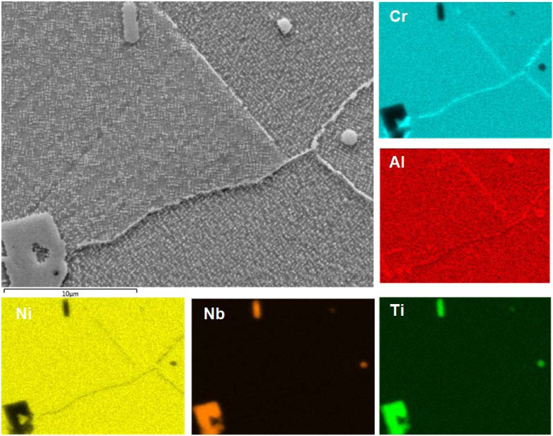
Figure 10 shows the high magnification EDX mapping performed on a large MX precipitate and the adjacent GB. It is clear that at this temperature, the development of G phase (enriched in Si and Nb) is favoured at MX site.
SEM image and elemental maps for the 1H specimen showing a Ti-rich MX precipitate, Cr-rich M23C6 precipitate developing at the MX site, and Nb-rich G phase developing at the MX site
The phases identified were therefore: a matrix of γ grains; a uniform dispersion of γ′ particles; large globular primary Ti- and Nb-rich MX formed in the microstructure prior to aging; a population of M23C6 nucleated at GBs, twin boundaries, on MX particles and intra-granularly; initiation of Si-rich G phase, suggested by Si- and Nb-rich regions of MX particles. Figure 11 shows selected examples of the microstructural constituents’ morphology in the H-aged samples.
a Low magnification SEM image showing the grain structure of H-aged; b grain boundary containing M23C6 and possibly G phase; c large Ti-rich MX (dark contrast) transforming into G phase and M23C6; d high magnification of Ti-rich MX transforming into G phase and M23C6, surrounded by γ′ particles e, f γ′ particles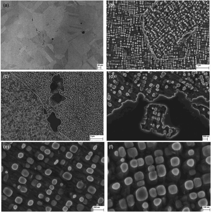
Summary of phases present in 1 and 1H aged samples

Phase quantification and IPS analysis
Phase quantification results as well as IPS and average particle size, obtained for both aged specimens

Conclusions from the comparison between IN740H and IN740H microstructures
The extent of the G phase encountered in specimen aged-H appears to be lower than that of specimen ref-aged. The compositional change of specimen aged-H also causes the fraction of γ′ to decrease. The dimensions of γ′ are coarser than those found in specimen ref-aged but this is mostly caused by the difference in aging time.
It is important to highlight the suppression of any visible η formation and the lower G phase fraction observed in specimen aged-H, in comparison to its parent IN740 alloy. Beside the lower γ′ content, this appears to be one of the best improvements in alloy IN740H. It is also very possible that the fraction of G phase will undesirably increase with increasing aging time. Longer aging times should be investigated in order to assess the effects on the microstructure of alloy IN740H.
IN740H has been successfully developed to achieve better high-temperature performance by decreasing the fraction of phases which are detrimental to the creep properties of the alloy. In particular, acicular η phase (growing at the expenses of γ’) is connected to higher creep susceptibility by making crack initiation and propagation more favourable.
Footnotes
Acknowledgements
The investigation is part of the collaborative project ENER/FP7EN/249809/MACPLUS, funded by the EU within the FP7 framework. Special thanks to S. McCoy (Special Metals) for providing the samples.