
Abstract
Titanium nanocomposite (TiTR) was synthesised from Triumfetta rhomboidea leaf extract and used as corrosion protection of mild steel (MS) in 1 M hydrochloric acid. The corrosion inhibition process of the nanocomposite was explored using weight loss, electrochemical impedance spectroscopy (EIS) and potentiodynamic polarisation (PDP). The corrosion inhibition performance of TiTR is a function of concentration and temperature. The results revealed that TiTR was 84.27% efficient in limiting MS corrosion at 1.0 g L−1 and 3 h. The TiTR acted as a mixed-type corrosion inhibitor and Langmuir isotherm indicated that TiTR was adsorbed on mild steel through physisorption by forming a monolayer of film. Surface morphology study of MS visually confirmed the protection of MS by TiTR through film formation.
Introduction
It is an established fact that mild steel (MS) plays a major role in most industries due to its superb mechanical properties, versatility and economical cost of production but its low resistance towards corrosion constitutes a great challenge. The multipurpose structural applications of MS in metallurgy, mining, construction, automobile, oil and gas, fabrication, process and petrochemical industries made it necessary to constantly be in contact with hostile conditions in which it experiences corrosion attacks [1]. For example, some industrial processes, such as pickling, descaling, etching and oil-well acidising, utilise large volumes of highly corrosive inorganic acids, either in a steel bath or flows through steel pipes, for cleaning and getting rid of impurities, and in the process, the structural reliability of the metal is compromised. One of the most efficient approaches for mitigating the corrosion of metal is the use surface coatings as well as the additives known as corrosion inhibitors [2–5]. Synthetic organic and inorganic corrosion inhibitors are the most common industrial corrosion inhibitors used till date. Likewise, nanoparticles of metal and metal oxides and those combined with other corrosion inhibitors as composites have also been applied mainly as surface coatings while others were directly added [6–13].
Nanoparticle materials have a particle size of 100 nm or less with excellent mechanical and chemical properties [14]. As a result of the extremely small particle size and large surface area, nanoparticles prevent corrosion reaction at metal/solution interface by blocking the active sites of the metal [15]. They readily form bonds with molecules of organic compounds [16] and this ability makes them better suited for self-healing the flaws in organic films formed, during the process of corrosion inhibition, on the metal's surface. They also improve the surface resistance of metals to corrosion currents of corrosive media by blocking tiny pores in protective films formed by organic inhibitors when used as composites. Further, they provide hardness as well as stability for the organic inhibitors thereby enhancing their efficiencies [17].
Some techniques used for synthesising nanoparticles include atomic layer absorption, thermal deposition and chemical vapour deposition but these techniques have been deemed potentially unsafe and hazardous [18]. Therefore, investigation into the possibilities of utilising different parts of plants as safe and green route for synthesising nanoparticles for corrosion inhibitors had gained momentum over the past few years. Hence, plant materials are the latest precursors for the synthesis of corrosion inhibitors as shown by many researchers [19–22]. Likewise, the potential effect of the use of biomaterial-metal nanocomposites to reduce corrosion of mild steel in aggressive media has also been reported [23–26]. Plants’ materials became precursors in the synthesis of nanocomposites for possible application as corrosion inhibitors of metals in corrosive media because these materials are promising, non-toxic, eco-friendly, versatile and good substitute for inorganic corrosion inhibitors [27–30].
Triumfetta rhomboidea Jacq. (Family: Tiliaceae) is commonly known in English as Diamond burbark, Burbush, Burweed or Chinese burr. It is an agricultural weed widely distributed in the tropics and subtropics. It is a competitive shrub species that disperses by means of its epizoochorous burs, and can become invasive in pastures and disturbed areas where it can form dense stands. A number of medicinal properties have been described for different parts of the plant. In Eastern Africa, it is used in folk medicine for its abortive properties on the gravid mammalian uterus. In traditional medicine, the stem and leaves of the plant are used for the treatment of diarrhoea and dysentery [31]. Iroha et al. [32] investigated the effect of the aqueous leaf extract of Triumfetta rhomboidea on the corrosion resistance of carbon steel in 1 M HCl solution and approximately 87% efficiency was reported.
Although nanoparticle composites of various metals have been synthesised from many plant materials, there is no report on the synthesis of titanium nanocomposite from Triumfetta rhomboidea or any report on the research into such nanocomposite as corrosion inhibitors of metals. Hence, the present work reports the green synthesis and characterisation of titanium nanocomposite by utilising methanolic leaf extract of Triumfetta rhomboidea as a precursor. The nanocomposite was investigated as a corrosion inhibitor of mild steel in 1 M HCl solution.
Materials and methods
Green synthesis of Titanium nanocomposite nanocomposite
Triumfetta rhomboidea (TR) leaves were collected from Lagos, Nigeria and identified in the Department of Crop, Soil and Pest Management of the Federal University of Technology Akure, Nigeria. The leaves were washed to remove dirt, dried for 21 days, ground, screened and stored in air-tight containers. Approximately 250 g of powder samples were extracted for 72 h in 2.5 L methanol, filtered with Whatman filter paper, concentrated with rotary evaporator, further dried and stored.
Nanocomposite of titanium and T. rhomboidea was synthesised by bio-reduction of titanium salt using a modified method described by Essien and co-researchers [19]. A 1.0 mL of 99.5% anhydrous TiCl4 (Lobachem) was added in drops to a constantly stirred mixture containing 10 mL T. rhomboidea extract and 80 mL distilled water. The mixture was continuously stirred for an hour at room temperature with a magnetic stirrer. A visual change in the colouration of the mixture from green to dark brown indicated nanocomposite formation. The resultant product was thereafter centrifuged for 30 min at 4000 rpm. The residual solid was subsequently washed with distilled water several times, dried in a thermostatic oven at 60°C and stored in a desiccator for characterisation and corrosion tests.
Characterisation of Titanium nanocomposite nanocomposite
The representative functional groups of the synthesised nanocomposite and its precursor (crude extract) were analysed using a PerkinElmer 100 Fourier transform infrared (FTIR) spectrometer. Colloidal solutions were made from the specimens and evaporated to dryness. The dried samples were mixed with KBr pellets to obtain solid residues used for the FTIR analysis. The spectra were recorded in the range of 400 and 4000 cm–1 at a resolution of 4 cm–1.
Additionally, the powder X-ray diffraction of the synthesised TiTR nanocomposite, before and after calcination in a muffled furnace at 600°C for 3 h, was also carried out to identify the crystallite nature and calculate the crystallite size. The analysis was carried out on a Rigaku MiniFlex II X-ray diffractometer operating at 40 kV and 30 mA using Ni-filtered Cu Kα radiation of λ = 1.5406 Å at a 0.02 scan step.
The grain patterns and surface characteristics of TiTR nanocomposite were investigated by observing the sample under a 15 kV Phenom ProX (Thermo Fisher, USA) scanning electron microscope. A few drops of the colloidal solution prepared from the nanocomposite were placed on aluminium grid sample holder, dried, and analysed. Similarly, high-resolution micrographs of TiTR nanocomposite were obtained and studied with a Philips CM 120 transmission electron microscope. A dispersion of small amount of TiTR was first made in 10 mL ethanol and later sonicated for 10 min at 10 kV. The dispersed samples were subsequently formed on a Leica cryo-microtome which was positioned on a 100 × 100 mm2 carbon–copper grid and observed with the microscope.
Furthermore, a nitrogen gas adsorption analysis was performed on TiTR nanocomposite with a TriStar II 3020 2.00 volumetric analyzer (Micromeritics Corp.). A TiTR portion weighing 0.58 g was first degassed at 90°C for 6 h. Afterwards, adsorption and desorption isotherms were obtained by passing N2 gas in and out of the sample at a constant temperature of 77.2 K and relative pressure (p/po) range of 0.004–0.996. The Barrett–Joyner–Halenda (BJH) and Brunauer–Emmett–Teller (BET) models were used to estimate the average diameter, the average size of the pores and the specific surface area of the sample.
Corrosion inhibition experiments
Preparation of MS samples and test solutions
Composition of mild steel used for corrosion tests.

Gravimetric study
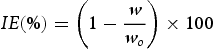
Previously weighed coupons were immersed in a 1 M HCl without and with the addition of different amounts of TiTR for 3 h at 298 K. The coupons were thereafter retrieved, immersed in NaOH solution to neutralise residual acid solution, rinsed in acetone, dried, and re-weighed. Each corrosion measurement was conducted in triplicate and the average weight-loss values were used for the calculation of corrosion rate (CR in mg cm–2 h–1) and inhibition efficiency (IE in %) with Equations (1) and (2), respectively.

Electrochemical measurements
Potentiodynamic polarisation (PDP) and electrochemical impedance spectroscopy (EIS) were used to investigate the electrode potential and frequency responses of MS in solutions of 1 M HCl only and those containing a selected amount of TiTR nanocomposite for inhibitor. A VERSATAT 4 potentiostat was used for all experiments at room temperature. The cell set-up was a three-electrode system that comprised a reference electrode (saturated Ag/AgCl), a platinum auxiliary electrode and polished MS coupons as the working electrodes. The electrolytes are 100 mL solutions of 1 M HCl acid with and without the selected concentrations (0.2, 0.6 and 1.0 g L–1) of TiTR nanocomposite. Before each measurement, a potential sweep was applied at a scan rate of 1 mV/s for 30 min to determine the stable, open-circuit corrosion potential (OCP) between –250 mV and +250 mV. Dynamic polarisation of the potential responses of MS coupon in each electrolyte to the potential sweep was thereafter recorded at the OCP. Furthermore, the instrument was regulated to appropriately apply an alternating current (AC) of 5 mV amplitude, at the stable potential that was initially obtained, through the electrolyte between 10 mHz and10 kHz frequency range. The response of each measured system to the frequency modulation was recorded as a sinusoidal spectrum. Each experiment was repeated three times to check the reproducibility, and the average values were used.
Surface morphology
Steel coupons of dimensions 10.3 × 10.5 × 10 mm3 were immersed in test solutions without the inhibitor and with the optimum TiTR concentration determined during weight loss study for 24 h. The specimens were later retrieved, thoroughly rinsed with distilled water to remove residual acid, dried in warm air current and preserved in a desiccator for 8 h. The surface characteristics of the steel coupons were thereafter examined with a scanning electron microscope coupled to an X-ray dispersive spectrometer (Phenom ProX). The instrument was operated at 15 kV potential to determine their surface morphology and elemental compositions.
Results and discussion
The FTIR analysis
The functional groups present in the synthesised TiTR nanocomposite were investigated by using FTIR spectroscopy and compared with that of T. rhomboidea extract. Figure 1 shows the comparison between the spectrum of TiTR nanocomposite and that of T. rhomboidea leaf extract and the significant characteristic peaks obtained during the analysis. The broad absorption peaks observed in the range 3600–3050 cm–1 for both TiTR and its precursor extract are attributed to the O–H stretching of alcohol or phenolic rings. For TiTR nanocomposite, the observed peak is more intense and the reason could be due to the strong binding affinity of the phytocompounds of the leaf extract to the titanium nanoparticles through the Ti–OH bonds. The peaks at 2933 cm–1 for TiTR and 2927 cm–1 for the crude extract are those of the C–H stretching saturated hydrocarbons while the stretching of C = O of conjugated alkanones is responsible for the absorption peak observed at 1710 cm–1. The spectrum of TiTR nanocomposite and that of the leaf extract also show peaks at 1624 and 1607 cm–1, respectively. These are credited to the infrared absorption of either the stretching of C = C bond or the N–H bending of amines. The stretching of the C–O bond of esters is evident from the peaks at 1176 and 1167 cm–1 for TiTR and T. rhomboidea leaf extract, respectively [33]. In addition, there are characteristic absorption peaks for TiTR nanocomposite at 815 and 610 cm–1. These signals appear as a result of absorption by Ti–O–Ti, O–Ti–O and Ti–O bonds [22,34].
FTIR spectra of TiTR nanocomposite and the methanolic leaf extract of T. rhomboidea.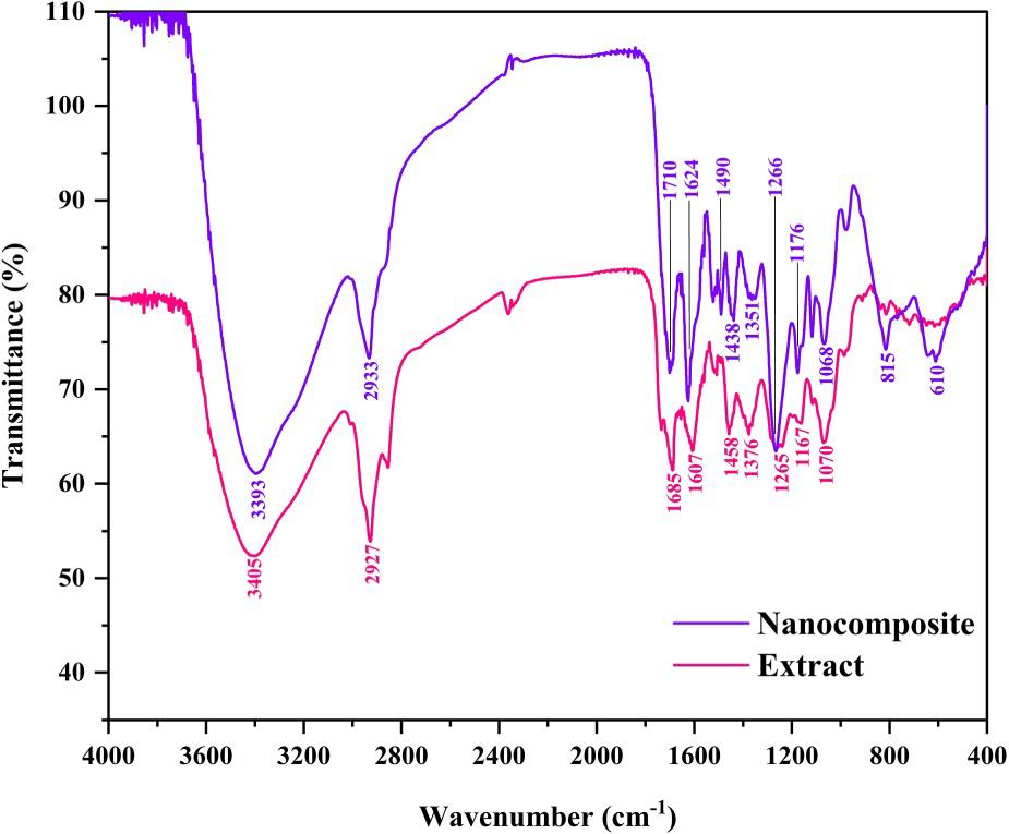
From the FTIR spectra, it is observed that the nanocomposite retained the major functional groups and bonding systems that were initially present in the extract (precursor) but with strong intensity, as highlighted by the strong peaks of nanocomposite spectrum. However, there are two prominent peaks at 610 cm–1 and 815 cm−1 in the nanocomposite. The presence of these characteristic absorption bands confirms the formation of nanocomposite of titanium with the extract and that the bonding is via linkage to O atoms.
XRD analysis
The crystalline structure of TiTR nanocomposite, before and after calcination, was probed by X-ray diffractometry (XRD). The diffraction results are shown in Figure 2. The diffraction patterns obtained for the sample before calcination did not show characteristic strong peaks that could be attributed to a specific crystalline structure. This pattern could simply be credited to the phytochemical structures of the leaf extract from which the nanocomposite was synthesised. However, several diffraction patterns were obtained from TiTR nanocomposite after it was calcinated for 3 h. The peaks appeared after the organic matrices covering the titanium particles were calcined. The diffraction peaks at angles 25.32°, 37.88°, 48.02°, 55.24° and 62.72° correspond to the tetragonally arranged TiO2 anatase. The diffraction angles of the peaks matched with the JCPDS 21–1272 file which describes the Miller indices of the anatase phase as (101), (004), (200), (211) and (204) [35,36]. This is an indication that the titanium nanoparticles in the nanocomposite were mainly bonded to oxygen atoms of molecules from the leaf extract during synthesis. The difference in the recorded patterns is as a result the leaf extract completely encapsulating the titanium nanoparticles in its organic matrices.
Diffraction patterns of the synthesised TiTR nanocomposite before and after calcination.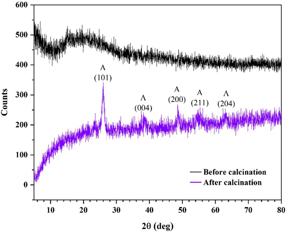
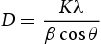
Moreover, the mean diameter of the TiTR crystals obtained after calcination was estimated using the Scherrer's expression (Equation (3)).
SEM and TEM observations
Powder samples of TiTR before and after calcination were observed under scanning electron microscope for surface morphology. The SEM micrographs of the observations are illustrated in Figure 3. From the images, it is observed that both the samples of TiTR before and after calcination are irregular, nonporous and aggregate particles. This is probably due to the strong coordination between titanium atoms and the molecules of the leaf extract in the composite. It is also observed that the TiTR sample before calcination appear as small irregular clusters without crystalline grains. However, crystal grains are clearly visible after the sample was calcined but with the same irregular shape and aggregate feature.
SEM micrographs of TiTR nanocomposite (a) before and (b) after calcination.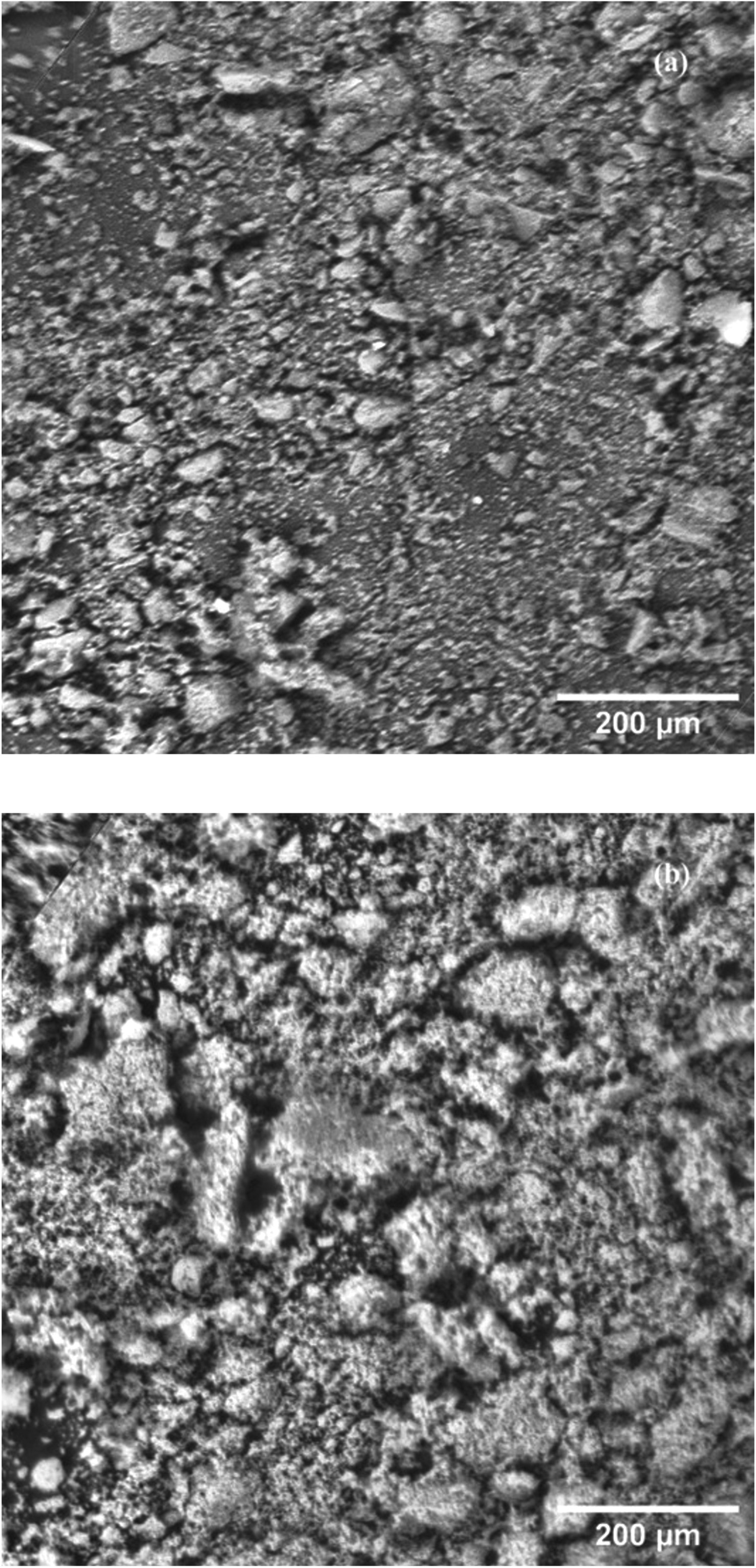
TiTR nanocomposite particles were observed in higher resolution with the use of a transmission electron microscope. TEM was used to study the dispersion, shape and size of the sample. The images (Figure 4) confirmed the results from SEM observation as the particles are clearly not dispersed in the medium used for the microscopic technique. Clusters (aggregates) of irregular-shaped particles with a size range of 4.35–8.67 nm could be observed from the images. The clustering probably occurred during preparation for microscopy analysis when the sample was dispersed in ethanol [12]. The aggregation of nanoparticles is generally the thermodynamically enabled particle-particle interactions in a colloidal system, which leads to the formation of strong covalent bonds [37]. Hence, the aggregation of TiTR particles resulted from the strong interactions between the phytoconstituents of the leaf extract and the titanium particles during synthesis. This particle size range measured from the HRTEM analysis corroborates the results obtained from XRD analysis.
High-resolution TEM images of TiTR at differentresolutions.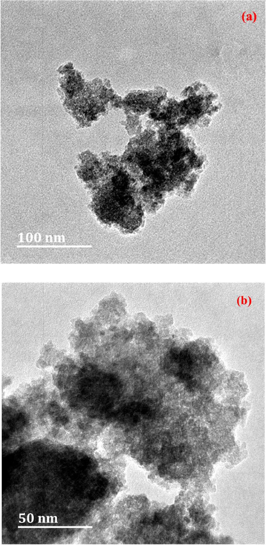
N2 gas adsorption measurement
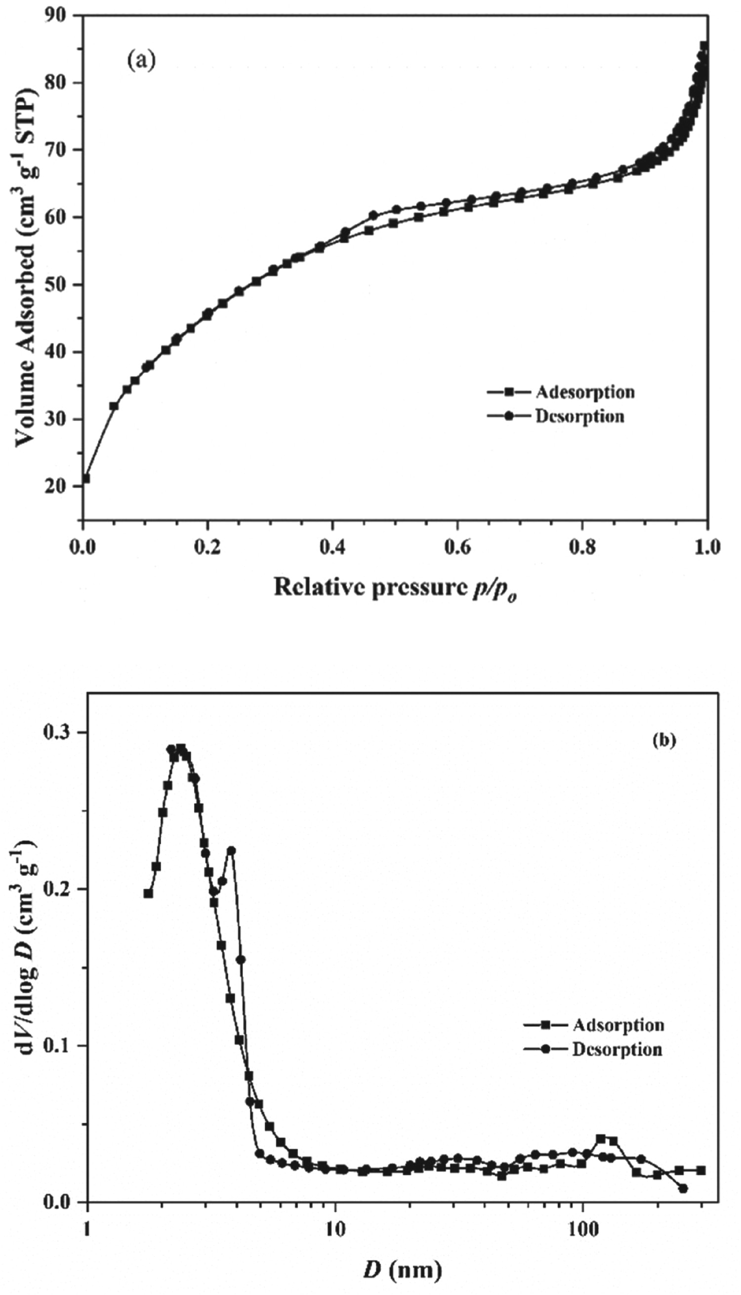
The volumetric adsorption/desorption of nitrogen gas was carried out on TiTR to evaluate its specific surface area and particle, as well as its pore size and volume size, using BET and BJH models, respectively [38,39]. Figure 5(a) shows the adsorption and desorption isotherms of TiTR nanocomposite particles. The hysteresis loop formed from the overlap of the curves is a Type IV hysteresis loop, which indicates that TiTR is a mesoporous material [40]. The (a) N2 gas adsorption/desorption and (b) BJH plots for the TiTR nanocomposite.
Corrosion-inhibition studies
Gravimetric measurements
The performance of TiTR nanocomposite as an inhibitor of mild steel (MS) corrosion in acidic medium was investigated by gravimetry. The dependency of the corrosion rate on the TiTR concentration is illustrated in Figure 6. The results obtained indicate that the presence of TiTR in the corrosive solution substantially lowered the rate at which MS corrodes compared to when the inhibitor is absent. The ability of the inhibitor material to effectively minimise the corrosion extent of MS also directly translates to a good performance, especially as the quantity of TiTR in the corrosive solution is increased. This is largely due to the presence of more inhibitor molecules in the solution to form stronger and better protective-film layers on the metal surface. An IE value of 84.26% was obtained for the optimum concentration (1.0 g L–1) at the temperature of 298 K compared to 87% reported by Iroha and Madueke [32]. The disparity is a result of the immersion time for which the experiments were conducted. The process of inhibition by corrosion inhibitors requires an induction time for an effective protective film to form on a metal surface [41]. Longer immersion time allows an inhibitor to be properly adsorbed on the surface of a metal substrate which consequently leads to high efficiency. The performance of TiTR recorded herein for all concentrations at 3 h immersion time is quite significant given that corrosion attack of the acid on the steel substrate is expected to be at its peak. Similarly, the particle size of titanium nanoparticles bonded to the phytoconstituents of the extract contributed to the enhancement of the protection ability of the inhibitor by filling the porous structures in the organic matrices to prevent corrosive ions from migrating to the metal's surface. Moreover, the large surface area of the nanocomposite, as determined from the N2 adsorption, enabled it to easily spread over the metal surface, thereby limiting the contact of the acid solution with the surface.
Dependence of corrosion rate and inhibition efficiency on the concentration of TiTR nanocomposite.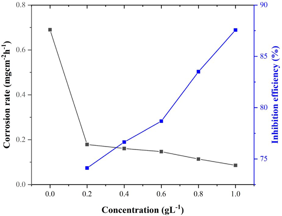
Additionally, results show that the inhibition action of TiTR against MS corrosion is also affected by temperature. Figure 7(a) shows the important relationship between the ability of TiTR to inhibit MS corrosion in 1 M hydrochloric acid solution and temperature. The increase in corrosion rate is experienced as temperature increases and this relationship is obvious at all TiTR concentrations. This observation is characteristic of the desorption of some adsorbed molecules of the inhibitor from the surface of the metal due to the increase in the solution turbulence because of the increased evolution of hydrogen gas [42]. In other words, the molecules of TiTR are held to the MS surface by a weak force. As a consequence, elevation of temperature easily breaks the adsorptive force between the inhibitor and the metal surface, thereby reducing the potency of the inhibitor, as shown in Figure 7(b). It is noteworthy that a relative magnitude of about 4% reduction was only recorded for IE values between 298 and 328 K for all concentrations. This suggests that, despite temperature increase, TiTR still possessed the ability to inhibit the corrosion of MS as a result of the presence of titanium nanoparticles in the composite which enhances the inhibition ability of the inhibitor and probably forms metal–metal and metal–oxygen coordination with the MS surface.
Temperature-dependence of corrosion rate and inhibition efficiency on the concentration of TiTR nanocomposite.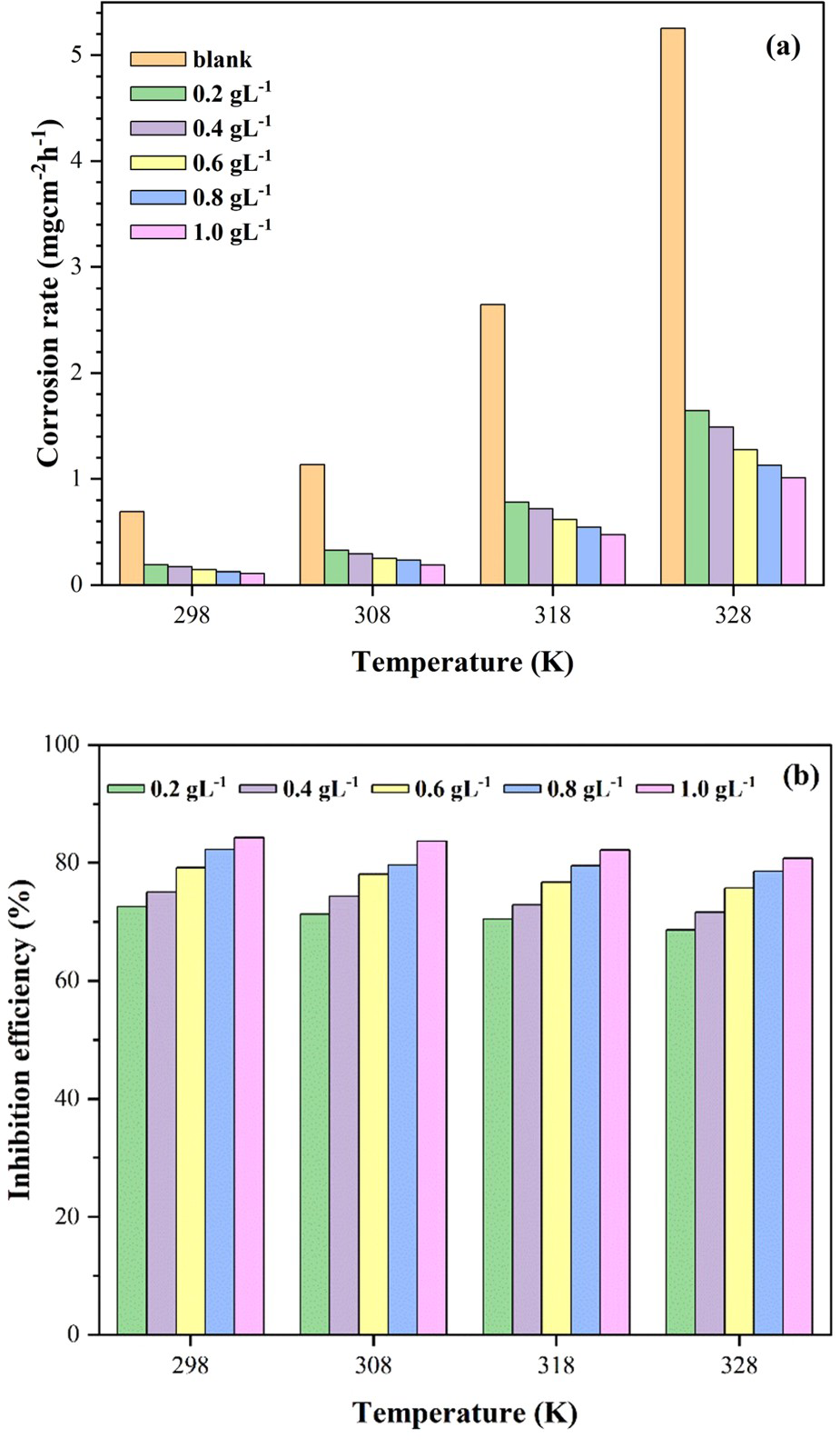
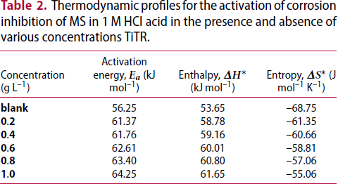
Activation parameters
The activation criteria for the corrosion process of MS in the presence and absence of TiTR nanocomposite in hydrochloric acid are considered from the analysis of the temperature-dependence results obtained by gravimetry. The activation energy ( Arrhenius curves for the corrosion of mild steel, in the presence and absence of TiTR nanocomposite, in 1 M HCl acid. Thermodynamic profiles for the activation of corrosion inhibition of MS in 1 M HCl acid in the presence and absence of various concentrations TiTR.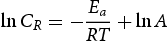
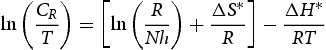
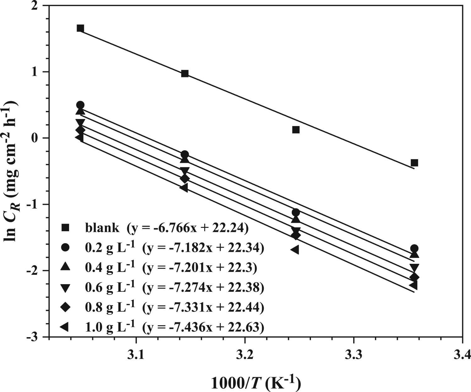
The plots in Figure 9 represent the plots of ln
Adsorption isotherms
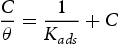

Calculated parameters of Langmuir model for TiTR at different temperature values.

Transition state plots for the corrosion of MS in 1 M HCl solution both in the absence and presence of TiTR nanocomposite.
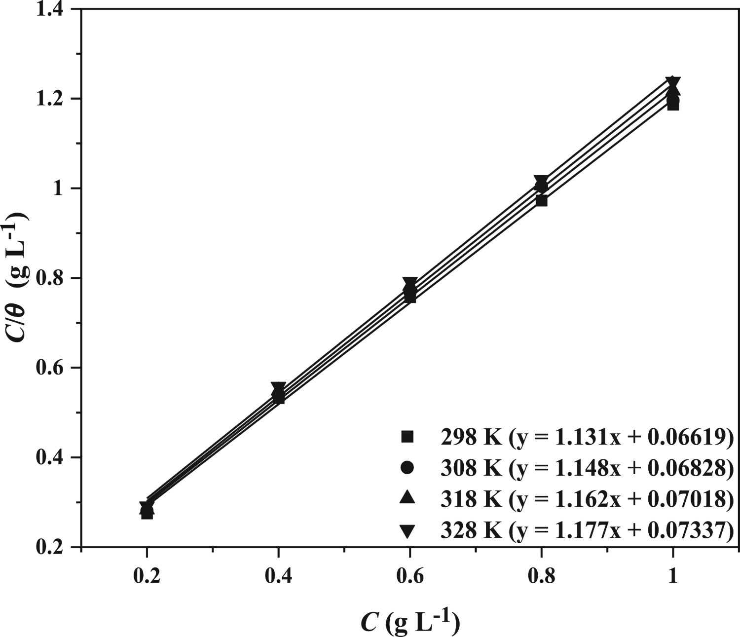
Langmuir curves for TiTR nanocomposite at different temperature values.
Potentiodynamic polarisation study
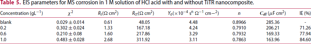
Since corrosion process is a redox reaction which simultaneously occurs at the cathode and anode, PDP measurements were conducted to evaluate the effect of using selected concentrations of TiTR nanocomposite to improve the corrosion resistance of MS in 1 M HCl solution. Polarisation curves (Figure 11) for the selected concentrations were obtained at 298 K and parameters such as Ecorr (corrosion potential), Icorr (corrosion current density) as well as βa and βc (respective cathodic and anodic Tafel slopes) were acquired by extrapolating the linear sections of the anodic and cathodic regions of the curves. The polarisation parameters are recorded in Table 4 along with inhibition efficiency, which was evaluated using Equation (9).
Polarisation profiles for mild steel corrosion in 1 M HCl with and without TiTR nanocomposite. Potentiodynamic polarisation parameters for mild steel corrosion in 1 M HCl solution in the presence and absence of TiTR.
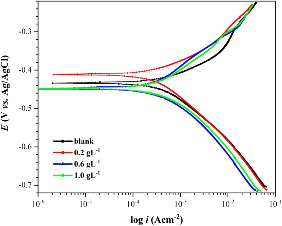
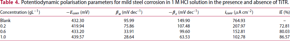
A shift in the corrosion potential is observed for the inhibited solution which is towards a more negative Ecorr with an increase in the inhibitor concentration. This is an indication that, even though TiTR is a mixed-type inhibitor, it prominently inhibits cathodic corrosion more than anodic corrosion [47]. The largest shift in the Ecorr for the inhibited solutions, relative to the uninhibited solution, is also related to the classification of any corrosion inhibitor. A difference of ±85 mV describes the inhibitor as either anodic or cathodic while any value beyond categorises the inhibitor as a mixed type [9]. Herein, the largest shift recorded for the corrosion inhibition of MS by TiTR is 12.36 mV, hence, the inhibitor may be generally classified as a mixed-type.
EIS measurements
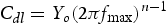
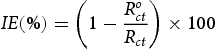
The impedance responses to frequency variations of mild steel specimen in the absence and presence of TiTR are illustrated by the Nyquist and Bode spectra (Figure 12). In Figure 12(a), the plots shaped in semicircles are due to the predominantly charge-transfer controlled corrosion process [51], the departure from perfect semicircle is due to the roughness of the metal surface and the inconsistent build-up of charges at the metal/inhibitor interface as a result of frequency dispersion [52]. Similarly, the diameter of the Nyquist plots for the inhibited acidic solution increased with concentration compared to those for the uninhibited systems; this is probably due to the resistance to charge transfer at the metal/inhibitor interface set up by TiTR upon adsorption onto the mild steel surface.
(a) Nyquist curves and (b) Bode plots for the corrosion of mild steel in 1 M solution of HCl acid in the presence and absence of TiTR.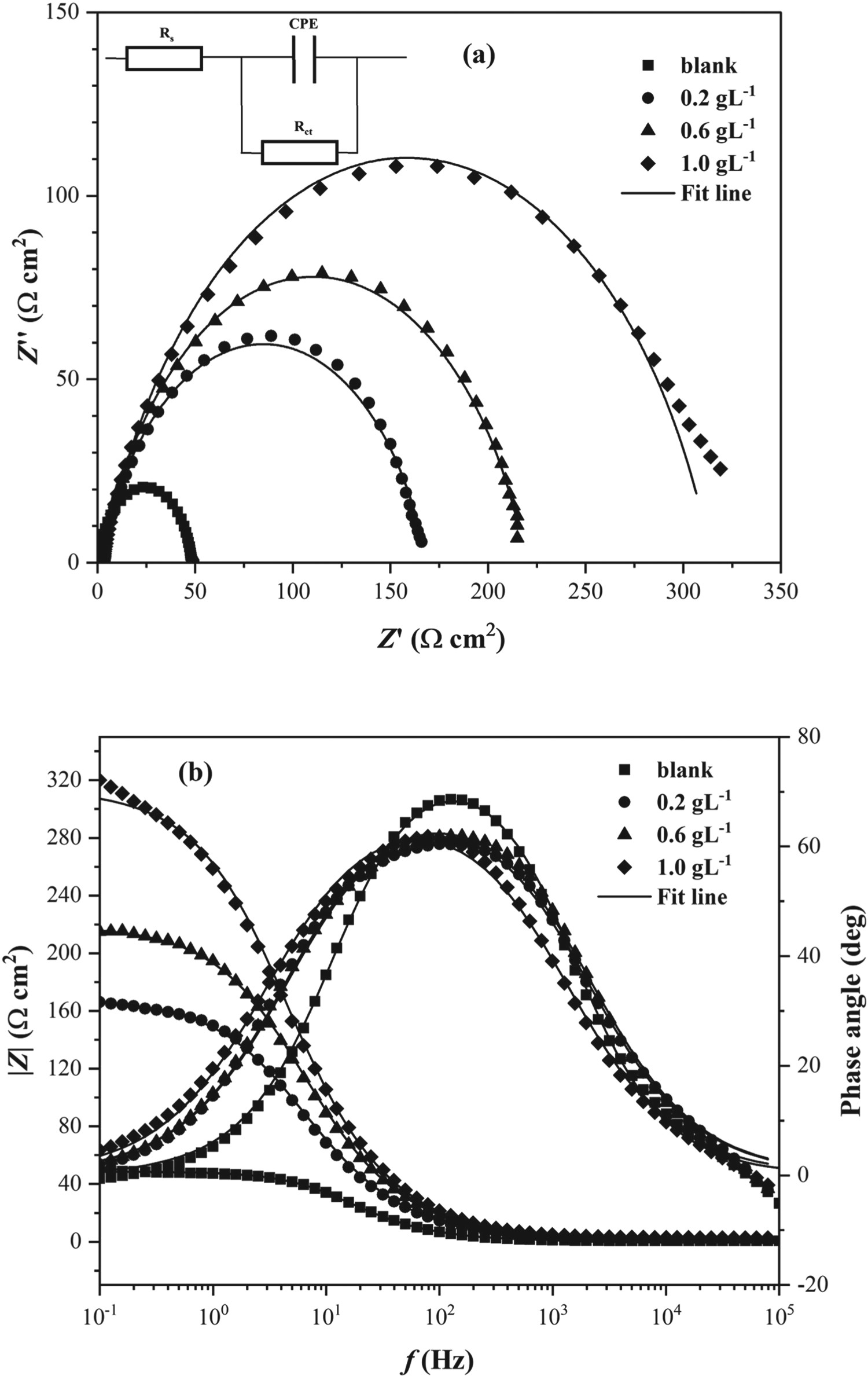
EIS parameters for MS corrosion in 1 M solution of HCl acid with and without TiTR nanocomposite.
Surface morphology
The SEM micrographs of MS specimens immersed in the acid medium with and without the optimum TiTR concentration for 24 h at 298 K are illustrated in Figure 13 at different resolutions. As revealed in Figure 13(a,c), the exposure of MS sample to the uninhibited corrosive medium resulted in significant material loss, which is evident from the jagged surface. In contrast, the presence of TiTR substantially attenuated the effect of corrosion attack on the metal, as observed from the smooth surface in Figure 13(b,d). The smooth morphology obtained with the addition of TiTR is a confirmation of a protective layer formed by the composite on MS surface through adsorption. This layer effectively minimised the migration of ions at the metal-solution interface. Likewise, the titanium nanoparticles encapsulated within the plant extract matrix made the composite impervious to corrosive ions that ordinarily could have migrated through the porous structure of the plant extract.
The SEM micrographs of mild steel after 24 h immersion in the absence (a, c) and in the presence (b, d) of 1.0 g L–1 TiTR at different magnifications.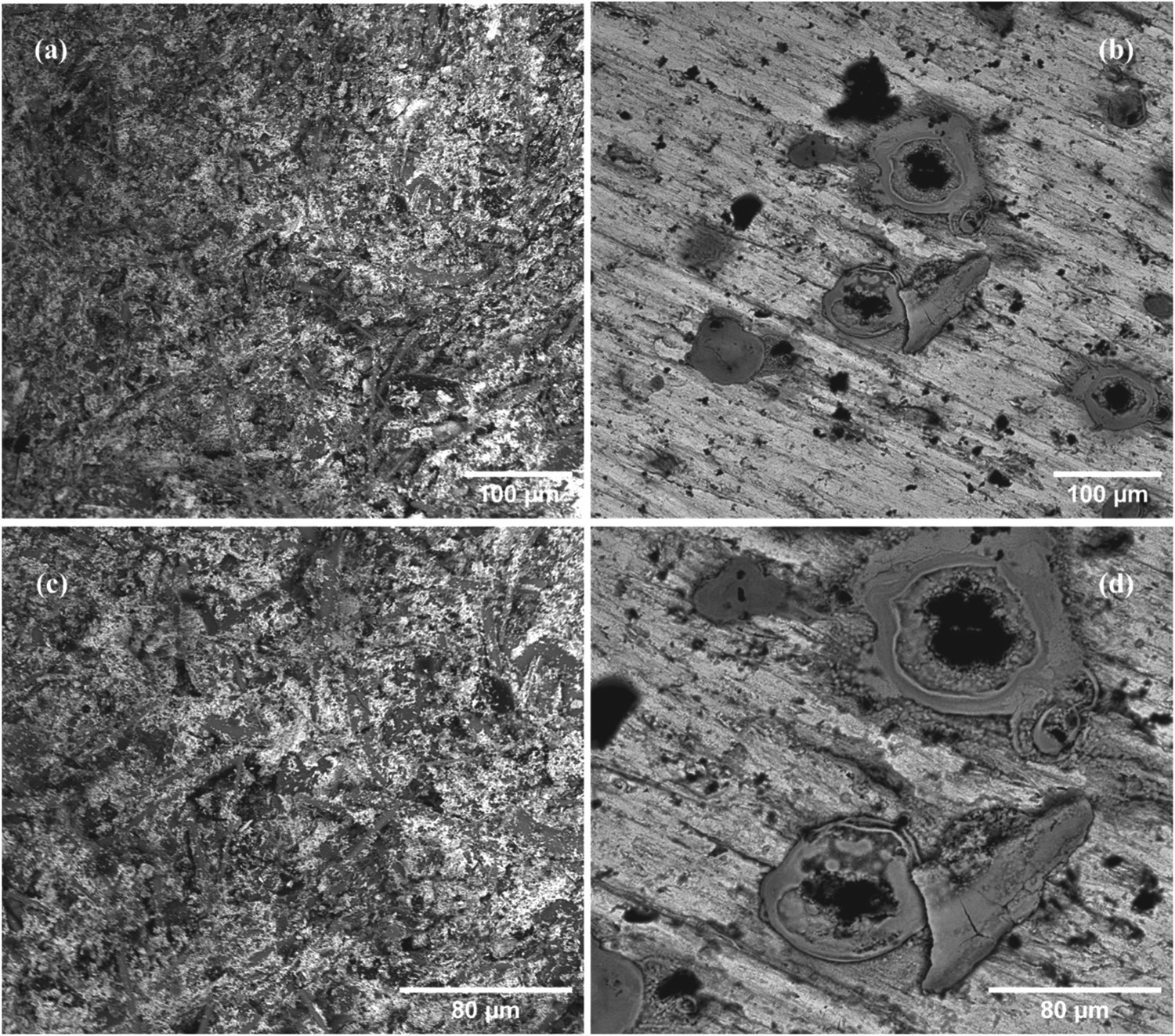
The EDX spectrum in Figure 14(a) represents the unfettered oxidative dissolution of MS by the acid, with a resultant low Fe composition (1.80%). Conversely, the presence of TiTR greatly preserved the Fe content of the MS through complex-formation at the metal-solution interface (Figure 14(b)). The absence of Cl− and low intensity of O from both spectra is associated with the rinsing of metal after retrieval from the acid medium.
EDX spectra of mild steel (a) in the absence and (b) presence of 1.0 g L–1 TiTR after 24 h of immersion. Inset: composition of steel surface by wt-%.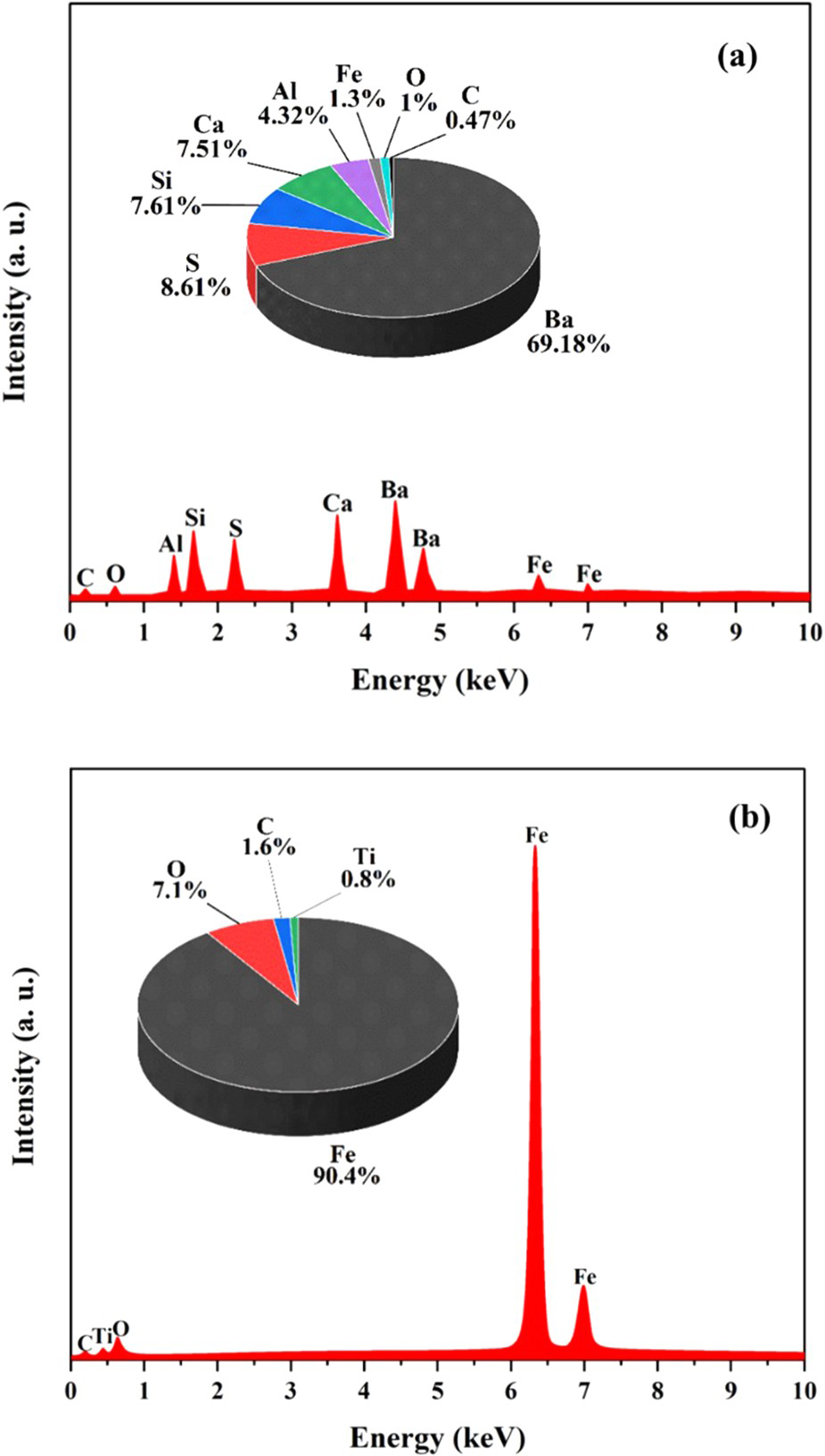
Conclusion
Based on the results of the investigation in the present work, the following conclusions are drawn:
Titanium-Triumfetta rhomboidea (TiTR) nanocomposite was synthesised from leaf extract of Triumfetta rhomboidea and titanium. The composite was characterised using Fourier-transform infrared spectroscopy, Xray-diffraction, scanning electron microscopy, transmission electron microscopy and gas adsorption techniques. The characterisation results revealed that the TiTR retained most of the functional groups of the phytocompounds of the leaf extract. Furthermore, the data showed that the nanocomposite is made of crystalline anatase with nanosized structure and pore dimensions. Gravimetric measurements into the application of TiTR as possible green corrosion inhibitor of mild steel in hydrochloric acid showed that the weight loss of steel was reduced by 84.27% at 1.0 g L–1, which is better than its corresponding leaf extract. The data from electrochemical studies revealed a reduction in the rate of charge transfer from the metal into the aggressive solution with the application of the nanocomposite inhibitor which formed a protective layer on the metal. Adsorption isotherms showed that TiTR was adsorbed on steel surface by forming a monolayer adsorption film through physical adsorption. Surface analyses of the metal validated the results of the gravimetric and electrochemical studies, confirming the preservation of the metal in the aggressive solution by the nanocomposite inhibitor.
Footnotes
Disclosure statement
No potential conflict of interest was reported by the author(s).