
Abstract
ABSTRACT
Undoped ZnO and Mg-doped ZnO (MZO) thin films were synthesized by sol–gel dip-coating technique. The effect of Mg dopant on the structural, surface-morphological, and optical properties together with the photocatalytic activities of the films was studied. Morphological study shows that incorporation of Mg increases the porosity of the films. Structural investigation reveals that Mg doping declines the crystallinity of the films. The optical analysis demonstrates that the Urbach energy of the films increases due to Mg doping, suggesting the formation of localized energy states. Photocatalytic activities of the films were evaluated by studying the degradation of methylene blue under the ultraviolet light irradiation. MZO demonstrates better photocatalytic efficiency compared to ZnO films. The enhanced photocatalytic efficiency of MZO can be attributed to the charge trapping by localized states, and porosity-induced surface area enhancement due to Mg doping.
Introduction
In recent years, water pollution has become a global challenge as it possesses serious consequences on human life and natural ecosystems. Discharge of untreated effluents as well as heavy metals into the water resources from industries is the major source of water pollutions. Hence there is a pressing need to develop technologies to decontaminate the industrial wastewaters so as to reduce their impact on human health and environment. Significant research efforts have, therefore, been put forward to find an efficient, cost-effective, and sustainable route for decontamination of water [1,2]. Recently, photocatalysis has emerged as a green technology that can remove toxic organic materials, bacteria, and environmental pollutants from water. Photocatalysis is an advanced oxidation process in which photocatalyst materials generate electron–hole pairs by absorbing photon energy. The photo-generated electrons and holes take part in the oxidation process and eliminate pollutants from water [3,4].
Zinc Oxide (ZnO), a wide band gap metal oxide semiconductor, is extensively used for the decontamination of water by photocatalysis. Pronounced photosensitivity, high oxidizing power, and nontoxicity have made ZnO a popular photocatalyst [5,6]. ZnO can decontaminate water by removing different types of dyes, including methylene blue (MB), rhodamine B, acid red 14, eosin Y, and different sorts of bacteria [4-6]. However, for pure ZnO, the photo-generated electron–hole pairs that create during photocatalysis recombine very quickly which reduces the photodegradation efficiency [7]. The improvement of the photocatalytic performance of ZnO by retarding the charge carrier recombination has, therefore, become a key motivation of ZnO based photocatalysis research.
Doping with metal ion is one of the most effective approaches to improve the photocatalytic efficiency of ZnO [8,9]. Metal doping can enhance the photocatalytic performance of ZnO by tuning the band gap, introducing localized states, and modifying the surface morphology of ZnO. So far, a number of metal atoms including, Al, In, Ag, Cu, Mg, Fe, Mn, Ni, Sr, Li, Na, K, Ce, Cd, etc. [8-16] have been used as a dopant to improve the photocatalytic efficiency of ZnO. Among them, Mg is considered as an appropriate choice as dopant, because the ionic radius of Mg+2 (0.78 Å) is similar to that of Zn+2 (0.83 Å) so they can be easily substituted [17]. Moreover, the nontoxicity and easy availability of Mg makes it a suitable choice as a dopant for ZnO.
Enhancement of the photocatalytic performance of ZnO nanoparticles by Mg doping has been reported in several articles [16-19]. Abed et al. synthesized Mg-doped ZnO (MZO) nanocrystals by sol–gel method and evaluated their photocatalytic activity by studying the photodegradation of Rhodamine B [16]. They showed that Mg doping creates charge traps in ZnO which improve the photocatalytic efficiency of the nanocrystals. Lu et al. synthesized MZO by the modified sonochemical synthesis method [18]. They showed that Mg doping increases the optical band gap of ZnO nanoparticles which in turn improves their photocatalytic efficiency. Etacheri et al. synthesized MZO nanoparticles through co-precipitation method and use them to study the photodegradation of MB dye [19]. They demonstrated that Mg doping widens the band gap of ZnO and provide superior textural properties that allow efficient electron–hole separation which enhance the photocatalytic performance. In most of the previous work, the MZO photocatalysts were in the form of nanoparticles. The nanoparticles need to be separated from aqueous solution by sedimentation or ultrafiltration after photocatalysis. This requires extra time and cost which limits the applicability of powder samples. An effective approach to overcome this limitation is to deposit the photocatalyst materials onto a substrate in the form of thin film. A detailed study about the effect of Mg doping on the photocatalytic performance of ZnO thin film is still inadequate. Therefore, the main goal of this study is to evaluate the effect of Mg doping on the structural, morphological, and optical properties together with the ultraviolet (UV) light-assisted photocatalytic performance of ZnO thin films.
The deposition technique plays a major role in controlling the surface morphology, structural and optical properties of the thin films. A number of techniques are being used for the fabrication of ZnO thin films namely spray pyrolysis, sol–gel, drop-casting, chemical bath deposition, pulsed laser ablation, sputtering, etc. [11,20-23]. Among them, sol–gel dip coating is one of the most versatile, economic and dominant techniques that offer excellent compositional control, better homogeneity, and lower crystallization temperature together with large area deposition of thin films [11,24]. Owing to these advantages, we have chosen a sol–gel dip-coating technique for the synthesis of pure and MZO thin films and their structural, morphological, optical properties together with UV light-assisted photocatalytic activity are reported in this article.
Material and methods
Synthesis of ZnO and MZO thin films
Sol–gel dip-coating technique was used for the synthesis of ZnO and MZO thin films. Zinc acetate dehydrate [Zn(CH3COO)2.2H2O] and Magnesium sulphate dehydrate [MgSO4.7H2O] were used as the precursor materials for Zn and Mg, respectively. Ethanol and Di-ethanol amine (DEA) were used as solvent and stabilizer respectively. To synthesize ZnO thin films, the precursor solution was prepared by dissolving [Zn(CH3COO)2.2H2O] in ethanol followed by stirring with a magnetic stirrer for 2 h. Then DEA stabilizer and deionized water were added to the solution and the combined mixture was stirred at room temperature for 2 h yielding a homogeneous sol. To deposit thin films, clean microscopic glass substrates (10 mm × 15 mm × 1.5 mm) were dipped into the precursor solution followed by withdrawal at a speed of 10 mm/min. The dip-coated thin films were then dried at room temperature followed by calcination at 350 °C for 1 h to remove organic compounds from the films. To synthesize MZO thin films, sols with different concentrations of Mg (0, 2, 4, and 6 wt-%) were prepared by dissolving an appropriate amount of [MgSO4.7H2O] and [Zn(CH3COO)2.2H2O] in ethanol. The rest of the procedure was kept the same as the pure ZnO film synthesis process.
Characterizations of ZnO and MZO thin films
The structural properties of the thin films were studied by an X-ray diffractometer (3040XPert PRO, Philips) using monochromatic CuKα radiation (λ = 1.54 Å). X-ray diffraction (XRD) data were recorded at a scanning speed of 2 degree/min, between 20° and 70°. The surface morphology of the films was investigated by a field emission scanning electron microscope (FESEM) (JSM 7600, Jeol). The optical properties of the films were studied by recording the absorbance and transmittance spectra of the films within the wavelength range of 200‒1000 nm using a UV-visible spectrophotometer (Dynamica HALO DB-20S). A homemade four-point collinear probe setup was used to measure the DC electrical resistivity of the thin films.
Photocatalytic activity measurement
The photocatalytic activities of the thin films were evaluated by studying the photodegradation of aqueous solution of MB dye under UV irradiation. The MB solution was kept in dark for 60 min to achieve adsorption–desorption equilibrium [11]. To perform photocatalytic experiment, ZnO or MZO thin films were dipped inside a beaker containing 100 mL aqueous solution of MB. A 12W UV light source was kept 20 cm above the beaker and was irradiated. The irradiated MB solution was collected at a regular interval of time, and their absorbance spectrum was recorded using the UV-visible spectrophotometer.
Results and discussion
Surface morphology of ZnO and MZO thin films
The surface morphology of ZnO and MZO thin films were studied using field emission scanning electron microscopy (FESEM). Figure 1 shows the FESEM micrographs of ZnO and MZO thin films. It is observed that the ZnO film has a compact structure composed of nanoflakes distributed uniformly over the substrate. Few pores are observed on the surface of the ZnO thin film. With the increase of Mg dopant concentration, the pore-density and the pore-size increases. The roughness of the film surface also increases with the increase of Mg dopant concentration. These phenomena can be attributed to the difference in ionic radius between Zn and Mg. Owing to their dissimilar size, when doped, Mg introduces lattice distortion into the ZnO lattice, resulting in stress that causes pore into the thin film. With an increase of dopant concentration, the stress and consequently the pore-density increases [25]. It is important to note that the porosity in the films increases the accessible surface area for the materials and are suitable for catalytic and gas sensing applications.
FESEM micrographs of (a) pure ZnO (b) 2 wt-% MZO (c) 4 wt-% MZO and (d) 6 wt-% MZO thin films. Arrow indicates the positions of pores on the surface of the films.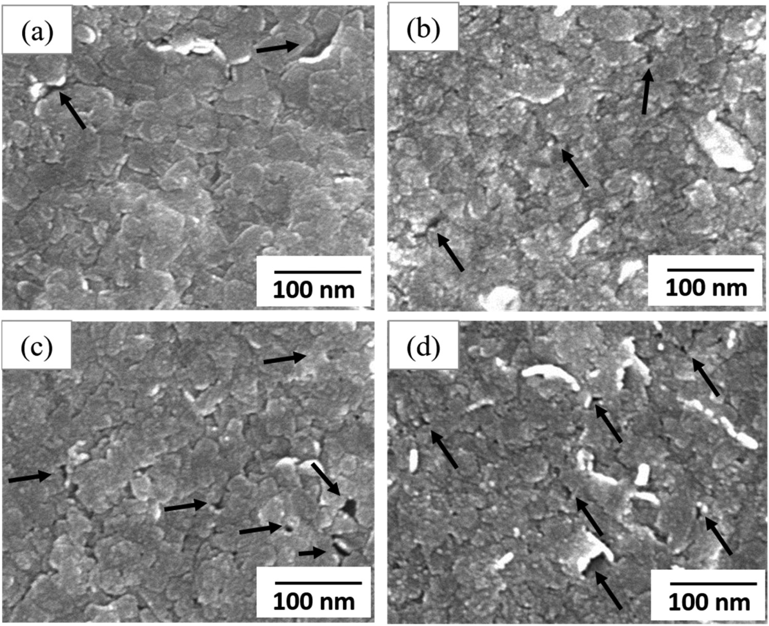
Crystalline structure of ZnO and MZO thin film
XRD analysis was performed to evaluate the crystal structure and different structural parameters of the ZnO and MZO thin films. Figure 2 shows the XRD patterns of ZnO and MZO films synthesized by sol–gel derived dip-coating method. The undoped ZnO exhibit diffraction peaks correspond to the (100), (002), and (101) crystal planes at 2θ values of 31.69°, 34.18°, and 36.31°, respectively. The observed diffraction peaks matched with the standard JCPDS card no. 36-1451. From the XRD pattern, it is observed that the peaks are very sharp which corresponds to the highly crystalline nature of ZnO thin film. With the incorporation of Mg, a small shift in XRD peaks to the lower angle is observed. Such a shift could be attributed to the smaller atomic radius of Mg ion (78 pm) compared to that of the substituted Zn ion (83 pm) [17]. The shifting of diffraction peak signifies the incorporation of Mg ions into the ZnO lattice due to doping [11]. The intensity of the diffraction peaks decreases with the increase of Mg-doping concentration suggesting deterioration of the crystallinity of the films due to doping. During doping, defects are introduced into the lattice sites which may decline the crystalline quality of the films [11].
XRD pattern of the undoped ZnO and MZO thin films.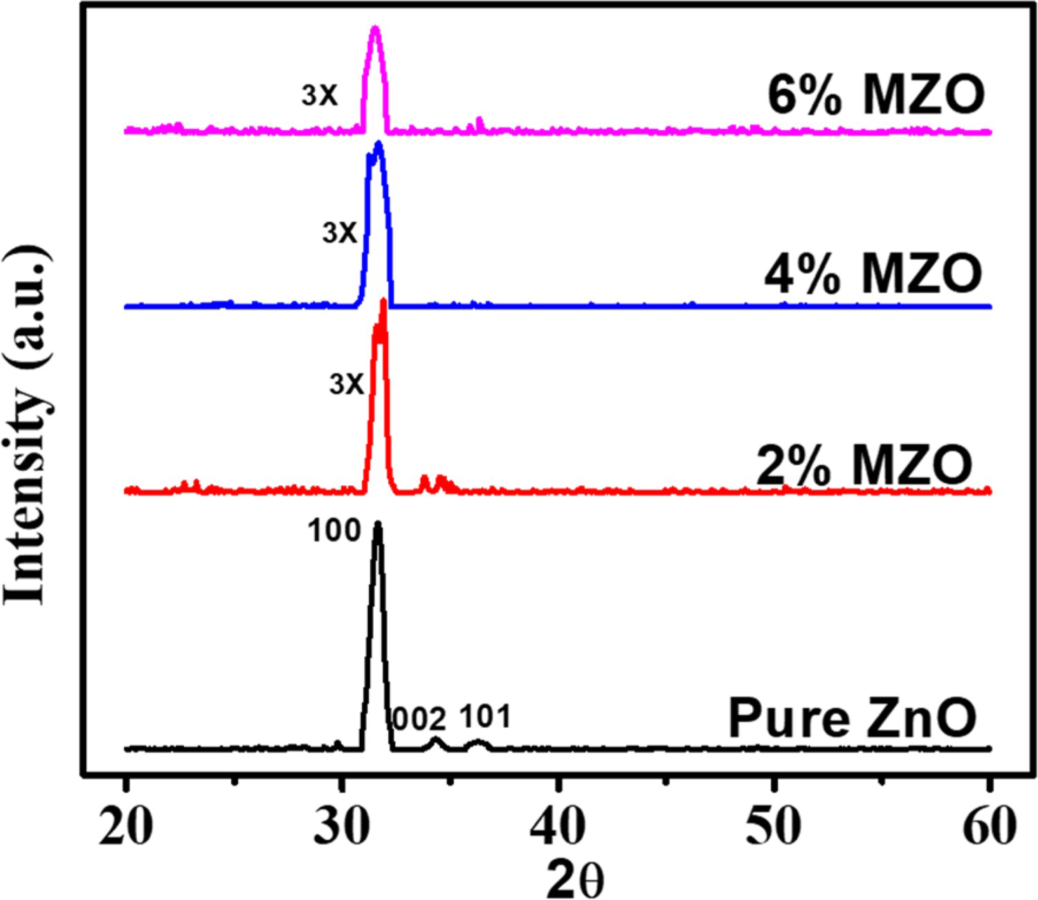

Structural parameters of MZO thin films.
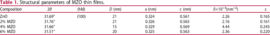
The microstrain of the films was calculated using the relation [28],,

Optical properties of ZnO and MZO thin films
The optical transmission spectra of as-prepared thin films were investigated using UV-VIS spectrophotometry at wavelength range of 200‒1000 nm. Figure 3(a) shows the transmittance spectra of the ZnO and MZO films as a function of wavelength. All the samples show sharp cut-off at the UV region together with uniform transmittance within the visible spectrum. The cut-off frequencies for pure ZnO, 2 wt-% MZO, 4 wt-% MZO, and 6 wt-% MZO are 368, 362, 370, and 365 nm, respectively. The cut-off at UV region indicates a high optical band gap of the films. In the UV region, the transmittance of all the thin films is less than 20%. Transmittance as high as ∼85% are obtained in the visible region for the 2 wt-% MZO and 4 wt-% MZO thin films, and are higher than that of the ZnO films. The higher transmittance of the MZO films can be attributed to the crystalline nature and porosity of the films [30,31]. Figure 3(b) shows a plot of the absorption coefficient of the films as a function of photon energy. Lower absorption coefficient is observed in the visible region due to the higher transmittance of thin films in that region. The absorption coefficient depends on the concentration of Mg-dopant and the highest value of the absorption coefficient was observed for 6 wt-% MZO thin films.
(a) Transmission spectra and (b) absorption coefficient for ZnO and MZO thin films for different amount of Mg dopant concentration.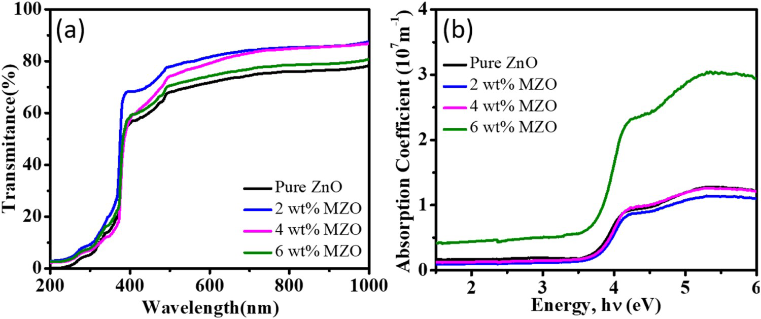
The optical band gap of ZnO and MZO thin films were calculated from the Tauc relation [20]
(a) Tauc plot for estimating the band gap of the ZnO and MZO thin films, (b) variation of optical band gap as a function Mg-doping concentration in ZnO thin films.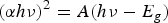
Such an increase in band gap could be attributed to the Burnstein‒Moss effect [32]. When semiconductor is heavily doped, the donor electrons occupy the states at the bottom of the conduction band, and the Fermi level moves towards the conduction band. As a consequence, the electron requires more energy to move to the unoccupied state of the conduction band resulting in an increase of band gap [32,33].
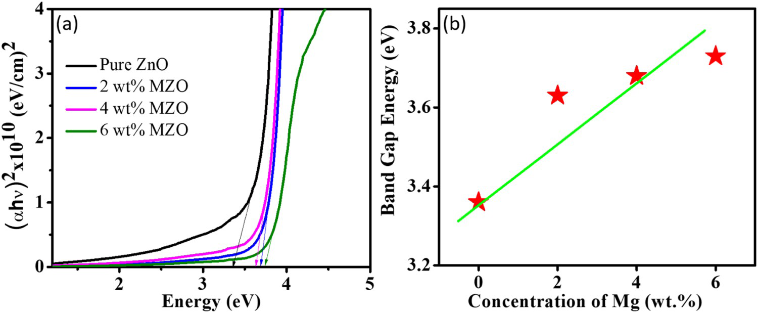
In optical transition, an electron from the valance band absorbs a photon and move to the conduction band across the band gap. During this transition, the electron may experience disorders caused by defect centres and/or thermal vibrations. These disorders create a density of states tailing into the forbidden energy gap. The width of this tail, called the Urbach tail, is an indicator of the presence of defect levels in the forbidden gap between the valence and conduction bands. The energy associated with this tail is referred to as the Urbach energy and can be calculated by the relation [34]:
(a) Plot of ln (α) vs energy to evaluate the Urbach energy of ZnO and MZO thin films, (b) variation of Urbach energy as a function Mg doping concentration in the films.
Electrical properties of ZnO and MZO films
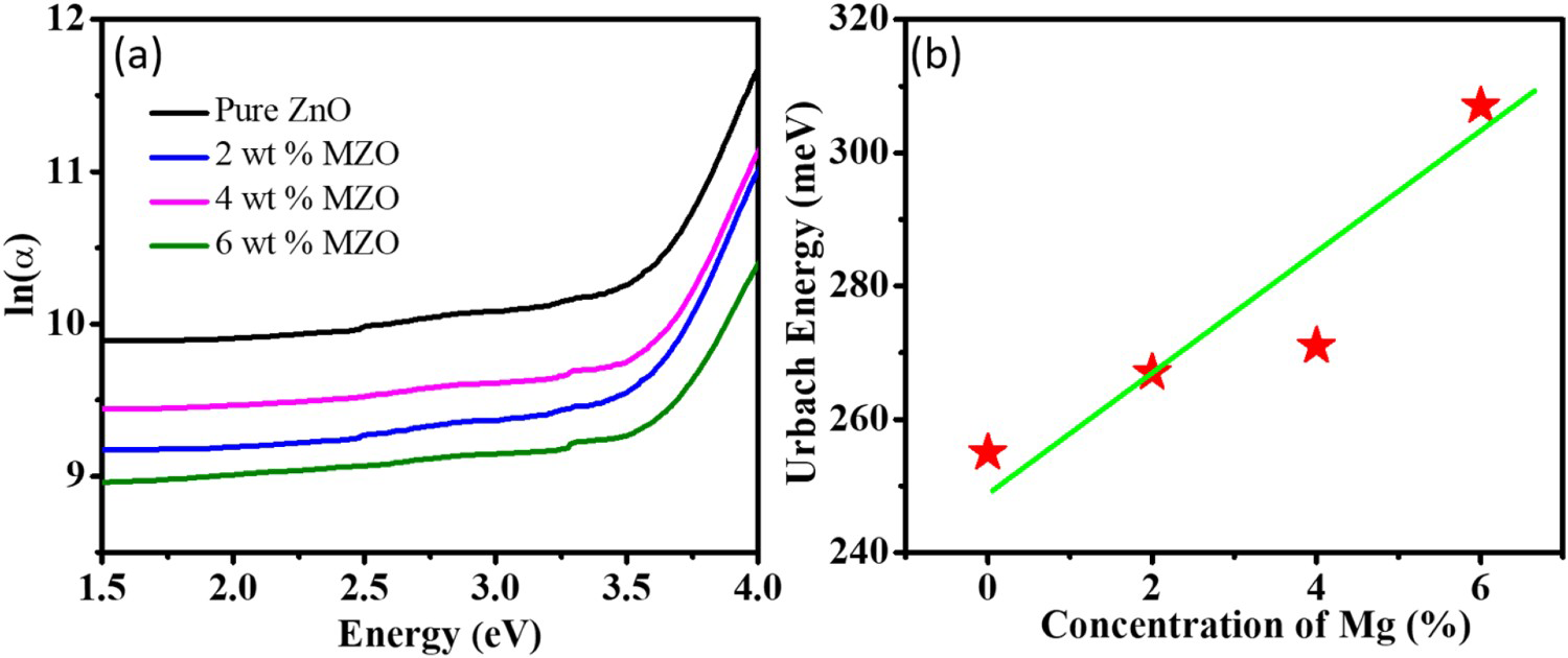
The DC electrical properties of the ZnO and MZO films were studied by van der Pauw four-point collinear probe methods. The electrical resistivity, ρ of the thin films were estimated using the formula [27]
Variation of the electrical resistivity of ZnO thin film as a function of Mg doping concentration.
Photocatalytic activity of ZnO and MZO thin films
The photocatalytic activity of ZnO and MZO thin films were evaluated by studying the disintegration characteristics of MB dye under UV light illumination. To achieve this, a solution containing MB and photocatalyst was illuminated by UV light and the corresponding changes in the intensity of the absorbance peak at ∼664 nm were monitored from the absorbance spectrum of MB solution in a regular time period.
Figure 7 shows the variation in the intensity of the absorption spectrum of MB dye with ZnO and MZO films for different duration of UV illumination. It is observed that the intensity of the absorption peak reduces with the illumination time for both ZnO and MZO thin films. However, with the presence of MZO film the peak intensity of the absorption spectrum of MB reduces at a faster rate than that of the ZnO film. For a quantitative analysis of photocatalytic activity, the peak intensity (at 664 nm) at any time (C) is divided by the initial intensity (C0
) and the values of C/C0
are plotted against corresponding irradiation times. Figure 8(a) shows the plot of C/C0
as a function of irradiation time for MB, MB + ZnO, and MB + MZO. From figure, it is obvious that the photocatalytic efficiency of MZO film is higher than that of the ZnO films. It is also observed that MZO film degrades 70% of the MB dye after 5 h of UV illumination, whereas ZnO film degrades 55% of the dye for the same duration of illumination.
The effect of (a) ZnO and (b) MZO thin film on the absorption spectra of MB solution for different reaction time under UV light illumination. Plots of (a) C/C0
and (b) ln(C/C0) as function of UV light irradiation time for the degradation of MB dye with the presence ZnO and MZO thin film as catalyst.
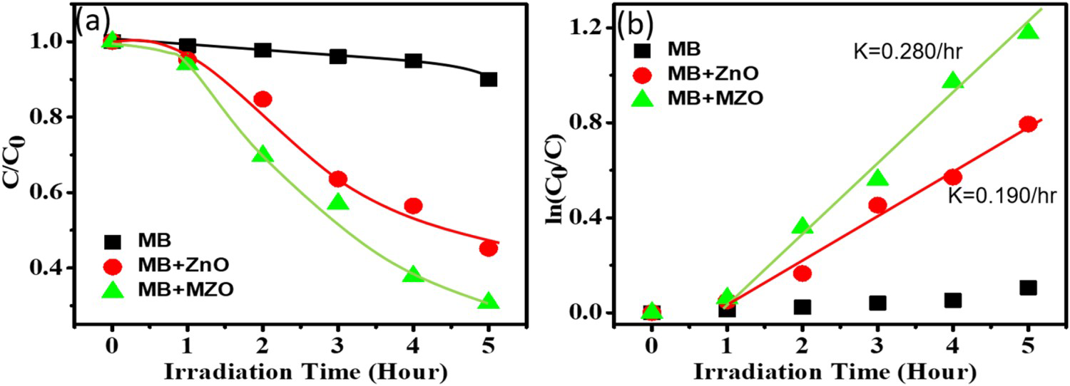
To understand the characteristics of the photocatalytic process, the degradation kinetics of ZnO and MZO films were investigated by the Langmuir–Hinshelwood (L-H) model [39,40]. According to L-H model, the reaction rate, R, is proportional to the fraction of the surface covered by the reactant, θ and can be expressed as

When the initial concentration of the organic compound is very low (KC<<1), the above equation reduces to a pseudo-first-order reaction:

Mechanism for the photocatalytic degradation of ZnO and MZO thin films
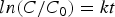
Figure 9 represents a schematic diagram illustrating the photocatalytic degradation mechanism of ZnO and MZO films. When UV light illuminates, electron (e−) from the valance band of ZnO move to the conduction band leaving behind a hole (h+) in the valance band. This e− (h+) reacts with the water molecule (atmospheric oxygen) and produces hydroxyl radicals ·OH (superoxide radicals ·O2
-). The radicals oxidized and thereby degrades the organic dye [11,16].
Proposed schematic diagrams to explain the effect of Mg doping on the photocatalytic activity of ZnO thin films.
The improvement of the photocatalytic performance of ZnO due to Mg doping could be attributed to a number of factors. For pure ZnO the photo-generated electrons in the valance band move quickly to the conduction band, recombine with the holes, resulting in a reduced rate of photocatalytic degradation. The optical analysis (Figure 5) showed that Mg doping introduces localized electronic states in the energy band gap. These states may act as trap centres which trap the electrons from the conduction band and thereby reduce the probability of electron–hole recombination [40]. Furthermore, an increase in the optical band gap due to Mg doping causes higher redox potential of the photo-generated electron/hole pairs, which significantly increases the catalytic efficiency of the MZO films [41]. Moreover, with Mg-doping, the porosity of the film increases, this in turn increases the surface area of the films that makes the films exposed to more organic pollutants [25]. Additionally, the porous structure of the film can greatly increase the scattering of incident UV light [42]. As a result, more UV light is absorbed by the MZO thin films which help to improve the photocatalytic efficiency of the thin films.
Conclusions
In summary, undoped and MZO thin films were deposited by the sol–gel dip-coating method and the effect of Mg doping on the photocatalytic performance of the films was studied. Mg doping was found to introduce pores in the surface of the films and deteriorate the crystallinity of the films. The optical band gap and the Urbach energy of the films increase with the increase of Mg dopant concentration. The photocatalysis experiment showed that Mg doping significantly improves the photocatalytic efficiency of the films. The improved photocatalytic activity of the films could be attributed to the retardation of the photo-generated electron/hole recombination rate due to the creation of charge traps together with increased surface area caused by pore-formation due to Mg doping. Finally, the MZO thin films synthesized by a simple, effective, and economic method may open up a versatile route for efficient water purification by photocatalysis.
Footnotes
Disclosure statement
No potential conflict of interest was reported by the author(s).