
Abstract
This review describes biofilms and the need for a materials science/engineering approach to solve the problems in the medical field. In particular, biofilm problems are closely related to infectious diseases. Most chronic diseases and the hospital-acquired infections could be attributed to biofilms. Biofilms usually form on materials. They may be biomaterials such as implants, catheters, stents and others. These films can also form on items outside of the human body. They include beds, service tables, medical knives, needles for injections, etc. Even though materials obviously affect biofilms formation and growth, there have been few studies using the materials science approach. In this review, we summarize the concept of biofilms from the viewpoint of materials science. Topics include the interaction between biofilms and materials (especially metallic materials), evaluation techniques, a bird's-eye analysis of previous investigations, and a discussion about the future direction for developing anti-infectious metallic materials.
Introduction
Since the pandemic of COVID-19 began, people have been keenly concerned about infection. Infection (from bacteria and viruses) is mainly spread from humans to humans. It may also spread from various animals and by contaminated materials. Even though information is still lacking about viruses, bacteria (especially those protected by biofilms) are responsible for many infections.
Technical (biological) terms often used in Biofilm related topics.
In this review, we start by summarizing the concept of biofilms from the viewpoint of materials science. Various topics are discussed including the interaction between biofilms and materials (especially metallic materials), a bird’s-eye analysis of previous investigations, and a review of practical materials used in medical fields (based on the mutual interaction between metallic materials and bacteria/biofilms). Finally, we present the future direction of investigation for the development of anti-infectious materials.
Materials and biofilms
Biofilms are formed by bacterial activities. Since bacteria exist everywhere, biofilms are likely to form everywhere. However, they need nutrition that often exists on substrates. Substrates may be abiotic (all kinds of physical materials) or organic tissues/organs. Biofilms form at the interface between environments and substrates. From this viewpoint, substrates (materials or tissues) affect the formation of biofilms in various ways.
Figure 1 shows schematically how biofilms form on materials and tissues. The process starts with the attachment of planktonic bacteria on the substrate surface. The planktonic bacteria are those existing individually. We have thought that bacteria live as a singular floating existence in most cases. However, this arbitrary presumption was wrong. It is true that bacteria are floating freely in some cases. These floating bacteria are called ‘planktonic bacteria’. They are different from the bacteria in biofilms. Their differences will be discussed later. Planktonic bacteria are generally seeking nutrients. For bacteria, their nutrients should be carbon compounds, which exist as conditioning films [30–46] on materials’ surfaces. These films are a kind of inhomogeneous film-like matters (Figure 1(a)). Therefore, bacteria tend to attach to materials’ surfaces to get nutrition (Figure 1(b)). Bacteria have to overcome many kinds of barriers to attach to materials’ surfaces [47–52]. Basically, two different phases having their own electric double layers would act repulsively due to high osmotic pressure, when they would overlap each other. From the viewpoint of surface static charges, metallic materials generally have a negative charge. Bacterial surfaces usually have charges too. Therefore, it is possible that they might be repelled from each other by electrostatic forces. However, other forces and actions would occur to overcome the repulsive potential barrier. Such an attachment and detachment process would continue at the beginning stage. After a period of time, the attachment action would overtake the detachment one and the number of bacteria on the material’s surface would begin to increase (Figure 1(c)). When the number of bacteria reaches a threshold value in the local area of materials’ surfaces, a phenomenon relating to a signal deduction process called ‘quorum sensing’ would occur [53–99]. The signal proteins released from the bacterial cells would increase with the concentration of the bacteria. Then they would re-enter the bacterial cells and react with DNA. As a result, a phenotype change would occur in order to excrete polysaccharides outside of the bacterial cells. This phenomenon is quorum sensing and a kind of signal deduction process by proteins. A certain signal chemical(s) for bacteria is used to increase the concentration of bacteria in a restricted local area and then the bacteria in the area begin to excrete polysaccharides. Sticky biofilms form on materials’ surfaces, as shown in Figure 1(d). We presume that polysaccharides could make a great contribution to increase stickiness in the region of materials’ surfaces. However, bacteria originally secrete various kinds of proteins around them. Portions of bacteria are constantly destroyed (bacteriolysis). Therefore, biofilms must contain not only polysaccharides but also proteins, nucleic acids and lipids [7,100]. These polymers could form bridges that result in inhomogeneous film-like matters (such as hydrogels), and biofilms form on materials’ surfaces as a result. This is the ideal biofilm at the beginning stage. However, a biofilm could collapse due to the lack of nutrition within it and by enzymes working to break it apart (Figure 1(e)). Or the other physical factor could make contribution to the collapse, such as flow etc. [101,102]. In any case, bacteria released from biofilms return to the environment as planktonic bacteria. They float freely again and look for other substrates to attach to. Once bacteria attach to new sites, the same process occurs to produce other biofilms. In such a way, biofilms would cover many parts of materials macroscopically. Schematic illustration for biofilm formation, growth and rupture.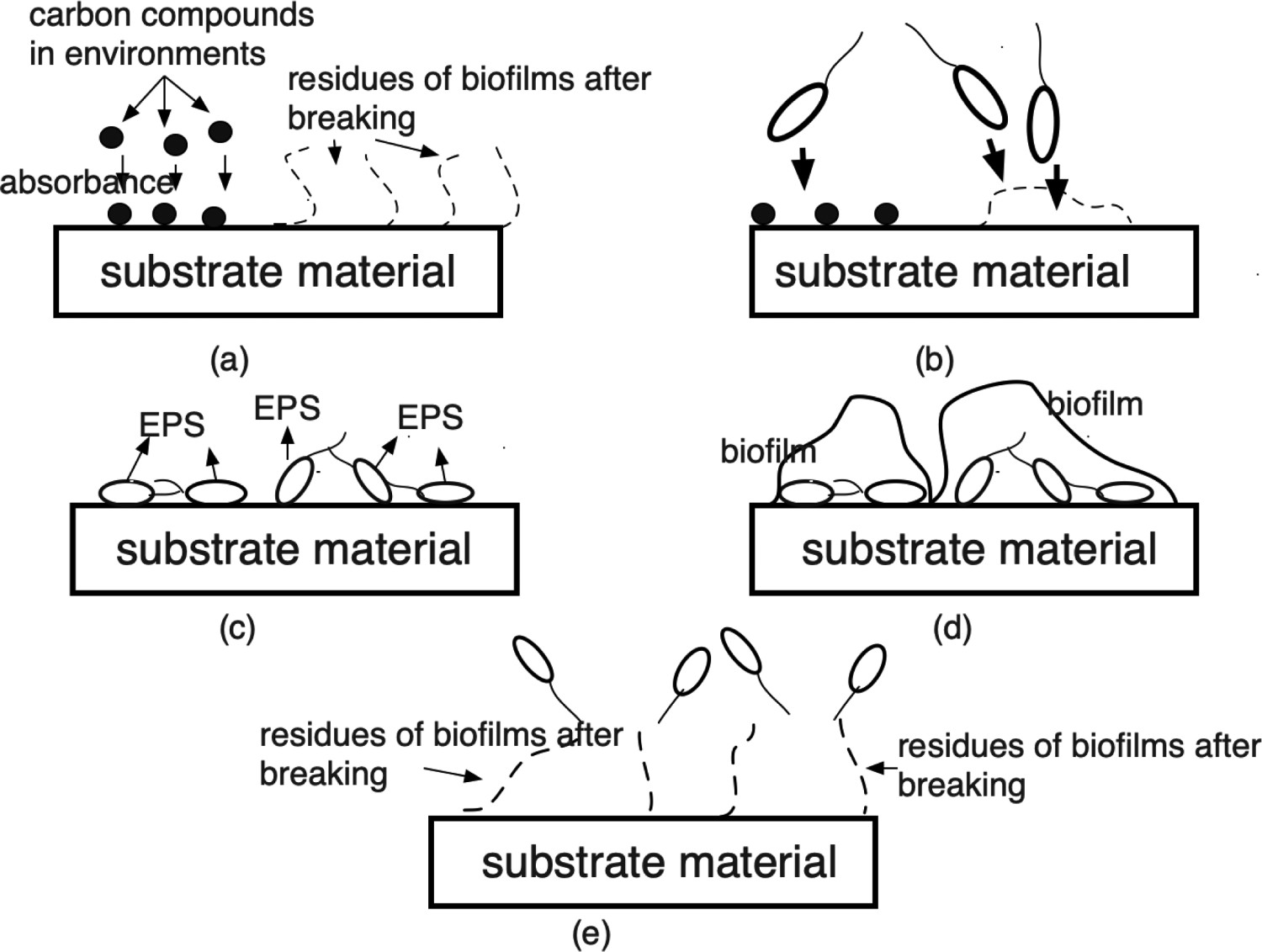
Without materials (substrata), there would be no biofilms at all in most cases. Biofilms always form at the interface between a liquid–solid phase, a vapor–solid phase and liquid–liquid interfaces. Similar processes described above must occur at these interfaces. When thinking about biomaterials, biofilms formed on industrial materials should also be considered. As shown in Figure 2 schematically, each phase has its own electric double layer. When the two different phases are overlapped, the repulsion would occur due to the high osmotic pressure. Therefore, bacteria must overcome the repulsion (due to some other forces), in order to form biofilms. At this point, the substrate materials would make a great contribution to biofilm formation and growth. As described in the previous section, there are already some theoretical proposals to consider the effect of substrates. However, we would like to propose our simple guidelines from the practical viewpoint as materials scientists. The guideline may not be applied to some cases successfully, even though there are many complicated factors involved in the formation and growth of biofilm. However, one could reach a better solution based on this practical guideline. Repulsion forces between different phases when they approach each other.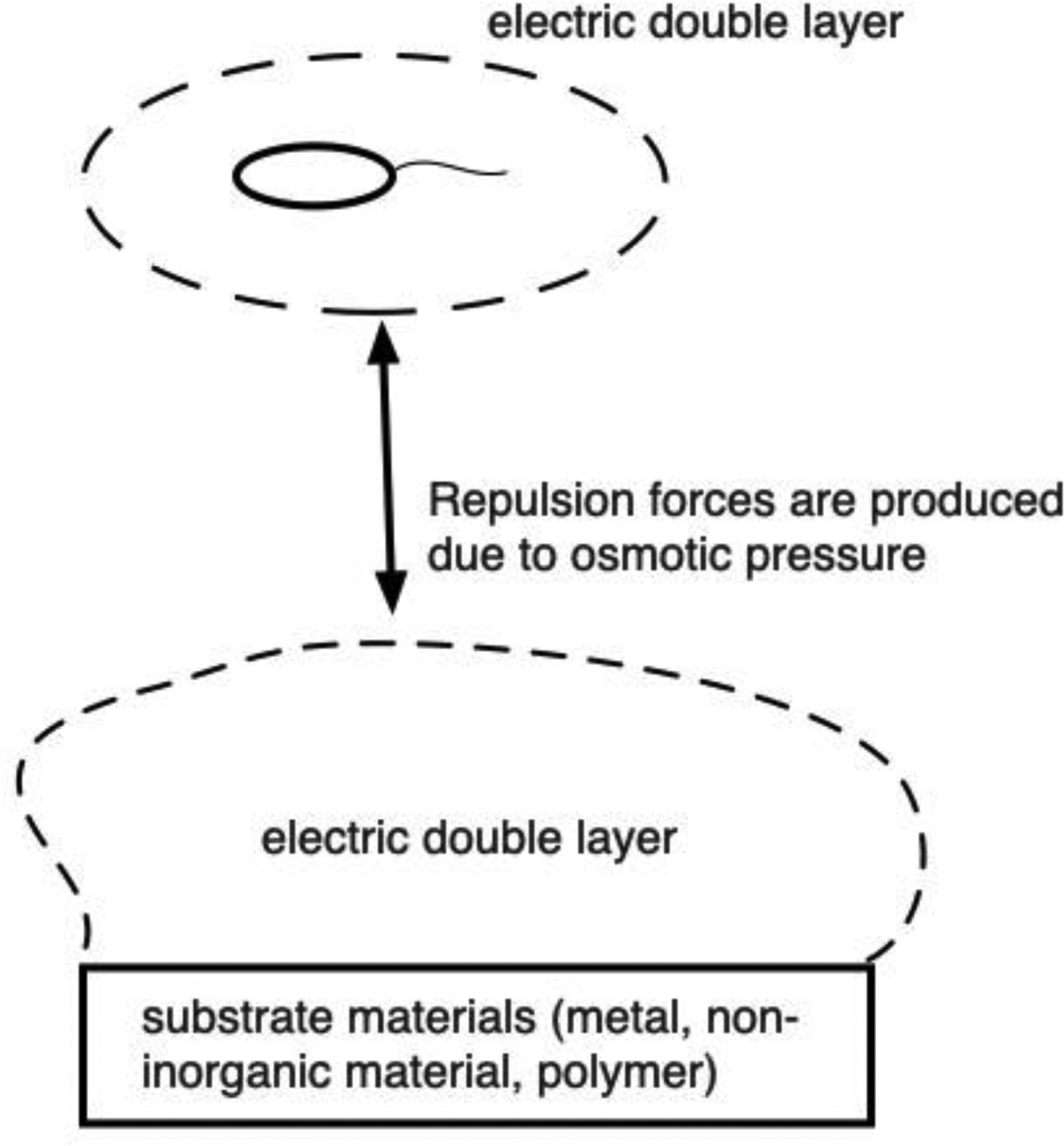
In regards to the forces between bacteria or biofilms and substrate materials, we first classify materials in the following three categories: metallic materials, non-metallic inorganic materials (ceramics, etc.) and polymers. As shown in Figure 3 schematically, the interaction between bacteria/biofilms and materials depends on which steps are to be considered. Therefore, it is difficult to solve the biofilm problems from the viewpoint of materials. However, this approach is needed to solve the problems properly, even though it has been lacking so far and not fully understood. Initially, a material’s surface must be conditioned, so that bacteria could overcome the repulsion and attach to its surface. When the material would be virgin, some carbon compounds could adsorb on the surface. At this stage, the interaction between carbon compounds and materials’ surfaces could be initiated. At the second stage, the interaction between materials and bacteria or their excreted proteins might begin. And at the third stage, the interaction between materials and extracellular polymeric substances (EPS) in biofilms would be the key factor for making a biofilm stable or unstable. For all of these cases, the biofilm control depends on materials and their interfacial properties. Various interactions between materials and bacterial environments depending on the growth stage of biofilms.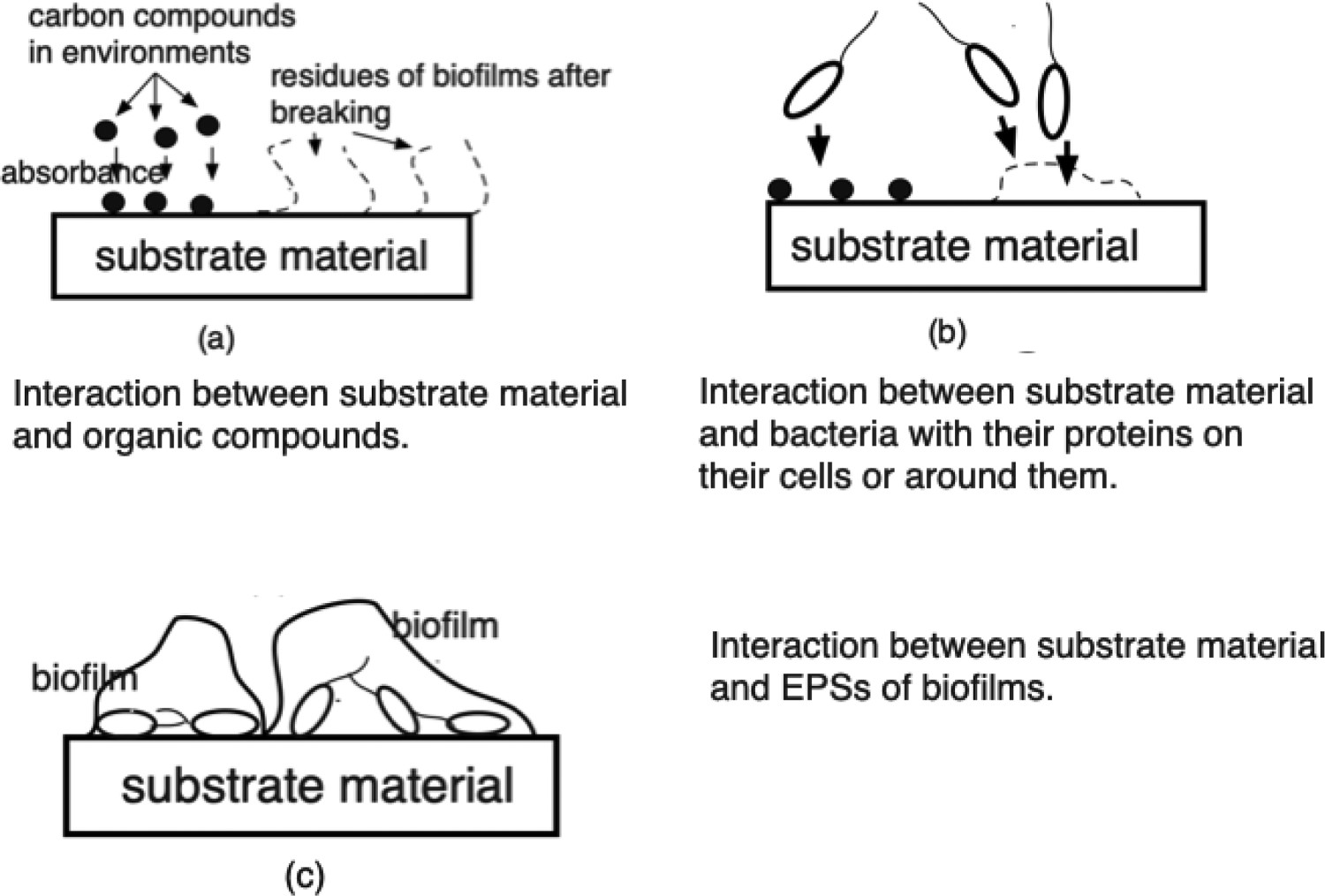
Biofilms, infection, and chronic diseases
Biofilms form by bacterial activities. The process could occur both outside and inside of our bodies. Figure 4 compares schematically how biofilms form in both cases. In extracorporeal cases, bacteria generally attach to the solid materials (Figure 4(a)). On the other hand, bacteria attach to tissues of organisms in intracorporeal cases. Bacteria attach to a surface and the number of bacteria increases in order to excrete polymeric substances. Then the biofilms begin to form and to grow, overcoming the resistance of various antibody responses (Figure 4(b)). In both cases, the increase of bacterial number is the common and most remarkable characteristic. In the extracorporeal case, the increase of bacterial number is actually defined as infection, since ‘infection’ could be expressed as the case when pathogenic microbes enter the bodies of organisms to increase bacterial numbers and to cause disorders. From the viewpoint of infection’s definition, biofilms might be almost equal to the concept of infection. Some surveys say that most chronic diseases (more than 80%) [103] or many diseases (more than 60%) [104] are caused by biofilms. However, some doctors have said that biofilms would almost be infection itself. In particular, chronic diseases are caused by biofilms, since bacteria have high resistance to antibiotics. It clearly shows that diseases caused by bacteria residing and hiding in biofilms, would retard one’s recovery as a result. When biofilms form, bacteria exist in them. The ‘biofilm bacteria’ are different from planktonic bacteria which exist in the environment individually. Within a biofilm, nutrition could be shared by bacteria. Also due to some reasons, it is hard for biocides and antibiotics to kill bacteria in biofilms. Biofilms Growth in extracorporeal (a) and intracorporeal (b) environments.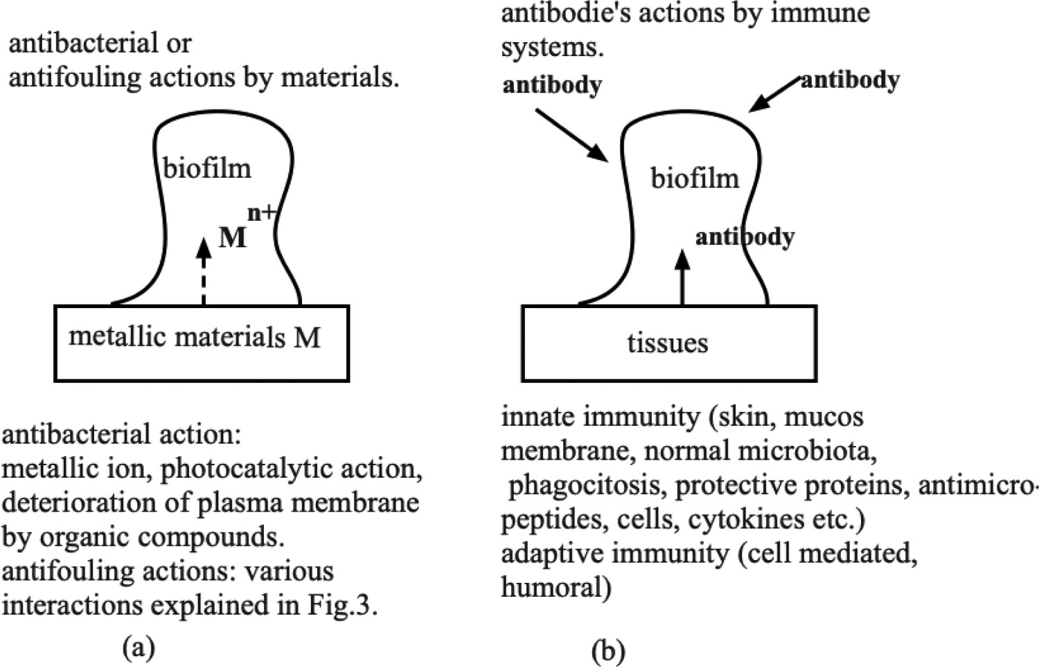
Why do bacteria in biofilms have high resistance to antibiotics? There may be many reasons for that. According to some investigations so far, the following three reasons and mechanisms could be mentioned at this point [105–113].
One of them is the filtering effect of biofilms. Actually, biofilms are composed of many kinds of extracellular polymeric substances. Some of them may be hydrophobic and others may be hydrophilic. In such an environment, the repulsion between antibiotics and EPS would occur and would be an obstacle for antibiotics to penetrate through biofilms.
The second possibility is the production of enzymes which could decompose some antibiotics. An example for this type of mechanism is beta lactamase in biofilms by Pseudomonas aeruginosa which would hydrolyze beta-lactam antibiotics such as penicillin [114,115].
Biofilms and chronic diseases (some typical examples).
General concept of biofilm and its relationship with metallic materials
The attachment of bacteria to materials’ surfaces would be affected by many factors such as flow rate, pH, temperature, etc. Many researchers have clarified various factors so far. However, bacterial attachment could be affected by factors on the material’s side. What kind of factors could we postulate for the material’s side?
We could classify the artificial materials into three categories: metals, non-metallic inorganic materials and polymers, in order to consider the affecting factors. This way, a correlation between biofilms and materials could be considered more in detail. Since materials in each category show different interactions with bacteria and EPS, the materials’ groups need different explanations for biofilm formations. In this review, we would like to focus on metallic materials.
As for metallic materials, metallic ions dissociated from metals in aqueous environments play an important role, as shown in Figure 4(a). As described above, biofilms contain lots of polymers composed of proteins, polysaccharides, lipids and nucleic acids. They might form a bridge among themselves. However, many metallic ions might be a part of the bridge among polymers, or they might bind to proteins as a chelate [120]. This means that metallic materials might bind to proteins as signal matters. Also they might stick to the proteins (existing on the bacterial membranes), which originally work as receptors or transporters. It is very hard to determine which kind of metallic ions work for a certain purpose and the process would be too complicated to fix. However, the experimental results must be reflected by such a series of complicated phenomena between proteins or other polymers and metallic ions [121].
Metallic materials are generally ionized at the vicinity of surfaces at equilibrium states to some extent. When equilibrium states are broken to polarization status (due to some reasons), then corrosion would occur to produce ions in the aqueous solutions. The phenomenon is related to corrosion resistance. And it may be related to the toxicity of the metallic materials, since metallic ions would bind to the surfaces of human cells leading to harmful toxicity. However, biofilm formation characteristics are also related to such an ion dissolution like corrosion resistance, since all of these are promoted by the dissolution of metallic elements into their ions. Figure 4 shows the concept of interactions between metals and biofilm formation. Bacteria excrete and scatter proteins around them in environments, when they float freely as planktonic bacteria. When they approach metallic materials’ surfaces, they must have strong repulsion due to the overlap of electrical double layers. However, some metallic elements such as iron, one of the essential elements for organisms, have high attraction forces with bacteria, since some proteins around bacteria might be reactive with iron ions. In addition to that, many bacteria have binding sites on their outer membranes as ‘transporter’. Due to these reasons, some metallic ions would attract bacteria, which would lead to biofilm formation. Therefore, the good corrosion resistance would lead to the control of biofilm formation very often. From this viewpoint, the interaction between metallic ions and polymeric substances derived from bacteria and biofilms must be the key to control biofilm formation and growth.
In fact, the investigation of cases for iron and steel have been advanced. Bacterial cells need iron to survive, since iron plays an important role to convey oxygen and to make contributions for various Redox reactions. However, it is hard for soluble divalent iron to exist under the current atmospheric conditions on earth. Iron usually exists as a trivalent ion. Therefore, bacteria generally have difficulty incorporating iron into their cells. Usually, bacteria incorporate iron into their cells as a complex using siderophore, a low-molecular compound derived from bacteria (Figure 5(a)) [122,123]. This process is well-known for E. coli. However, the incorporation mechanism due to the formation of metallic iron-polymeric molecule complexes seems to be applied to more general cases [124]. Also except for iron, aluminium [125], calcium, copper, zinc, manganese and chromium [126] could be selected to form metallic complexes. According to the tendency, biofilms could be formed on those metallic materials, even though some of them could prevent the growth due to their anti-bacterial effect. Table 3 summarizes the siderophore and binding metals for some practical metal elements. The uptake mechanism of metallic ions. The iron case was mentioned as an example. Siderophores and binding metals.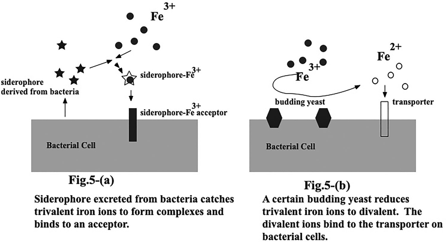
There is another uptake mechanism for metallic ions. That is the incorporation of metallic ions through transporters on cell membranes. Transporter could be defined as an acceptor composed of proteins to bind many kinds of matter and to incorporate them inside cells [135–143]. Various metals are incorporated into bacterial cells through the metal-binding transporters. As for iron, the haem transporter [144–148] is very well-known to ‘catch’ iron ions and to incorporate the metallic ion into the cells. Generally, divalent metallic ions have their own transporters on bacterial membranes (Figure 5(b)). Both mechanisms would work to realize the strong interactions between metallic materials and bacteria to form biofilms and overcome various repulsive factors. There have been few investigations from this viewpoint so far. However, developments in this direction are expected soon.
The difference between biofilm problems and antibacterial ones
Antibacterial effect
Some concepts of antibacterial effects.
For all of those cases, bacteria growth would be suppressed. However, the extent and the suppressed ways are basically different. When we come to think about the mechanism to kill bacteria or to control bacterial growth, materials could be classified into three types.
The first class corresponds to antibacterial metallic materials. The concrete examples are silver and copper. Those metallic materials are ionized and those ions would attach to a certain site of the surface of bacteria (for example, transporters) and make the role of proteins ineffective. As a result, the growth of bacteria might be retarded or controlled significantly.
The second class is some ceramics having photocatalytic properties [151–160]. In most cases, they would be oxides such as titanium oxides. In this case, the irradiation of light would lead to the generation of active oxygen due to the photocatalytic reactions. The active oxygen would kill bacteria or make the metabolic reaction inactive.
The third class is organic chemicals attacking and impairing plasma membranes. Basically, there are many kinds of organic chemicals for the purpose and they are developed not by the R&D of materials, but by biocides and antiseptic drugs.
Among those detailed concepts, Kohkin (A Japanese word corresponding to ‘the control of bacteria number on materials surfaces’) has been a universal technical term in English and has been applied to manufactured products. Materials having Kohkin effect do not require the killing or complete elimination of microorganisms as do other classifications of sanitization. Generally speaking, the decrease down to one-hundredth of the original bacteria number corresponds to the appearance of the Kohkin effect. When we come to think about the original concept of Kohkin, the materials in the first and second classes belong to the categories for the Kohkin effect, obviously. They are generally environmentally friendly and safe for our human bodies, while the small amount of use would lead to Kohkin. As for metallic materials, silver and copper have been representative elements. As for photocatalytic materials, titanium oxides are representative ones. Actually, most of the proprietary materials are those matters.
When discussing metallic ions and bacterial attachment, the interaction between metallic ions and bacteria becomes important again. In this case, the attachment of metallic ion to bacterial surfaces would play an important role to control the bacterial growth. Antibacterial effects of metallic materials must lead to a decrease in the number of bacteria on materials’ surfaces. Therefore, this effect contributes to antibiofilm properties ((b) in Figure 3). In light of that, antibacterial effect of metallic ions would lead to antibiofilm properties of materials. However, biofilms are not evaluated only by antibacterial effects alone, since the killed and collapsed bacteria could be one of the components in biofilms. This is the reason why the number of living bacteria could not precisely evaluate the antibiofilm properties of materials.
Biofilm and biofouling
The two technical terms very often appear simultaneously and it might lead to misunderstanding and confusion. Therefore, we would like to clarify the difference and also to explain how they would affect the biomaterials.
As explained repeatedly, biofilms are heterogenous film-like matters formed at the interface. Particularly, our case in this review is restricted to biofilms at materials’ surfaces. Therefore, biofilms are matters themselves as bacterial products. As already explained, biofilms are composed of bacteria (including both living and dead bacteria), EPSs and water. The components and those ratios would generally change with time and substrates. Bacteria would attach to mucosal membranes, tissues or some other human biological substrates, while they would attach to biomaterials inserted into our human bodies as implants, etc. For the former, natural defense systems (immune systems) work to control bacterial growth and biofilms. However, they usually don’t work in the case of the latter. Therefore, biomaterials might develop biofilms on them without any controls, which might lead to serious infections. It is generally called Biomaterials associated infectious diseases (BAI). However, we would like to refer to it in this review as Biofilm Associated Infectious diseases, since both terms are almost equal.
On the other hand, biofouling is the process where bacteria would attach to materials and every phenomenon involved with the attachment. When we restrict the process to biomaterials related matters, there are many kinds of biofouling as processed in our bodies. For example, catheters, stents and various implants would have biofouling by biofilm formation/growth processes. The biofouling process contains the stage of a conditioned surface and also that after the breakdown of biofilms. Therefore, using the word ‘biofouling’, we always try to consider the biofilm-related phenomena as a whole to propose problems or to solve them holistically [161–166].
Biofilm associated infectious diseases
What are biofilm-associated infectious (BAI) diseases
As already described in the previous section, biofilms would be almost the same as infection and they would cause diseases, while forming on implants or components used in hospitals. Conventionally, BAI means biomaterials associated with infectious disease in many cases so far. However, it could be called ‘Biofilm Associated Infectious Diseases, since biofilms formed on biomaterials would lead to chronic diseases or hospital-acquired diseases. As already explained, bacteria residing inside biofilms formed on biomaterials have high resistance to antibiotics and a natural defence system such as phagocyte, NK cells, etc. In the next section, we introduce some concrete examples for BAI [167–171].
Concrete examples for BAI of metallic materials used biomaterials
From practical viewpoints, stainless steels [172–175], cobalt-chromium alloys [176–181], titanium alloys [182–186] (including TiNi alloys) and dental alloys [187–192] are important. They could be used as biomaterials as implants.
Stainless steels are applied to intervascular stents to broaden blood vessels. Since the infection has been rare for intervascular stents so far (about 0.3% morbidity) [193], stainless steels have a relatively high resistance to biofilm formation, while blood environments within artificial or natural blood vessels do not contain so many bacteria due to the function of the immune system.
Biofilm formation/growth characteristics have been investigated by a number of researchers. For example, Gristina et al. [194] postulated this anionic, extracapsular, polysaccharide slime produced by bacteria protects them from antibiotics and sequesters critical ions from the surface of biomaterials. They showed the minimum bactericidal level of tobramycin for bacteria in the biofilm formed on stainless steels. Adachi et al. [195] show the resistance of stainless steel against biofilm formation/growth, compared with titanium. Oga et al. [196] indicated that the biofilms by Staphylococcus epidermidis under laminar flow and the formation capabilities were compared among metallic materials including stainless steels and polymers. According to their results, stainless steels could control biofilm formation much more than polymers did.
Co–Cr alloy systems are also used as biomaterials. They are classified into four types according to ASTM. Cast CoCrMo alloy (F75) [197], wrought CoCrWNi alloy (F90) [198], wrought CoNiCrMo alloy (F562) [199] and wrought CoNiCrMoWFe alloy (F563) [200]. They were developed for dental materials at first. Then they were applied to artificial joints. Actually, the artificial joint generally has relatively high morbidity from the viewpoint of medical treatments. It ranges from 0.5% to 1.0% [193]. Chromium tends to form thin and dense protective oxide films and therefore the toxic elements do not work to mitigate the biofilm formation. Their biofilm formation/growth characteristics have been investigated so far [201–205]. Urushibara et al. compared biofilm formation capabilities among some dental alloys, Titanium, Co–Cr, Ag–Pd–Au etc. and decided the order of resistance.
Titanium and its alloy (Ti–6Al-4V alloy) are also often used as a biomaterial, since they have good mechanical properties and also good corrosion resistance [206–217]. The metal and its alloys also form thin and dense protective oxide films on it, which lead to high corrosion resistance. Therefore, they would have relatively high resistance to biofilm formation. When we think about their good biocompatibility, titanium alloys are used much more practically. They are used mainly for artificial blood vessels as intravascular devices and for artificial joints as extravascular devices. We also investigated the biofilm formation of the titanium and titanium alloy system by using some appropriate biofilm evaluation methods on the laboratory scale [218]. In our case, we found that pure titanium had a high resistance to biofilms by E. coli (K12, G6). However, the resistance against S. epidermidis (ATCC 35984) was not so high. As this example shows, the resistance seems to depend on the combination of bacteria and substrate materials (Figure 6). Comparison between antibacterial effect and antifouling effect.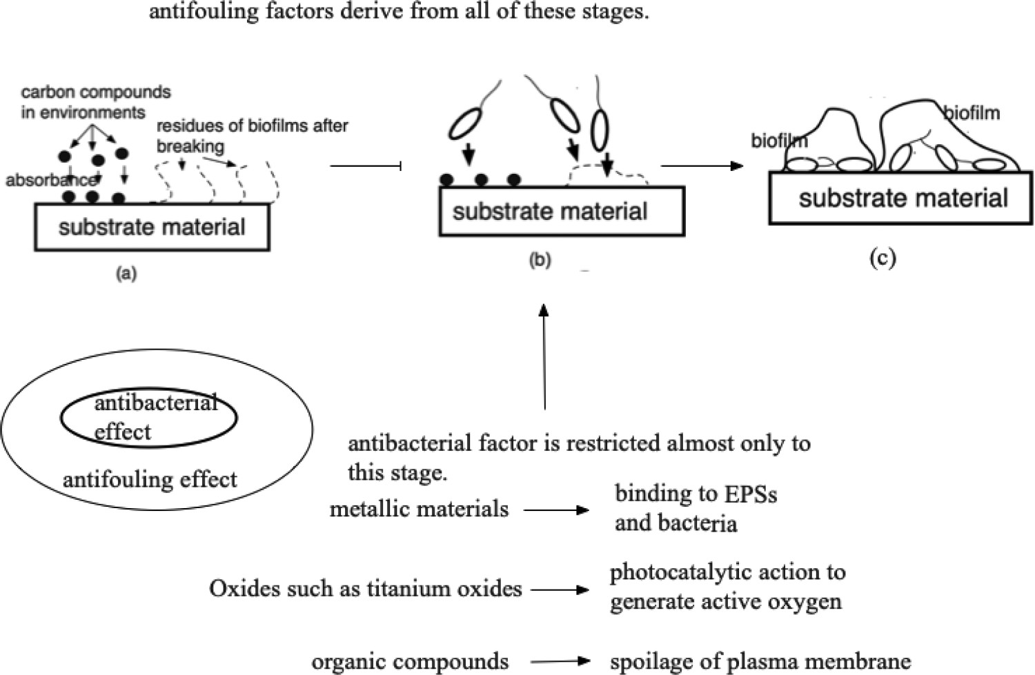
Other possibilities of metallic materials in the medical fields
In the previous section, we focused on the application of biomaterials applied inside our human bodies. However, there are many other applications of metallic materials outside bodies in the medical fields. They also lead to the nosocomially acquired infection (hospital infection). Since the type of infection through matters had not attracted people’s concern in the past so much, there were few data about it, the human resource about the investigation was lacking etc. As a result, the information at this point is not enough to describe thoroughly at this point. According to Hayashi in Kitasato medicine [219], he summarized the pathogens into three types. The first class contains pathogens intervening between human to human directly. The second class is composed of pathogens conveying human to human through materials. And this category was classified into three sub-groups, depending on the important ratio between humans and substances. The third group is composed of pathogens playing an important role for the infection through materials. The results are summarized and shown in Figure 7, schematically. Table 5 summarizes those bacteria on materials conveying bacteria to other humans and the materials’ sources. Infectious sources and bacteria. Infectious sources of materials in hospitals and related pathogens.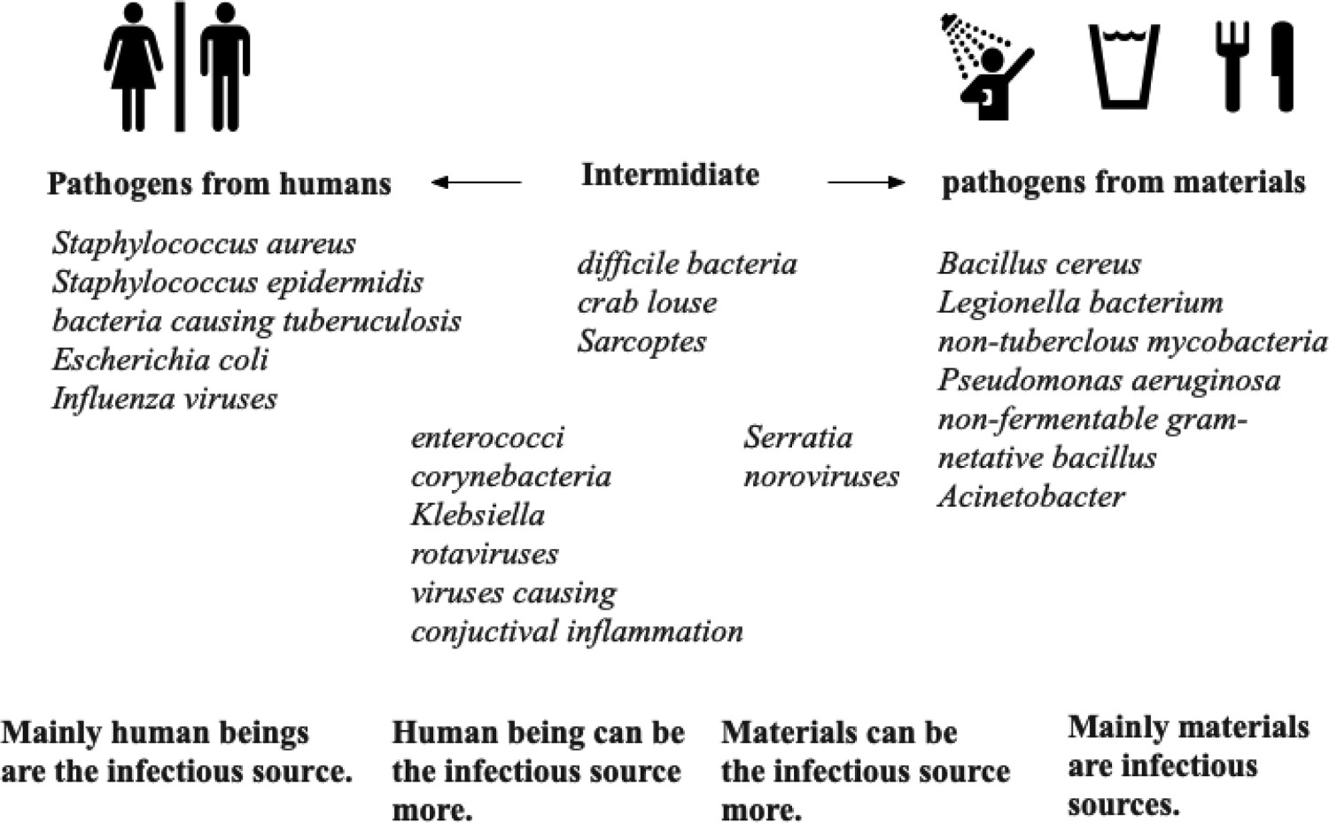
Being restricted to metallic materials, two kinds of materials, stainless steel [220] and copper alloy [221], seem to be used very often for the purpose of antibacterial effect. The copper element originally shows high antibacterial effect. Therefore, we could expect the antibiofilm properties, since the bacterial growth would be controlled. However, the affinity of copper with polymeric substances derived from bacteria is strong. And the dead bacteria would be also the component of biofilms. Therefore, the effect of antibiofilm is still controversial. On the other hand, silver has had shown high antibacterial effect historically [222]. Therefore, silver has been used for alloy components or coating materials for those general metals.
Stainless steels containing silver or copper have been developed around the turning of the century [223,224]. And various silver coated and copper coated materials have been developed [224]. The application cases fell short of expectations in the past. However, the rapid increase of antibacterial markets (including antibiofilms and antiviruses ones), people’s strong concerns all over the world may get a chance to utilize the accumulated knowledge base of antibacterial materials, so that the research and development in the past will serve a lot to make Good Health and Well Being World come true [225]. From the academic viewpoint, the suitable evaluation method must be established. We will write about this topic in the following section (see Section Biofilm characterization for material developments).
Biofilm characterization for material developments
Conventional characterization for materials – the merits and demerits
It is important to observe and evaluate the biofilm behaviours and the formation capabilities on practical materials in the medical fields. Of course, in vivo evaluation is naturally very important. Materials have been inserted into mice or other laboratory animals to check for biofilm formation/growth capabilities, under the collaboration of medical scientists, doctors and biologists [226–236]. Particularly, Lebeaux et al. [234] in the Institute Pasteur provide us the detailed review of in vitro and in vivo evaluation methods.
It is very important for us to check the materials’ anti-biofilm formation properties in vivo. The appropriate evaluation method should be established from the viewpoint of materials science, since materials scientists and engineers need to develop them in vitro at the initial stage of their R&D activity. Being apart from those biological evaluation methods, some instrumental analyses familiar to materials science researchers and engineers are desirable for the future developments. In light of this need, there are still a few evaluation methods available. The movement for the future will be explained in the next section. At this point, we could mention some microscopic techniques.
The simplest and introductory instrumental way to observe biofilms is with an optical microscope. The optical microscope uses a light as a probe to irradiate materials’ specimens and the reflected or transmitted light forms an observation image. Metal microscopes, stereomicroscopes, biological microscopes would be available too [237–241]. The optical microscope irradiates light from above the sample, and the light reflected by the sample is observed. Therefore, it is also called a reflection microscope. On the other hand, for the biological microscope, a light source is placed under a sample. The sample is irradiated from below, light is emitted, and the light passes through the sample and is guided to our eyes. Therefore, this system can be called a transmission optical microscope.
In general, in the case of an optical microscope, the probe is visible light, and the signal is reflected light or transmitted light. Since the probe is visible light, the resolution is necessarily influenced by the wavelength of visible light as the probe. Actually, the resolution (d) is determined by the following equation.

The resolution (d) can be calculated for an optical microscope by using Equations (1) and (2). Resolution is the shortest distance (in micrometers) between two points that can be distinguished by the observer. However, the resolution is micrometer order usually, when we use optical microscopes. And when the size of objects would be close to the minimum limitation, the artefact would be inevitably involved.
Some representative fluorescent pigments and their excitation/emission wavelength.
The confocal laser microscope played a successful major role in the structural analysis of biofilms [242–247]. The measurement principle is as follows. A laser beam from a pinhole (point light source) is projected onto the sample. Further, a pinhole and a detector (photomultiplier tube) arranged at the image position of the sample are arranged. Here, when the focus is on the sample and the pinhole on the sample is also focused, most of the reflected light can pass through the pinhole and be received on the detector.
A confocal laser microscope exhibits great power when used in combination with a fluorescent dye (fluorescent confocal microscope). The reason is high contrast and resolution. A resolution much higher than that of a normal microscope can be obtained in all directions in the XYZ directions. Therefore, it seems that it became possible to observe uneven biofilms that are difficult to observe with a normal microscope.
Scanning Electron Microscopy is very often used for material analyses. The electron is used as a probe and the analysis is performed in a high vacuum chamber in order to project an electron beam onto a sample’s surface without being scattered. If the degree of vacuum is low, gas molecules collide with the electron beam and are scattered, which significantly reduces the observation accuracy. When the analytical method would be applied to biofilm research, a special procedure to make samples is needed. Since biofilms would generally contain lots of water components, it is replaced with alcohol gradually, frozen and evaporated in a vacuum. However, the original form was lost and some hydrated components might disappear. At this point, some artefacts might be involved in the results. Recently, low vacuum pressure SEM (E-SEM) has been newly developed. When this would be applied to biofilm research, new findings can be expected in the future [248–251].
Focused Ion Beam (FIB)-SEM is another promising way to observe biofilms. Ions released from the ion source are focused on target specimens by a condenser lens (an electrostatic lens). An electrostatic deflector makes the ion beam scanned on specimens. Then the secondary electrons are scattered from the biofilm on specimens and the image is obtained. Since the microfabrication and trimming in the chamber is possible, three-dimensional visualization of local areas (‘slice and view’) is possible. Therefore, many investigations to visualize biofilms have been investigated so far.
Using the same electron as a probe with conventional SEM, TEM (Transmission Electron Microscopy) makes it possible for us to observe the interference figures produced by the transmitted electron beams. Since TEM uses the electron as a probe, the dissolution is much higher than that of optical microscopy. While the latter’s would be micrometer order or submicron one, TEM could cover the target on nano-order scales. At this point, there are three problems that need to be improved for this analysis. The first one is the need of a dehydration process to make specimens. In this case, the TEM would share the same problem with the SEM. The second problem is how to prepare thin specimens, since electron beams must penetrate the specimens. The third problem is how the artefact could be avoided through hydration etc., since the TEM also needs a high vacuum to analyse specimens. Even though the hard problems must be solved in advance, TEM could give us useful information which other methods could get. Since the size of the bacteria is around 1 μm, as an example of biofilm observation using TEM, it is possible to analyse the distribution of elements on the surface of bacteria and the uneven distribution. For example, when metal ions interact with bacteria, data such as which part the metal ions are acting on can only be visualized using TEM. In this sense, the observation method using TEM is mainly the observation of the movement of bacterial cells or elements rather than the biofilm itself, but these data are based on the microstructure of the biofilm or the interaction between the bacteria and the material. As a powerful tool that is indispensable to know the action, it is expected that it will be further expanded in the future [252–256].
Raman spectroscopy [257–260] and FTIR [261–264] could be used to evaluate biofilms, since they could detect EPS as a component of biofilms. When we come to think about the interaction between biofilms and materials’ degradation, it is much more realistic for us to focus not on bacteria, but on EPS. From this viewpoint, we think that these methods are practical and future-oriented methods. Both methods use light as an electromagnetic wave. When the light enters the specimens’ surfaces, energy is irreversibly absorbed by vibrations of molecules depending on the chemical structure, and the signal obtained from the specimens could be analysed. As for Raman spectroscopy, the Raman scattering would be analysed, while FTIR would analyse the transmitted infrared light. When the special attachment would be used, one could analyse the reflected infrared lights (FTIR-ATR). Raman spectroscopy and the FTIR method should be used in a complementary way. FTIR is good at analysing asymmetric polarizable materials, and Raman spectroscopy is better at analysing nonpolarizable materials. Materials containing water can be analysed more accurately by Raman spectroscopy than by FTIR. On the other hand, Raman spectroscopy often suffers from fluorescence disturbances, but FTIR does not have such drawbacks. These advantages and disadvantages are mutually exclusive, and more accurate analysis becomes possible by using them in a complementary manner. We authors have used these methods very often to evaluate materials’ biofilm formation/growth properties successfully [265–272].
In addition, Atomic Force Microscopy (AFM) [273–283], Scanning Tunneling Microscopy (STP) [284–288], Mass Spectroscopy [289–295], Nuclear magnetic resonance spectroscopy (NMR), UV-VIS, etc [296–303]. have been also proposed, and actually used commercially to some extent. For the future, all of these methods should be used in a complementary way as described, and combined with some beneficial materials’ microscopic observations. Such a combination of these methods might be desirable from the viewpoint of materials science.
Some representative instrumental analyses were mentioned above and explained. These instrumental methods have played an important role to clarify the structure of biofilms, and to visualize them concretely in the light of qualification analyses. However, most of them were weak at analyzing biofilms quantitatively.
Quantitative evaluation of biofilms using a biological method
Being apart from those immaterial scientific methods, there is an industrial and more instinctive way for the evaluation. Since biofilms would form on materials’ surfaces by bacteria, when the bacteria number would exceed a certain threshold value locally, the antibacterial effect would be related to the evaluation, as described above. Therefore, the evaluation for antibacterial effect could be one of the effective evaluations. The antibacterial effect of products could be tested, using an international standard, ISO22196. The test is usually called the Film Covering Method and it provides us with the useful information needed for practical applications. The concrete procedure is as follows. The specimens were put in a plastic Petri dish, while the bacterial solutions were prepared according to a certain procedure. The bacteria were incubated in 10 mL of a nutrient broth for 24 h at 35°C, and then diluted two–thousand folds with sterilized water and established as a bacterial solution. The diluted bacterial solution was applied to the specimen (16 micro litter per centimetre) and then a polymer film was laid over the solution. The sample was kept in an incubator for 24 h at 35°C. After the incubation, a solution of 10 mL of sterilized water containing 200 micro litter of Tween 80 (a nonionic surfactant and emulsifier) was introduced into the plastic Petri dish and the bacteria attached to the specimen and polyethylene film were washed into the aqueous solution. To determine the number of viable cells, serial decimal dilutions of the cell suspension were made, a 0.1 mL portion of which was uniformly spread on an agar medium. The plate was incubated at 35°C for 24 h and the colonies formed were counted. The viable cell count was represented as colony forming unit per milliliter (CFU/mL). The final colony formation number was measured to evaluate the antibacterial properties. This evaluation method is very suitable for concrete antibacterial goods. The efforts, achievements and versatile applicability to realize a reproducible and practical evaluation process to a standard should be estimated very highly. It is pretty easy for the process to be modified to practical applications for each case.
Since the antibacterial effect would not be equal to that of biofilms, other proper evaluation methods should be established. From the viewpoint of materials’ evaluation, there are still very few proper evaluation methods. For example, ASTM international already established some standards. However, they would be focused on the number of bacteria and it would not lead to the research and development of materials themselves, since the information would not help us to solve the interaction problems between materials and biofilms so much. In addition to that, most of the standards already established are oriented to the evaluation of chemicals, biocides and antibiotics. The standard of evaluation for materials is needed to develop the new antibiofilm and antibiofouling materials. A new standard that focuses particularly on biofilms and on the interaction between biofilms and substrate materials is needed from the industrial viewpoint.
New research and development – to International standardization
We mentioned the Film Covering Method as an industrial evaluation way as well as other methods by ASTM [304–307]. However, they are not enough to evaluate materials and components of practical produces, unfortunately. From this viewpoint, such a practical evaluation system available in industries should be developed much more. Now the authors are collaborating with a non-profit organization in Japan called SIAA [308] to develop a standardized method for biofilms [309]. To achieve the purpose, we adopted the staining method using crystal violet and are trying to demonstrate the availability using some instrumental analyses. We adopted this method, because crystal violet has been used to evaluate biofilms by many biologists [310–333]. However, the availability from the viewpoint of materials science has not been carried out so much yet. Since the cationic part of the chemicals would stain some parts of EPS in blue, the negative and positive ones should be investigated much more. In some application cases, the evaluation might be impossible or not available, since the chemicals would significantly stain the substrate itself [265,268].
The principle is shown in Figure 8 schematically. The material having biofilms on it is immersed in 0.1% crystal violet solution. Crystal violet (C25N3H30Cl) is an ionic compound composed of organic cation and chloride ions. The cationic part attaches to EPS in biofilms, stains them and shows a blue colour. Therefore, the stained parts must correspond to the quantity of biofilms. Based on this concept, stained biofilms on materials’ surfaces are extracted by alcohol or a certain surface- active agent such as dodecyl sodium sulfate. The light at the wavelength of 570 nm is irradiated to the extraction liquid. Since crystal violet absorbs the light selectively, the absorbance is measured to evaluate the quantity of biofilms. The use of crystal violet has been investigated by many researchers and it seems like this method has been widely recognized. Therefore, the method will be standardized in the future. The staining method will be combined with some materials scientific method(s) and will work well to analyse biofilms both qualitatively and quantitatively soon. Crystal violet assay to evaluate biofilms on materials.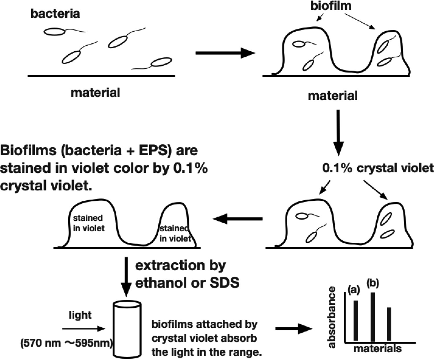
Future scope
Countermeasures should be proposed properly. Biomaterials have been selected among biocompatibility, mechanical properties and other special requirements (for example, an osteoconductive property). Due to certain reasons, the selection of materials has had a limitation from the beginning. Here we have other requirements from the viewpoint of biofilms. As we and others continue biofilm research in the future, new requirements may appear in light of an anti-infectious capability. Then we may have to make balances among all of them and try to solve the problem holistically once again. When we come to think about the balance, we may not adopt new biomaterials having high resistance against biofilm formation/growth so easily. In such a case, the surface coating seems to be the appropriate one. Actually, many coating methods have been proposed. However, they have not been commercialized. That is also one of the difficulties which we will encounter many times in the future.
In this review, we introduced the concept of biofilms producing infectious diseases. The concept and topic have been investigated in medical science and also environmental science so far. However, the approach to problem solving should be carried out more from the viewpoint of materials science, since biofilms always form at the interface between environments and materials. However, such an approach has been lacking so far. Therefore, we introduced the concepts for biofilms and described the interactions between biofilms and substrate materials. Also we proposed the concept about the interactions between substrate materials and bacteria/biofilms from the viewpoint of materials science. On a new perspective point of view, we will move to the new stage to hopefully solve the problems in the near future. We (the authors) sincerely wish that this manuscript will help materials scientists and engineers advance to the next new stage of R&D for new biomaterials.
Footnotes
Acknowledgments
We appreciate the Advanced Technology R&D Center of Mitsubishi Electric Co., Japan Food Research Laboratories (JFRL), and The Society of International Sustaining Growth for Antimicrobial Articles (SIAA) for their very useful advice and information. A part of this work was also supported by the GEAR 5.0 Project of the National Institute of Technology (KOSEN) in Japan.
Disclosure statement
No potential conflict of interest was reported by the author(s).