
Abstract
Peri-implant infection is rapidly becoming an – if not the most – important clinical challenge for indwelling medical devices. To alleviate the global rise in antibiotic use for the treatment of such infections, a plethora of biomaterials/bioengineering-based antimicrobial strategies are emerging to restrict or ideally to eliminate microbial adhesion and biofilm formation on implant surfaces. Yet, the development of such approaches faces specific challenges, like biocompatibility concerns, reduced antimicrobial effectiveness, long-term stability issues and antibiotic resistance development, which limit translation to the clinic. This review provides insights into the antimicrobial activity of current state-of-the-art biomaterial-based approaches to address the aforementioned issues. Translational research strategies and regulatory framework are also emphasised as key elements facilitating clinical implementation of anti-infective biomaterials. This review closes with the vision that the integration of computational tools and experimental databases using artificial intelligence (AI) would provide new insights for the accelerated development of next-generation biomaterial-based antimicrobial strategies.
Introduction
In the last half century, implantable medical devices (IMDs) have revolutionised healthcare and have progressively become an integral part of surgical therapies and procedures, such as orthopaedic joint replacement and bone substitutes, dental restorations, cardiovascular intervention or catheterisation, etc.[1–4]. Every year, the use of IMDs in all such treatment options improves the quality of, and even saves, the life of millions of patients worldwide [5]. In 2020, the global IMD market was valued at 112.3 billion USD and projected to reach 160.3 billion USD by 2026 [6]. Such projected growth is primarily due to the growing geriatric population worldwide and an increasing prevalence of – often age-related – chronic diseases. Moreover, current advances in in vivo diagnostic and therapeutic devices can be expected to tap into the IMD market segment for younger patients as well [7]. These devices allow monitoring tissue healing processes in situ or administering a localised sustained drug delivery. This increased use of IMDs also demands durability and longevity, which are often compromised due to a drastic increase in the incidence of medical device-associated infections (MDIs) – also called biomaterial-associated infections (BAIs) or peri-implant infections. For example, MDIs account for 12 and 25% of all hospital-acquired infections in China and the U.S.A., respectively [8,9].
Overview of the infection rate, most common pathogens, typical treatment protocols and associated cost estimation for various common implantable medical devices.
Notes: CoNS: coagulase negative staphylococci; GNB: Gram-negative bacilli; IMD: implantable medical device; MRSA: methicillin-resistant Staphylococcus aureus; MRSE: methicillin-resistant Staphylococcus epidermidis; MSSA: methicillin-sensitive Staphylococcus aureus; MSSE: methicillin-sensitive Staphylococcus epidermidis; sp.: species; spp.: species plural.
MDIs are initiated by microbial contamination of the IMD by opportunistic pathogens during the time of surgery, but also at a later time point by haematogenous spread or a continued contact with the external environment. The most common organisms associated with MDIs are commensal bacteria, such as Staphylococcus epidermidis and Staphylococcus aureus, which are naturally present on the skin and mucosal membranes. However, Escherichia coli and Pseudomonas aeruginosa are of similar concern, because of their multi-drug resistance [41]. Recently, the role of fungal species, such as Candida albicans, in MDIs has also been emphasised [42]. All these pathogens have the ability to adhere and to colonise on solid surfaces, while forming microbial aggregates embedded in abundant self-produced extracellular polymeric substance (EPS), so-called biofilms [10,35]. Within this biofilm environment, the ‘slimy’ EPS layer, consisting of exopolysaccharides, proteins, lipids and extracellular DNA, shields the microorganisms from external threats, making them considerably more resistant to host immune defences and administered antibiotics than their planktonic counterparts, as well as to many types of physicochemical treatment, such as heavy metals, acidity and UV light [43,44]. The biofilm status of microorganisms is also associated with persisters, slow-growing or growth-arrested bacteria with a reduced metabolic activity and decreased susceptibility to antibiotics [45]. Even if antibiotic treatment effectively eradicates most bacteria in a biofilm, there is a significant risk that a small fraction of persisters will survive and reconstitute the biofilm, once the antibiotic therapy is stopped [35,45]. Furthermore, horizontal gene transfer is accelerated in the dense microbial populations in biofilms, which increases the dissemination of antibiotic resistance [35]. Overall, this leads to a high antimicrobial tolerance in biofilms and thus, high recalcitrance of biofilm infections [43].
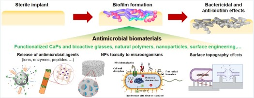
Indwelling medical devices are particularly sensitive to biofilm formation as they are inherently ‘foreign bodies’, which trigger an inflammatory reaction in reducing the immune response locally around the implant, while at the same time providing an ideal substratum for microbial adhesion [32]. Therefore, prevention of the initial microbial contamination is the first important step in the battle against MDIs. To this end, aseptic surgical protocols (laminar air flow rooms, strict hygiene, etc.), device sterilisation methods and the use of prophylactic antibiotics have already been successfully implemented and the effect of further measures in this regard is questionable [46,47]. As an adjuvant synergistic prophylaxis, anti-infective biomaterials have been considered as a valuable strategy to hinder pathogen dissemination and discourage biofilm formation. Over the years, a number of biomaterial-based antimicrobial approaches have been developed for implant surfaces, which can be broadly classified as surfaces that prevent attachment of microorganisms (antifouling), kill pathogens upon contact (contact-killing), materials incorporating antimicrobial agents, which are released locally around the implant (drug-releasing), or a combination of these approaches [42,48,49]. More recently, immunomodulatory anti-infective biomaterials, that do not dysregulate or even activate the host response, are gaining attention [50,51].
The pathogenesis of MDIs has already been addressed in various excellent review papers [10,35]. Some of these published reviews on antimicrobial biomaterials have captured the state-of-the-art either several years ago [38], or only focused on a single class of antimicrobial biomaterials (metal ions and nanoparticles (NPs) [52,53], polymers [54–58], materials with tuned surface topography [59], drug-releasing materials [60,61]) or a specific infection disease (orthopaedic and/or dental implant infections [62–65] and osteomyelitis [40,66] or infections on neuromodulation implants [67]) in the absence of an implant material [68]. Moreover, only limited data are available on the clinical performance of such antimicrobial implant materials [69]. A recent review by Kalelkar et al. critically analysed the translational potential of nano/microparticles, polycationic polymers or thermo-chemically responsive hydrogels as vehicles to deliver non-antibiotic antimicrobial therapeutic agents at the site of infection (in bone, lung, gastrointestinal tract, or eyes) [68].
In this review paper, we aim to discuss a wide spectrum of strategies based on antimicrobial biomaterials or biophysical stimulation for the treatment of implant-associated infections together with the mechanistic insights. We present here largely the most recent (<5 years) developments in this field and also present an international perspective on the current challenges that limit the clinical translation from bench-to-bedside as well as unexplored opportunities. The first focus in this review will be on the surface modification of metallic titanium (Ti) implant materials through nanoengineering approaches, such as surface texturing or by applying coatings of metallic NPs, possibly incorporated in diamond-like carbon coatings, or antimicrobial biomolecules via electrophoretic deposition (EPD). Within the class of ceramic and glass materials, special emphasis will be given to antimicrobial-releasing materials, such as crystalline and amorphous calcium phosphate (CaP) based systems as well as bioactive glass (BG) loaded with phytotherapeutics. With respect to polymeric materials, several natural polymers, including polyhydroxyalkanoates (PHAs), bacterial cellulose (BC) and collagen, will be discussed in reference to their antimicrobial properties. Lastly, other potentially translatable bioengineering approaches (e.g. electric/magnetic stimulation), which were never discussed in any of the published reviews, will be discussed for non-antibiotic antimicrobial applications. A number of unaddressed challenges including translating lab scale research to animal models and further to clinical studies, regulatory hurdles will be discussed, together with the emerging data science approaches. Taken together, this review provides critical insights for the implementation of the presented antimicrobial strategies for tomorrow’s medicine.
Nanoengineering of multifunctional titanium implant surfaces
The clinical performance of an implant is critically dependent on its interaction with the key components of the biological organism (i.e. tissues, cells, bacteria, blood, etc.), a property holistically defined as biocompatibility, which is directed by many properties such as surface topography and chemistry. As Ti alloys are widely investigated as orthopaedic and dental implants, much research is focused on the surface engineering of Ti alloys to encourage osseointegration, and to endow the implant surface with antimicrobial capabilities [49,62,63,65]. For example, the manipulation of biomaterial surface properties using nanoengineering approaches is deemed one of the most appropriate strategies to achieve the next-generation of multifunctional implantable metal-based biomaterials with improved osteoconductivity, osseointegration, and antimicrobial properties. This can be realised through precise control over the surface topography at the micro- and nanoscale or by altering the surface chemistry using nanoscale materials with antimicrobial activity. This section will cover recent advances in antimicrobial surface texturing of Ti biomaterials as well as chemical and biological nanocoating of Ti with metallic NPs, whether or not in combination with DLC coatings, or antimicrobial biomolecule coatings via EPD.
Surface texturing to enable a hierarchical micro-nanotopography
The substratum micro-nanotopography is recognised as one of the most important surface characteristics for the control of bacterial adhesion and prevention of biofilm formation [70,71]. The nanoengineering approaches to the design of antimicrobial surfaces have spurred the development of new surface patterns to significantly restrict bacterial adhesion and biofilm formation, in the absence of antibiotics [72–74].
Antifouling micro-nanostructured titanium materials
As presented in Figure 1, micro-nanostructuring of surfaces involve random, self-organised, or ordered surface features that result in three-dimensional (3D) topography of micro-nanoscale roughness, including both top-down (wet or dry etching) and bottom-up approaches (self-assembly) [72]. The fabrication methods for achieving nano-micro-rough topographies on Ti materials have been summarised in recent reviews [59,74–76]. In particular, the generation of micron-scale surface structures, such as those topographies found in nature on the surface of plant leaves (e.g. lotus leaves), and insect wings (e.g. cicada), have been explored for the purposes of antibiofouling, superhydrophobic surfaces [77]. Superhydrophobicity (a water contact angle >150° and roll-off angle <10°) of the surface can be achieved, when the micron-nanoscale protrusions on the surface lead to incomplete wetting of the surfaces (Cassie–Baxter wetting state) [78]. The entrapment of an air layer between the micron-scale features reduces the available surface area for bacterial attachment and, in air, impart low-adhesion properties that cause the rolling action of water droplets that collect surface contaminants (i.e. the lotus effect) [79,80]. In liquid, bacteria are unable to penetrate the liquid–air barrier due to the high interfacial tension of water [81]. For example, nanosecond laser structuring of Ti alloy surfaces to achieve a superhydrophobic topography was successful in delaying the formation of Escherichia coli and S. aureus biofilm, yet only for 48 h [82]. Indeed, it has been found that superhydrophobicity, imparted by surface structure, is metastable and may gradually transition to the fully wetted Wenzel state [77]. Irreversible transition from the Cassie–Baxter to Wenzel wetting states can occur due to the condensation or evaporation of water droplets, or external pressure. Thus, stable air-entrapment is highly important for durable superhydrophobic, antifouling properties of micro-nanostructured topographies [83]. Additionally, bacterial adhesion to a surface on the micron-scale, increases with surface roughness. However, bacterial adhesion on the nanoscale (10-100 nm) has been shown to be gradually inhibited [70,84]. For organisms smaller than the surface architecture, increasing roughness provides bacteria with greater attachment points, whereas dense, nanoscale features may reduce such sites for bacterial adhesion. Illustration of the fabrication of variable Ti mechano-bactericidal topographies and their mechanism of action. Hydrothermal etching, electrochemical anodisation, and plasma etching have all been used to develop antibacterial surface active patterns on Ti materials (patterns as pictured). The mechano-bactericidal mechanism can be explained as the nanostructure-induced rupture of bacteria by the mechanical forces imposed on the bacterial membrane as it adsorbs and stretches over high-aspect-ratio nanoprotrusions. The bacterial membrane rupture occurs at the point suspended between the nanopillars.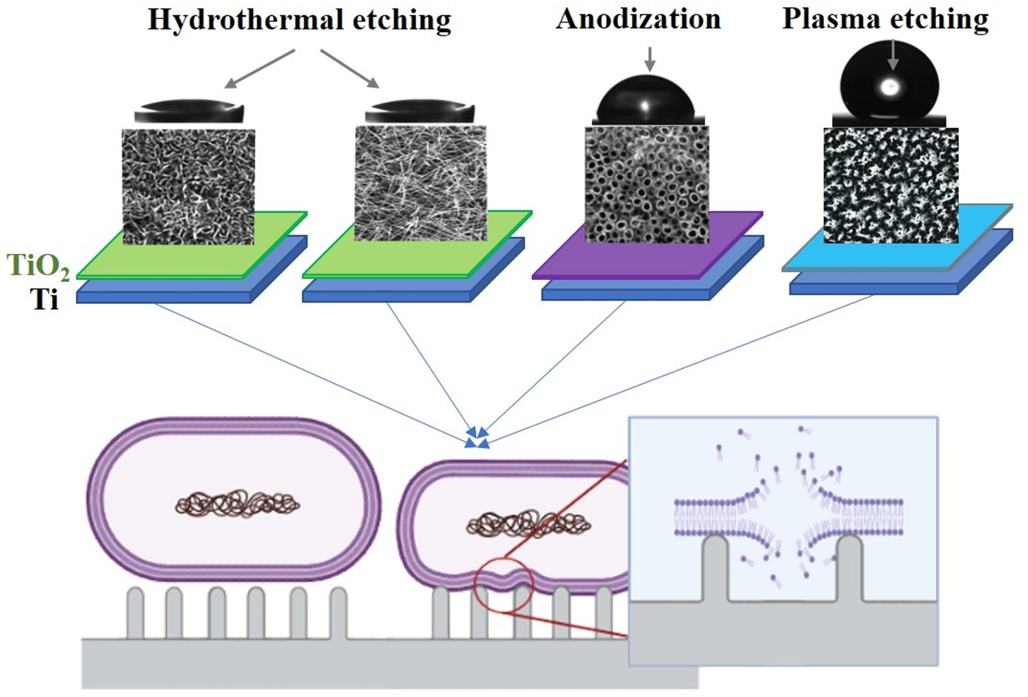
Mechano-bactericidal nanostructured titanium surfaces
Comparison of the bactericidal efficacy of different Ti nanotopographies, including nanowires, nanosheets, nanopillars and nanotubes, produced by hydrothermal etching, plasma etching, or electrochemical anodisation, which have been reported within the last 5 years. Data are presented as a percentage of non-viable cells.
Notes: Gram-negative bacteria are Escherichia coli (*) or Pseudomonas aeruginosa (**). Gram-positive bacteria are Staphylococcus aureus (†) or Staphylococcus epidermidis (††). ND: no data available.
Nano-coating of antimicrobial agents
Besides tuning the surface topography, chemical and biological surface modifications at the nanoscale offer an alternative nanoengineering approach to endow antimicrobial properties on the Ti implant surface [75,95]. We present here three promising routes to produce antimicrobial surface activity. Metal NP coatings can enable increased antimicrobial properties as compared to conventional materials due to their high surface area to volume ratio, while electrophoretic deposition allows a time-efficient coating yield of biomolecules, while preserving their antimicrobial activity.
Metal nanoparticle coatings
Metallic NPs, especially silver and gold NPs (AgNPs, AuNPs), display antimicrobial activity which is closely related to their particle size, high surface area, dispersibility and the ease of chemical modification by various surface functional groups [53,96]. Because of their multivalent nature and high surface area, these metal particles are capable of binding to different kinds of functionalised ligands, that specifically interact with receptors present at various target sites at the bacterial membrane. Once the NPs are internalised in the cell, they damage various molecules, including DNA and proteins, making it especially difficult for bacteria to establish resistance via an effective defence mechanism [97]. The NP toxicity may also result from NP interactions at the cellular membrane, even though they can be endocytosed [98]. The differences in NP toxicity depend on the different uptake kinetics and/ or on the specificities of the cellular target [99] (Figure 2). Their potential usage for the development of unique bioactive functionalised coatings on Ti implantable materials has been widely explored [75,100]. Metallic NPs can be coated on the Ti substratum via various deposition techniques, including EPD. Indeed, Ag additives to composite coatings on Ti materials have received the bulk of research attention; however, nontoxic and cytocompatible AuNPs, have shown uniquely advantageous broad-spectrum antibacterial activity against both pathogenic Gram-positive and Gram-negative bacteria [101,102]. In the recent past, triphenylphosphine (TPP)-stabilised AuNPs (TPP-AuNPs) have been explored as the precursor material for clinical applications, such as immunolabelling and therapeutics [103–107]. Furthermore, AuNPs stabilised by monosulfonated triphenylphosphine (TPPMS; IUPAC nomenclature: (m-sulfonatophenyl) diphenylphosphane) have been reported to exhibit in vitro antibacterial response. For example, Boda et al. analysed the antimicrobial activity of AuNPs stabilised with TPPMS in size range of 0.8–10.4 nm (Au0.8MS and Au1.4MS) [108]. Both Au0.8MS and Au1.4MS were shown to exhibit an antibacterial effect at 25 × 10−6 M Au against planktonic staphylococci with marked membrane blebbing and bacterial lysis in biofilm. The results indicated that AuNPs, conjugated with labile phosphine ligands alone, could show the potential bactericidal effects against staphylococcal infections [108]. Schematic illustration of NPs toxicity and thermally-induced toxicity by electrical and magnetic field caused by their potential interactions with bacterial cells. NPs and their ions and various external stimulus produce ROS inducing damage in bacterial membrane, proteins and DNAs, which eventually lead to bacterial death. NPs can kill bacteria cell by directly interacting with cell membrane and inhibiting the electron transport chain, which regulate the bacterial metabolic processes.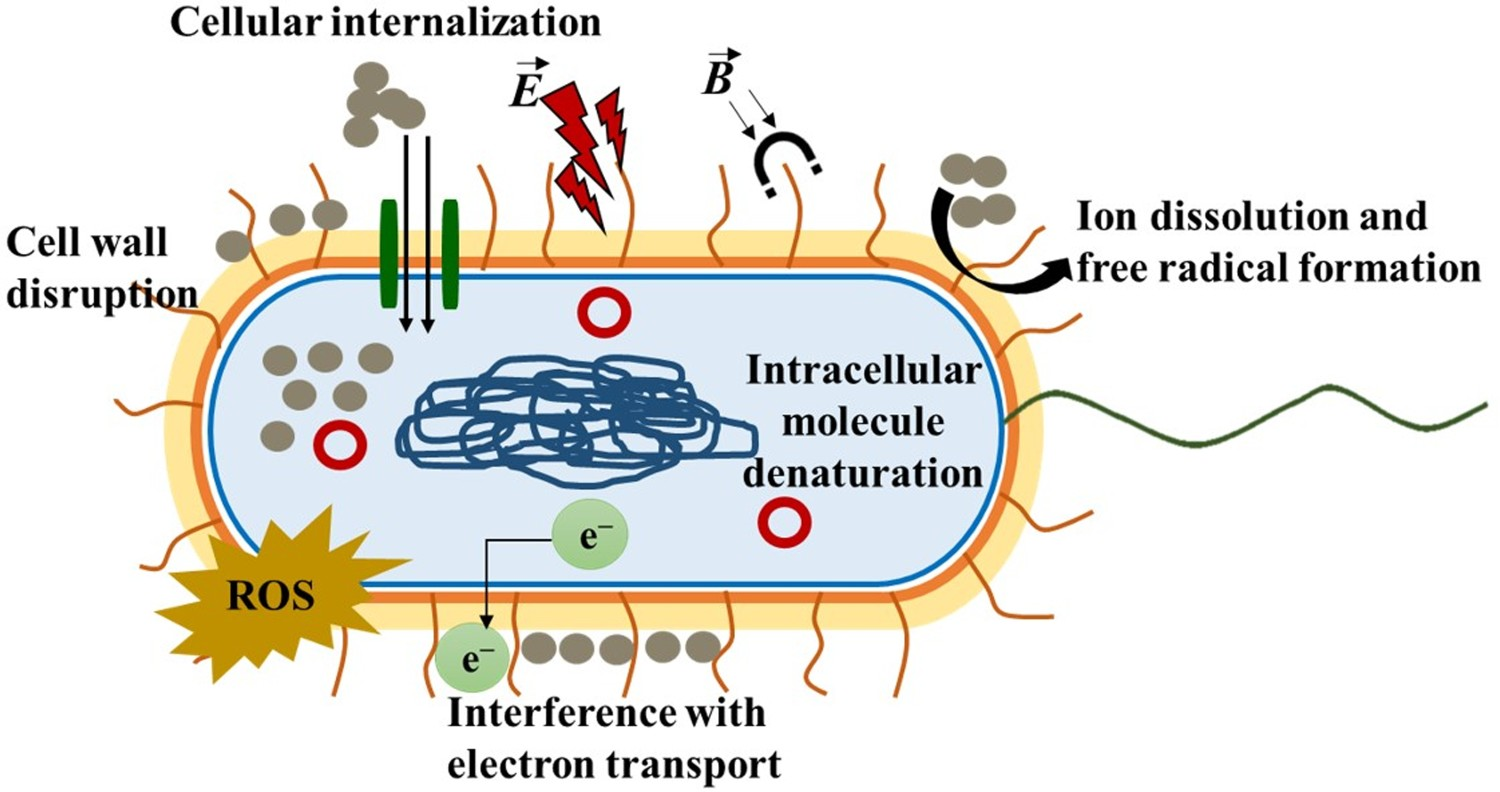
Special attention is also devoted to the modification of Ti using AgNPs incorporated in DLC coatings. DLC represents a major field of interest in the biomaterials community due to its antibacterial properties. The term ‘diamond-like carbon’, or ‘DLC’, was coined in the early 1970s to denote amorphous carbon films, which are expected to recapitulate those of genuine diamond in terms of density, strength and hardness [109]. Indeed, DLC films are characterised by high hardness (10-30 GPa) and high elastic modulus (100-300 GPa) [110]. Some of the key parameters for obtaining these ‘diamond-like’ properties are sp3 content and hydrogen content. In fact, DLC films contain intermediate hydrogen content (20-40 at%) and low overall sp3-bonded carbon content (up to 50%) [111]. The first one mainly controls the elasticity, whereas the second one determines the structure, passivates the dangling bonds and affects the internal stress of the film [112]. DLC coatings exhibit improved wear performance and compatibility with osteoblastic cells, in vitro [113–117]. It is known that DLC coatings allow cells to grow without inflammatory response. The addition of AgNPs to DLC further enhances its antimicrobial activity through the release of silver in its monoatomic ionic state (Ag+) (Figure 3), while limiting the toxicity towards human gingival fibroblast [112,118]. For dental implants, the use of AgNPs was reported to generate potential benefits, without causing cytotoxicity to periodontal cells [119]. Doped AgNPs on the surface of Ti-based alloys exhibited antibacterial activity in the infected peri-implant sites [120–123]. (a) Illustration of the release of Ag+ from Ag-DLC coatings and its inhibition (live/dead test) on Escherichia coli after 4 h of contact, (b) delamination of a DLC-based coating on 316L stainless steel (SS316L) due to high internal stress (most common challenge) and (c) desired release kinetic of Ag+ to enhance a controlled antibacterial activity for dental implants.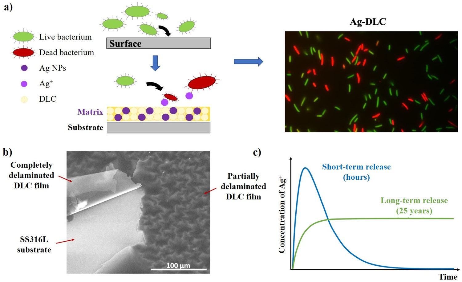
Summary of the literature available on the antibacterial activity and the potential cytotoxicity of Ag-DLC antibacterial coatings for biomedical implants over the last 5 years, including the substrates and the methodology used for their production.
Notes: CFU: colony forming unit; HVAP: high voltage anodic plasma; MAO: micro-arc oxidation; SaOS-2: sarcoma osteogenic; TVA: Thermionic vacuum arc.
Surface grafting of antimicrobial agents by electrophoretic deposition
Among the various surface coating techniques for Ti materials, EPD is frequently employed to produce biomedical coatings on metal, ceramic and polymer substrates with complex 3D shapes [133]. The advantages of using EPD include the possibility of depositing a wide range of additives and short processing times. Furthermore, the thickness and morphology of a deposited coating are easily tailored through simple adjustment of the deposition time and applied potential [134]. Tracing back to the first application of EPD for biomaterial coatings on Ti substrates, its use to produce simple coatings of hydroxyapatite (HA) has been extended to include multifunctional, composite and nanostructured coatings, that combine HA with other (bio)materials, such as biopolymers, graphene and graphene oxide, carbon nanotubes, as well as BGs and ceramic (nano)particles [135]. These combinations enhance the coating properties through the reduction of surface cracks, increased hardness, improved coating adhesion, corrosion resistance, thermal stability, improved biocompatibility, and antimicrobial function [135]. Recently, the use of alternating current (AC) fields, as opposed to the conventional direct current (DC) approach, has been introduced as this allows processing sensitive bioactive molecules from aqueous suspensions, while avoiding damage induced by water electrolysis (as seen in DC-EPD) and therefore preserving the biological activity (Figure 4) [136,137]. Antimicrobial coatings grafted on titanium by means of AC-EPD. (a) Schematic representation of the AC-EPD setup; where an asymmetrical triangular waveform is generated, amplified and delivered to the EPD electrolyte enabling selective deposition of biomolecules on the titanium electrode surface. (b) Caspofungin grafted on titanium by means of AC-EPD significantly reduces C. albicans biofilm formation on titanium [136].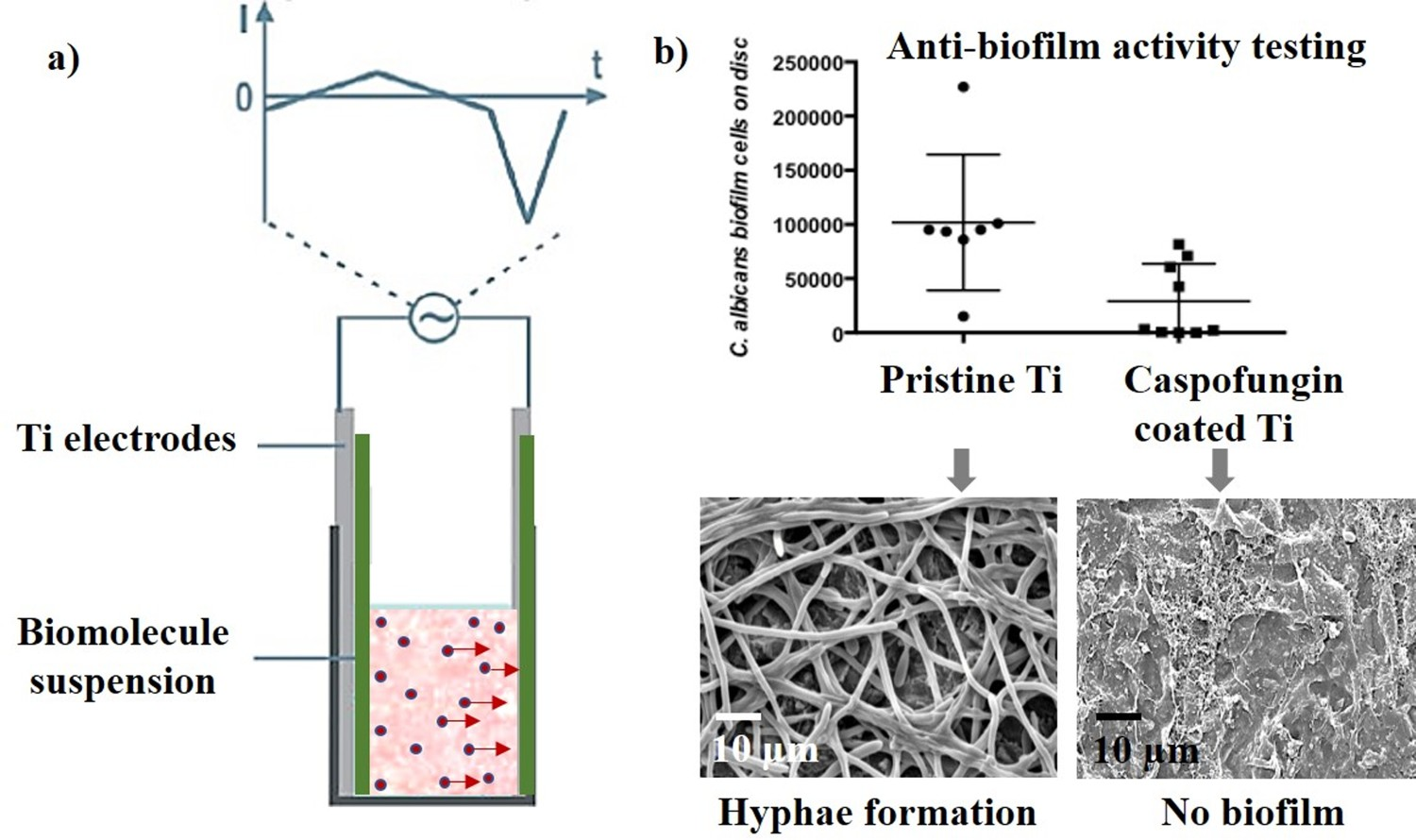
Summary of studies, published within the last 5 years, reporting the processing of antimicrobial coatings by means of EPD onto Ti substrata, including the antimicrobial activity and cytotoxicity (if available).
Notes: AgNP: silver nanoparticle; BG: bioactive glass; BSA: bovine serum albumin; CFU: colony forming unit; CLSM: confocal laser scanning microscopy; CS: chitosan; CuNP: copper nanoparticle; DNase: deoxyribonuclease; EPD: electrophoretic deposition; GO: graphene oxide; HA: hydroxyapatite; MSSA: methicillin-sensitive Staphylococcus aureus; rBMSCs: rat bone marrow-derived mesenchymal stem cells; MRSA: methicillin-resistant Staphylococcus aureus; NT: nanotubes; PEG: polyethylene glycol.
More recently, AC-EPD is also being applied to other antimicrobial compounds in order to establish non-releasing antimicrobial coatings, an approach which is thought to lower the risk of antimicrobial resistance (AMR) development in pathogenic bacteria. To this end, the titanium surface is first activated using linker molecules, such as silanes or polydopamine, which can covalently bind the antimicrobial biomolecules, thereby inducing antimicrobial activity [152,153]. Braem et al. demonstrated the applicability of AC-EPD for the deposition of the antifungal lipopeptide caspofungin onto silanised Ti substrata in order to accelerate general diffusion-controlled small molecule immobilisation processes [136]. It was shown that AC-EPD significantly reduced the process time for immobilisation, while producing high purity coatings which remained active against antifungal biofilm formation. Similarly, AC-EPD has also been applied for the time-efficient grafting of deoxyribonuclease I (DNase I) on polydopamine functionalised Ti surfaces [151]. This DNA-degrading enzyme targets eDNA, an important component of the biofilm matrix that contributes to surface attachment and cell-to-cell adhesion of bacteria. As such, AC-EPD DNase I coatings successfully inhibited the in vitro biofilm formation for S. epidermidis and P. aeruginosa.
Multifunctional antimicrobial-releasing bioactive ceramics and glasses
Given the increasing focus on novel materials that balance an antimicrobial effect with cell-stimulating functionalities, the research on bioactive materials that are suitable for delivering antimicrobial agents is rapidly expanding [154,155]. CaP compounds are particularly well investigated as bone regeneration materials, owing to their chemical similarity with natural bone mineral, their ability to accommodate a large number of bioactive ionic substituents and to adsorb (bio)molecular species [156]. Alternatively, BGs and related glass-ceramic biomaterials are well-known to promote osteogenesis, while being capable of controlling drug delivery from a mesoporous structure [155]. This section will focus on the most relevant approaches investigated in the field for bioactive antimicrobial-releasing surfaces, involving CaP-based systems as well as BGs.
Calcium phosphate-based systems
A large variety of CaP compounds can be prepared, whether crystalline or amorphous (Figure 5). Also, composite and hybrid biomaterials involving organic moieties and polymers have attracted the scientific community for modulating CaPs physical, chemical, mechanical, and biological properties. Whether crystalline or amorphous CaPs are concerned, several approaches have been explored for conveying antimicrobial properties and to progress toward smart responsive bioactive devices. Overview of CaP-based (crystalline and amorphous) strategies for antimicrobial approaches, including combinations with metal oxide particles and polymers. Concerning amorphous systems, their structural description [157] is based on the connectivity of the PO4 tetrahedra, using the Q[n] formalism, where [n] is the number of bridging oxygen atoms per PO4. Several domains can be distinguished depending on [n]: ultraphosphate glasses (low Ca content and cross-linked network of Q3 tetrahedra), metaphosphate glasses (long chains and rings based on Q2 entities), polyphosphate glasses (polymer-like materials with Q2 and Q1) and finally invert glasses (phosphate dimers and monomers respectively pyro- (Q1) and orthophosphate (Q0) anions).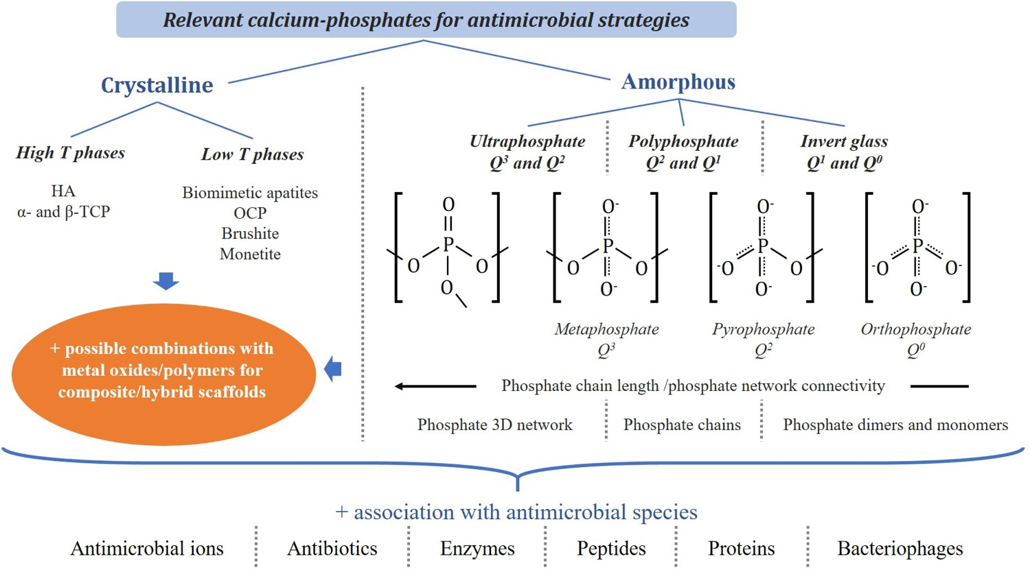
Crystalline CaP and related composites
Within crystalline CaPs, one needs to distinguish phases obtained at high temperature, such as stoichiometric HA, α- or β-tricalcium phosphates (TCP), which exhibit a high crystallinity and thermal stability, from phases obtained at low temperature such as hydrated CaP crystalline phases like nanocrystalline biomimetic apatites, monetite, brushite or octacalcium phosphate (OCP). Almost each of these chemical phases have shown promises in the preparation of antimicrobial biomaterials. Since CaPs do not have inherent resistance against pathogenic bacteria, significant research has been devoted to associate them with antimicrobial agents, including not only antibiotics for a controlled local release but also other entities like as metal ions, oxide NPs, enzymes, peptides, etc. (Figure 5). These compounds, when used at an amount lower than a critical limit, are not only non-toxic to cells but also generally possess broad-spectrum antimicrobial properties. For example, ion substitutions in CaP crystallographic lattice of antimicrobial ions, such as Ag+, Cu2+ or Zn2+, were reported for doped HA [158,159], β-TCP [160], and more recently for OCP [161]. Bacteriophages (bacterial viruses) are another family of antibacterial species that have attracted attention [162,163], and whose immobilisation onto CaP substrates (e.g. β-TCP [164] or nano-HA/alginate hydrogel [165]) have shown promising outcomes, for example for protecting osteoblast cells against S. aureus infection and refraining biofilm formation of multidrug-resistant E. faecalis. Phagotherapy could thus be seen as one appealing path to infection fight and should probably be further investigated.
Among CaP phases, biomimetic nanocrystalline apatites are of particular interest due to their greater reactivity (linked in part to the high mobility of their surface ions) as well as their capacity to adsorb a wealth of active agents [166,167], making it possible to envision stimuli-responsive approaches. Recently, core–shell microparticles of biomimetic apatites, possibly incorporated in freeze-cast polymer scaffolds, have been prepared to control the time sequence of ion release, e.g. with an antibacterial action (through Cu2+ or Ag+ doping) at the initial stage, followed by an osteogenic effect [168]. Another investigated strategy to induce antimicrobial properties is by association with active (bio)molecules/drugs; not only antibiotics (rifampicin, vancomycin …) [169,170], but also antibacterial enzymes [171], peptides [172] or relevant proteins (lactoferrin, etc.) [157], or molecular oxygenated species through a safe-by-design approach [173].
Besides, CaP-based composites with metal oxides have been explored. Among metal oxides, iron oxide (Fe3O4) and zinc oxide (ZnO) NPs are, for instance, approved by the U.S. Food and Drug Administration (FDA) [174]. The antimicrobial response of these NPs is related to the formation of reactive oxygen species (ROS), which damage the bacteria's cell wall and disrupt the cellular metabolism [175]. A broad spectrum of antimicrobial composites in HA-Ag, HA-ZnO and HA-Fe3O4 systems was developed to establish such efficacy [176,177]. For example, HA-Fe3O4 composites demonstrated bactericidal properties by rupturing the membrane of Escherichia coli [113]. Similarly, HA-Fe3O4 nanostructured coatings were reported to have better antimicrobial efficacy against different bacterial strains [176,178,179]. Also, CS with Ag-doped HA-magnetite NPs (up to the concentration of 400 μg ml−1) were reported to be haemocompatible and non-toxic towards NIH-3T3 fibroblast cells, and also inhibited the active growth of Escherichia coli and S. aureus [180]. Similar to these reports, ZnO-based composite is reported to slow down the respiratory electron transport system of both Gram-positive and Gram-negative bacteria. Such efficacy depends on morphologies (particle size and shape) and concentration of ZnO [99,181–185]. The inclusion of 25 wt% ZnO (<50 nm) reduced biofilm growth by ∼98% for S. aureus and 99% for Escherichia coli when compared to pure nanoHA [174]. Similarly, Zn-doped HA exhibited antibacterial efficiency against both S. aureus and Escherichia coli with a Zn doping level of 2.5 wt% and 1.0 wt%, respectively [186]. Wet-chemically synthesised rod-shaped ZnO-embedded in HA demonstrated bactericidal property against the pathogenic S. aureus strain [177]. Further, the synergistic action of the combinations of different antimicrobial agents was also an emerging strategy to enhance the bactericidal effect. For example, Ag-ZnO-Fe3O4 nanocomposites are reported to exhibit the inhibitory effect against the S. epidermidis and S. aureus [187]. Similarly, ZnO@Fe3O4 nanostructured material exhibited good antibacterial activity against S. aureus, H. pylori and Escherichia coli [188–190].
Amorphous calcium and phosphate-based materials
In parallel to crystalline CaP compounds, amorphous calcium phosphate-based materials have also been extensively investigated for bone substitution [157,191,192]. Two kinds of phosphate-based amorphous biomaterials are mainly investigated: phosphate-based glasses [192–194] and low temperature amorphous CaPs [195] (Figure 5). As phosphate glasses demonstrated tuneable degradation in a timeline ranging from hours to days [191], their composition/structure may allow the controlled release of major doping ions or therapeutic drugs. Due to the nature of the CaP matrix, such glasses were mainly used for bone regeneration in bacteria-related pathologies [196], although their applications for soft tissues, especially in wound healing, are expected to emerge in the future [197].
As for crystalline CaP compounds, several antimicrobial ions (Ag+ [198–200], Ga3+ [201–203], La3+ [204], Ce4+ [201,202,205], Zn2+ [206], Cu2+ [207,208]) are investigated to restrict the growth and viability of micro-organisms. As previously mentioned, the issue of bacterial resistance to antibiotics makes these strategies based on active ion-release most relevant [209].
The association of both molecular and ionic antibacterial agents to reach a synergistic effect is another relevant approach, which is, however, difficult to achieve in practice. Yet, two alternative promising ways could circumvent this difficulty. The first approach can be pursued via the synthesis of mesoporous phosphate-based glasses to act as carrier for both species, either in the glass network after fusion (ions) or into the pores (molecules). The implementation of this process for phosphate glasses is very recent [210,211]. The second approach is based on low-temperature-derived amorphous CaPs that can incorporate both types of active species during their synthesis. Phosphate entities can be either orthophosphate [195,212], pyrophosphate [213] or polyphosphate ions [192]. Among antimicrobial strategies, one can cite polyphosphate-based materials or coacervates [214–217] and amorphous calcium orthophosphates [218–221] with the possibility to associate both ortho- and pyrophosphates [222,223]. The tuneable ortho/(ortho+pyro) phosphate ratio could allow controlling the biological behaviour of these biomaterials, as it integrates benefits of both phosphate glasses (tuneable dissolution) and of amorphous CaP (synergistic effect of both molecular and ionic doping), allowing the direct combination with antimicrobial biomolecules or drugs.
Bioactive glass releasing phytotherapeutic molecules
Another interesting strategy to combat bacterial infections without compromising the integration of the implant with the surrounding tissues, is the development of biomaterials based on traditional herbal medicines and bioactive agents [224]. Nowadays, a large fraction of the world population still uses herbs as a primary form of medicine, owing to their bacterial resistance, bioavailability and cost-effectiveness [225,226]. Phytotherapeutics, for instance, are plant extracts that have been exploited to harness the deleterious effects of bacterial infections, since ancient times. Although the exact antibacterial mechanisms for most herbal medicines are still under investigation, it has been reported that phytochemical constituents could act as immunomodulators, enzyme inactivators and inhibitors of bacterial colonisation [227]. BGs, especially mesoporous BGs, as delivery vehicle for phytotherapeutics, can offer biodegradability, controllable resorption rate, biocompatibility and adjustable loading capacity, which warrant their functionality for transport and sustained release of the antibacterial herbal drug [228]. Apart from being used as delivery system for plant derived compounds, BGs themselves can act as sources of antibacterial activity due to their ion release ability upon immersion in body fluids, such phenomenon can enhance osmotic pressure and alkalisation of the surrounding environment [229]. Of particular note is the use of BGs in clinical practice as bone graft substitute materials to stimulate new bone growth and treat infections [230]. Nowadays, S53P4 BG (53.8 mol% SiO2, 21.8 mol% CaO, 22.7 mol% Na2O, 1.7 mol% P2O5) is the most widely used bioactive glass in clinical practice with applications against osteomyelitis, benign bone tumours and spondylodesis [231]. Its mode of antibacterial action is based on the pH increase that occurs close to the material due to cation release during BG dissolution, which makes it an ideal non-antibiotic candidate material for treatment of bone infections in times of evolving antibiotic resistance [232]. To increase the antibacterial activity of BGs, various active ions of biological relevance (e.g. Ag, Cu, Ga, etc.) can be doped in the BG structure to further improve the efficacy against microbial pathogens [233]. Such ions are released from the BG structure during dissolution and can act directly on bacteria. Thus, the benefit of the BG-herbal extract composites as innovative antimicrobial systems relies on the synergistic combination of the released biologically active ions from BGs and the therapeutic activity of the herbal metabolites (Figure 6). A plethora of phytotherapeutics, including coumarins, flavonoids, polyphenols, etc., have been conjugated with BGs with various compositions to mediate biocompatibility and bactericidal activity (Table 5). Several studies used metal oxides or synthetic antibiotics as benchmarks to prove the superior anti-infective properties and biocompatibility of plant-derived bioactive compounds, in combination with BGs [234,235]. It is worth noting that the key criterion to achieve a synergistic effect of the herbal extract and the BG ions is to supply a proper amount of each agent at the targeted site. Indeed, the therapeutic efficacy of the delivered agents has to be assured by performing a precise analysis of the synergistic mechanisms. Dual release of phytotherapeutics and therapeutic ions from the surface of bioactive glass particles, as well as from the mesopores of herbal drug-loaded bioactive glass carriers for antibacterial applications to combat both Gram-positive and Gram-negative bacterial strains. Summary of studies, published within the last 5 years, on the combination of herbal drugs and bioactive glasses with various compositions, including additional information on the form of application, performed antibacterial and cytotoxicological assays and most relevant outcomes. Notes: AgNP: silver nanoparticle; BG: bioactive glass; CFU: colony forming unit; CS: chitosan; DAPI: 4’,6-diamidino-2-phenylindole; HaCaT: cultured human keratinocyte; hDF: human dermal fibroblasts; PEEK: poly-ether-ether-ketone; PMMA: poly-methyl-methacrylate; NP: nanoparticle; MEF: mouse embryotic fibroblast; MIC: minimum inhibitory concentration; MBC: minimum bactericidal concentration, MTT: methyl thiazolyl tetrazolium, XTT: 2,3-bis-(2-methoxy-4-nitro-5-sulfophenyl)-2H-tetrazolium-5-carboxanilide.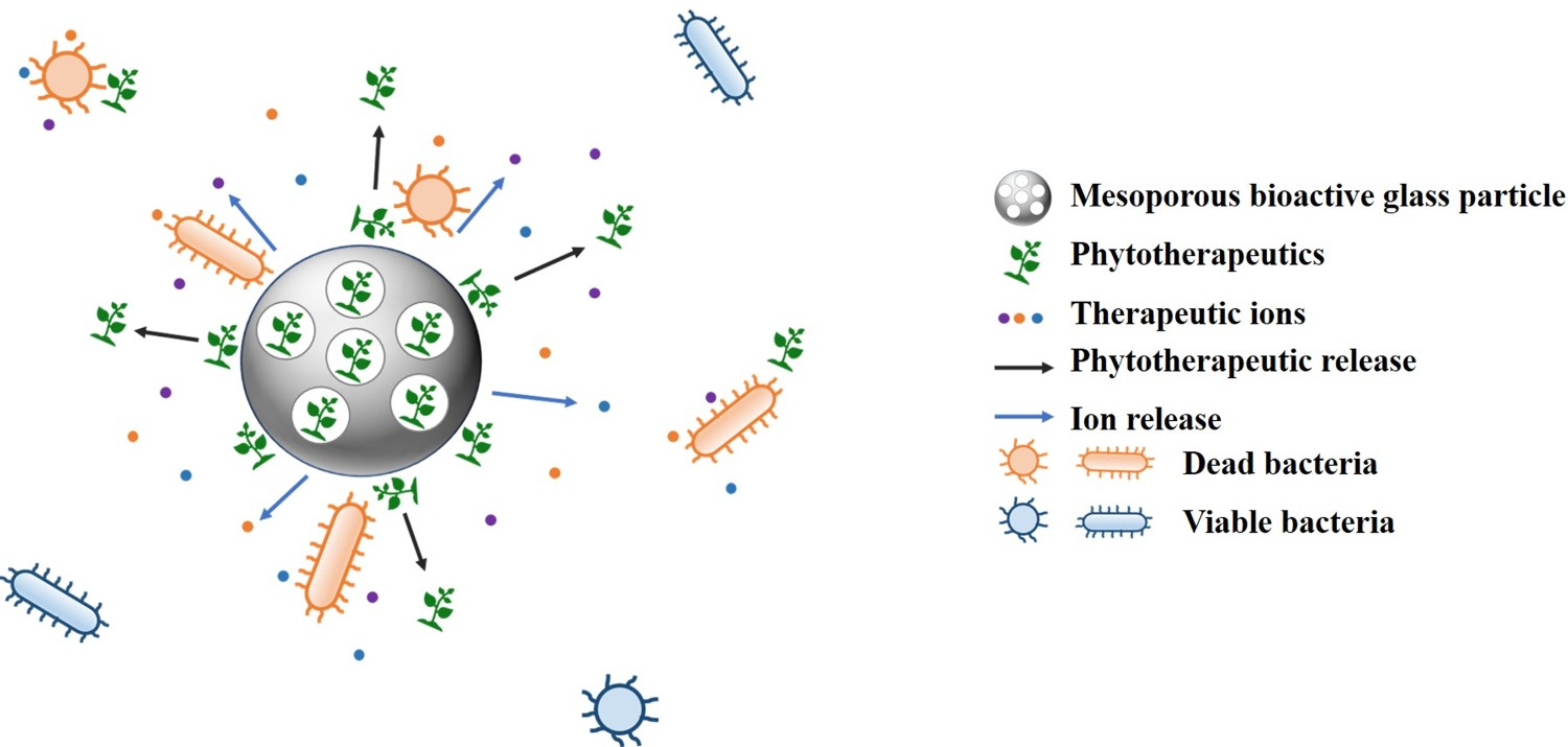
For load-bearing implant applications, however, the coatings of bioactive ceramics and BGs risk delamination during clinical use owing to their inherent brittleness. Moreover, coating systems require loading of the drugs prior to implantation, which often leads to an initial burst release of possibly toxic concentrations of the drug compound as well as premature depletion of the drugs, shortly after implantation. The latter is especially problematic in case of antimicrobial therapy as the exposure of microorganisms to sub-inhibitory concentrations, before biofilm eradication can lead to re-infection and AMR development. Recently, a drug-releasing Ti/BG system was reported by Kamarudin et al., where a mesoporous BG, contained within a microporous Ti structure, acted as a barrier for a more controlled release of chlorhexidine, an antiseptic used for treatment of peri-implantitis around dental implants [243]. In combination with a refillable internal reservoir, this composite allows fine-tuning the release of antimicrobial agents to therapeutic concentrations, i.e. active against biofilm formation, without initial burst and for a longer period of time [244,245]. This system opens a new insight into dental implant applications, where the preventive and curative approach against biofilm formation, and hence infection, can synergistically be implemented during implantation.
Natural polymers as multifunctional antimicrobial platforms
Polymers represent an important class of indwelling materials in the clinical setting, not in the least in applications prone to infection, such as catheters, ventricular shunts, etc. The versatile polymer chemistry and ease of processing, as compared to metals and ceramics, also offer opportunities for antimicrobial surface modifications of polymers [55], such as the incorporation of a variety of antimicrobials (antibiotics [54], metal ions and NPs [246], quaternary ammonium [247], halamines [248,249], enzymes [250], etc.) or the immobilisation of antifouling (polymer brushes, etc.) or bacteria-killing molecules (antibiotics, antimicrobial peptides (AMPs), cationic moieties, etc.) on the surface [57,251]. Moreover, polymers that undergo a noticeable physical or chemical alteration in response to (bacterial) environmental stimuli, are increasingly being explored for use as (antimicrobial) drug delivery devices as well as smart anti-infective coatings on other implantable materials. Therefore, ‘smart’ functionality represents a valuable strategy against AMR [58]. In this respect, the polymers with innate antimicrobial properties, such as synthetic antimicrobial polymers based on the template of natural AMPs (without the use of antibiotics) are being researched, some of which are effective against multiple-drug resistant pathogens [54,57,252]. Recently, several bioengineering strategies to address the infections of the urological implants are reviewed with a particular focus on developing biodegradable scaffolds and biostable alloplasts to inhibit the proliferation and colonisation of uropathogens [253].
Alternatively, natural polymers have gained a lot of interest in the last decades, as they possess high biocompatibility, bioresorbability, varied structural features and in some cases tuneable physical and mechanical properties [254,255]. Some can have an inherent ability to perform antimicrobial activity or can act as a polymeric matrix, including low molecular weight biocides and other antibacterial compounds [256]. In general, they provide non-toxic, environment friendly materials, offering long-lasting antimicrobial response [257]. Also, due to their bioresorbability, natural polymers offer the advantage of being removed once their function has been completed [254]. This section aims to describe polyhydroxyalkanoates (PHAs), bacterial cellulose (BC), and collagen – three of the most promising natural polymers in the biomedical field, focusing on their use as materials for antimicrobial applications.
Polyhydroxyalkanoates
Among the polymeric materials, PHAs, a family of biopolyesters synthesised by bacterial fermentation, are considered as promising candidates for medical applications due to their biocompatibility, bioresorbability, tuneable physio-chemical properties, and surface degradation properties [258–264]. The surface degradation properties, in contrast to bulk degradation, ensure the stability of the scaffold lasts, while tissue regeneration occurs. Several Gram-positive and Gram-negative bacterial strains are able to accumulate PHAs within the cytoplasm as energy storing compounds, when they are fed with a suitable carbon source under nutrient-limiting conditions. The type and structure of the produced PHAs mainly depend on the carbon source used for fermentation, and the bacterial strain [265].
Despite lack of antimicrobial properties and being non-antigenic in their natural form, several studies have demonstrated that degradation products of PHA hydrolysis can provide an inhibitory effect on microbial growth. The monomeric units of PHAs ([R]-3-hydroxyalkanoates, R-HAs) are hydroxyl-substituted fatty acids and they have been considered as antibacterial agents, due to their function as anionic surfactants. They can cause alterations in cell permeability that cause an inhibitory action, when adsorbed by bacterial cells [266]. The efficacy of a variety of R-HAs, such as (R)-3-hydroxy-n-phenylalkanoic acids [267] and (R)-3-hydroxycarboxylic acids [268], such as (R)-3-hydroxyoctanoic acid [269], has been assessed against both Gram-positive and Gram-negative bacterial strains. In several studies, biomolecules, such as AMPs, enzymes, plant-derived compounds, essential oils (EOs), polymers, and inorganic NPs, have been incorporated into PHA platforms and assessed against different bacterial strains [266,267,269–272] (Figure 7, Table 6). As AgNPs have attracted much interest due to their antibacterial properties and efficacy against bacterial biofilms, several studies have been conducted to demonstrate feasibility and effectiveness of PHAs/Ag platforms, inducing bacteriostatic / bactericidal effect against Escherichia coli and S. epidermis, while avoiding toxic effects [273,274]. Among other promising inorganic NPs and ions, molybdenum disulphide (MoS2) [275] and boron nitride [276] NPs have been encapsulated into a PHA/CS matrix and assessed at various doses against Gram-positive and Gram-negative bacterial strains. Marcello et al. proposed a novel antimicrobial material based on combination of P(3HO-co-3HD-co-3HDD) and poly-3-hydroxybutyrate, P(3HB) with HA [265]. Selenium ions have been used for substitution in the crystal structure of HA to obtain an antibacterial ceramic material for bone tissue engineering applications [265]. The antimicrobial behaviour of Cu-ions has been evaluated by incorporating Cu-doped 45S5 BG in different short-chain length and medium-chain length PHAs scaffolds [277].
Loading PHAs with plant-derived compounds or EOs is another approach that is gaining interest among scientists for antimicrobial purposes. Curcumin [278], cinnamaldehyde [279], and lime oil [280] have been encapsulated in PHA platforms and assessed at different concentrations against several bacterial strains. Additionally, antimicrobial peptides (AMPs) have been proposed as the best candidates for antimicrobial applications due to their activity against a broad spectrum of bacteria and decreased tendency to generate antibiotic resistance [281,282]. (a) The main stages of PHA production and the key approaches for the production of antimicrobial scaffolds. More specifically, a PHA hydrogel incorporating essential oil, a PHA membrane functionalised with AMPs and porous PHA scaffold functionalised with organic or inorganic nanoparticles. (b) Effect of PHA fibre meshes functionalised with Amhelin and Dispersin B on the bacterial adhesion [281]. (c) Analysis of the bacterial biofilms formed on the surface of neat PHA fibre mesh, PHA fibre mesh with NCO-sP(EO-stat-PO) and PHA fibre mesh with NCO-sP(EO-stat-PO), Amhelin and Dispersin B [281].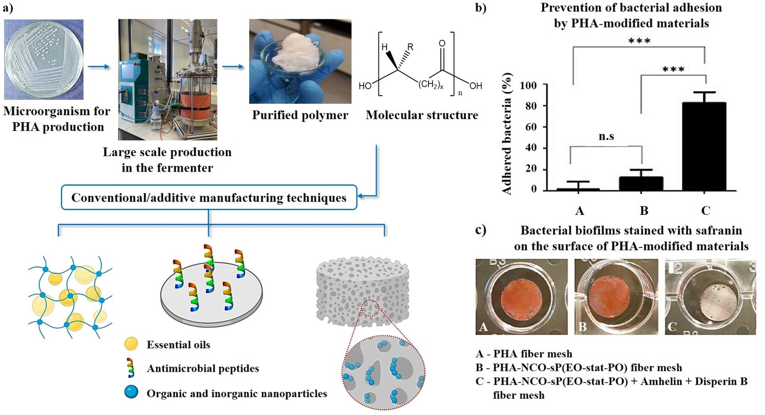
Bacterial cellulose
BC is another natural polymer that has gained interest among scientists [283]. BC is mainly produced by fermentation of Gluconacetobacter and Agrobacterium. The higher degree of crystallinity of BC compared to plant-derived cellulose results in better physical and mechanical properties, such as Young’s modulus, tensile strength, high thermal resistance and high retention of water. All these properties make BC a suitable material for biomedical applications. Over the last few years, increasing studies have been conducted on BC for medical applications, such as wound dressing, cancer treatment, bone and dental regeneration, and drug delivery. It is true that some of those applications require the biomaterial to be stable, however, the majority of them need the implant to degrade at an adequate rate without toxic byproducts, matching the tissue’s regenerating activity, and to be reabsorbed in the body [284]. BC is biodegradable by enzymes found in nature but the absence of those enzymes, in addition to the relatively high thermal, mechanical, and chemical stability of the material, limits its degradability in the human body [285]. Several factors affecting the rate at which BC can degrade have been identified, such as molecular weight, crystallinity, pH, hydrophilicity, morphology, its water uptake capability, and the presence of additives [284]. Consequently, different studies exploiting several strategies, such as periodate oxidisation [286–288], pre-irradiation [286], crystallinity disruption [289], incorporation of enzymes [290–292], proved that biodegradable BC-based materials are obtainable in order to target specific applications. A different approach where metabolic engineered bacterial strains were used to produce BC with linkages that are cleavable by inflammatory enzymes has also been studied [293,294]. Functionalisation and combination of BC with several compounds have been investigated to develop designed biomaterials with antibacterial properties (Figure 8, Table 6). AgNPs have been synthesised in situ and encapsulated in BC hydrogels [295,296] and pellicle (BCP) [297]. These biomaterials exhibited antibacterial activity against Gram-positive and Gram-negative bacterial strains. Among other inorganic compounds, ZnO NPs [260,298], CuO [299,300], and MgO [301] have been used as antimicrobial agents. Additionally, EOs, such as thyme [302,303], eucalyptus, and clove Eos [302], have been exploited to impregnate BC membranes and hydrogels, and the efficacy of EOs in affecting the cellular integrity of many bacterial strains has been assessed. Recently, chemical functionalisation has been investigated to modify BC membranes using 3-aminopropyltriethoxysilane (APTES). Its bactericidal effect is associated with the introduction of free amino groups along the BC polymeric chain. These biomaterials exhibited strong antibacterial properties against Gram-positive and Gram-negative bacteria strains, while being nontoxic to human fibroblasts [304,305]. (a) Bacterial cellulose (BC) production resulting in the formation of BC pellicles and (b) functionalisation of the BC membrane using two active agents, glycidyl trimethylammonium chloride and glycidyl hexadecyl ether, to introduce antibacterial functional groups. (c) BC hydrogel functionalised with AgNPs (AgNPs/BC) and the comparison of the antibacterial activity of neat BC (top) and AgNPs/BC (bottom) against S. aureus, B. subtilis, Escherichia coli, and P. aeruginosa [296]. In the graphs, antibacterial activity of non-functionalised and functionalised BC membranes agains S. aureus and Escherichia coli are shown [306]. Summary of studies, published within the last 5 years, reporting on PHAs, BC or collagen in combination with other antibacterial agents and materials, including antibacterial assays and most relevant outcomes. Notes: A.D.A.M.: antibiofilm dressing’s activity measurement; AgNP: silver nanoparticle; AuNP: gold nanoparticle; CFU: colony forming units; CS: chitsoan; BG: bioglass; EO: essential oil; GO: graphene oxide; MRSA: methicillin-resistant Staphylococcus aureus; MIC: minimum inhibitory concentration; MBC: minimum bactericidal concentration; NP: nanoparticle; P(3HB): poly-3-hydroxybutyrate; PHA: Polyhydroxyalkanoate; P(3HO): Poly(3-hydroxyoctanoate); P(3HB-co-12 mol-% HHx): Poly(3-hydroxybutyrate-co-12 mol-% hydroxyhexanoate); P(3HO-co-3HD): Poly(3-hydroxyoctanoate-co-3-hydroxydecanoate); P(3HO-co-3HD-co-3HDD): Poly(3-hydroxyoctanoate-co-3-hydroxydecanoate-co-3-hydroxydodecanoate); PHBV: Poly(3-hydroxybutyrate-co-3-hydroxyvalerate). As an on-site drug delivery system, collagen is an interesting natural polymer due to well-documented structural, physical, chemical, biological, and immunological properties [317,318]. It is one of the most abundant protein with approximately 30% presence in the body and more than 90% of extracellular matrix in skin, bone, and tendon [319]. Normally, it is readily harvested from marine, bovine, porcine, ovine, and equine sources. It has been established that its mechanical properties, adsorption volume of fluids or haemostatic activity differ, depending both on its source and processing (i.e. if enzymatically digested, degraded or dissolved) [317]. Moreover, collagen, as a fibrillary matrix, makes an excellent material with low immunogenicity, since it can degrade into physiologically well-tolerated products [320]. As a natural polymer, it provides advantages related to its inherent cell-signalling potential. In fact, type I collagen (bovine) is one of the most pro-angiogenic super polymers due to its suitability as a substrate for signalling and attachment of endothelial cells. Different compounds, including gentamicin [311], tannic acid [312], epigallocatechin gallate [313], CS [314], and AgNPs or ZnO NPs [315,316] have been incorporated to promote antibacterial activity of the collagen scaffold without compromising its cytocompatibility (Table 6). The release of an antibacterial agent can be influenced by modifying the matrix characteristics. The currently known methods to change local kinetics in on-site drug delivery using collagen-based matrices include cross-linking the matrix [321], changing the doping agent to make the drug less soluble [221], using a diffusion restrictor or a diffusion barrier [322], and composite with other materials [323–325], as shown in Figure 9. Nevertheless, some limitations are also reported, for example, exogenous crosslinking has shown to induce detrimental effects on cells and tissues, such as cytotoxicity, tissue calcification or even partial denaturation of collagen itself [320]. It is generally accepted that the control over defined molecular composition, mechanical integrity and drug release profiles is required in order to promote their clinical translation. Representation of modified collagen fibres as an on-site delivery system with incorporated antibacterial nanoparticles in the matrix and/or its combination with hydroxyapatite to modulate the release of the nanoparticles. Over more than a decade, significant attention in few research groups worldwide has focused on the use of various external stimuli-based therapeutic modalities together with implantable biomaterials as minimally invasive approaches for antimicrobial treatment [326–336]. Among the five categories of physical stimuli-based antimicrobial therapeutic approaches (photodynamic, ultrasonic, heat treatment, electrical and magnetic approaches), electrical and magnetic stimulation are recognised as promising strategies for enhancing antibacterial efficacy [326]. The conceptual framework involves the application of the intermittent delivery of electrical or magnetic stimuli to bacterial strains, when they are grown on electroactive or magnetoactive biomaterials. Some experimental studies were also conducted with bacterial cells in growth medium under magnetic stimuli. The experimental results have demonstrated significant promise, but the exact mechanisms of the antibacterial effects due to these bioengineering modalities are still under investigation. Nevertheless, it has been proposed that their modes of bactericidal action are by targeting the microbial membrane and intracellular molecules and by interfering with different microbial metabolic pathways [326], as shown in Figure 10. As such, electrical and magnetic stimuli-based approaches act on multiple targets, thereby lowering the probability of microbial resistance development. This section presents a summary of the published studies involving the electrical and magnetic stimuli to induce bactericidal properties on engineered biomaterials [327]. Schematic representation of various stimuli-based bioengineering strategies to enhance the bactericidal effects towards biofilm formation on a functional biomaterial surface. The possible mechanism of the mode of bactericidal action of electrical and magnetic field is to target the multiple locations of bacterial structure for destroying their membrane integrity and intracellular molecules and by interfering with their different metabolic pathway. The details of the experimental results, which contributed to developing our understanding, can be found elsewhere [104,321,337]. Based on the type of electrical signal, the electrical stimulation used for inhibiting the microbial biofilm formation is of different kinds. These include DC, capacitive, electromagnetic and inductive coupling stimulations, depending on the external source of wired or wireless power supply. The most common method of electrical stimulation used in the biomedical field to reduce the bacterial bioburden on infectious sites is by direct application of the electric field. The electric field strength, exposure time and the stimulation sequence are the most critical parameters related to the killing mechanism of action [175]. Together with the electrical stimulation, the functional properties of the biomaterials have also been reported to play a significant role in producing the synergistic effect on antimicrobial activity against a specific bacterial strain (Gram-positive or Gram-negative). Mechanistically, the electrical stimulation leads to the electrolysis of molecules, permeability and depolarisation of cell membrane (known as electropermeabilisation) on the surface of bacterial cells, which not only destroy the membrane integrity, but also result in the generation of toxic ROS and reactive nitrogen species (RNS) [327,328]. The elevated level of these toxic molecules interferes with microbial metabolism and increases oxidative stress, which finally kills the bacteria. The electropermeabilisation resulting from the application of low-strength electric fields has been reported to enhance the efficacy of antibiotics and biocides needed to kill the bacteria in biofilms [327]. In the recent past, several in vitro studies reported the electrical stimulus mediated bactericidal and bacteriostatic activity to address bacterial infection on different biomaterials [329–332]. For example, Jain et al. established the interdependence of field strength and exposure duration of a vertically applied electric field of 2.5 V.cm–1 towards bacterial growth inactivation on amorphous carbon substrates, in vitro [333]. Their study demonstrated that the bacterial inactivation is much more significant in the case of S. aureus bacteria, when grown for 4 h after electric field exposure. Additionally, they illustrated that applying a lower electric field for a long time or a higher electric field for a short time could equally deactivate growth of Gram-negative and Gram-positive bacteria [333]. Further, the antibacterial properties of biomedical materials with capacitive properties (titania nanotubes doped with carbon; TNT-C) have also been modulated well with both direct and alternating current (DC, AC), with the higher discharging capacity in the positive DC (DC+) group. The application of electrical current is reported to inhibit biofilm formation through cyclical charging [334]. Recently, Emanuel et al. reported a clear increase in bacterial growth reduction, due to increasing the current density (0.01 ± 0.005–5.2 ± 0.5 A.cm–2) in tested electric field strengths (1–4 kV.cm–1) [335]. They observed that an increased current density led to higher cell permeability and a larger bacterial cell volume [335]. The biological response of prokaryotes, especially the microbes, to the magnetic field is an emerging field of research. The variation in the influence of the magnetic field on bacterial survivability depends specifically on the intensity and exposure time, the type of bacterial strain and the culture condition [336]. The exposure of bacterial cells to magnetic field interferes with ion transport mechanism via the lipids and ion channel proteins, leading to intracellular osmotic imbalance and the conformational changes of intracellular molecules. The dysfunction of intracellular molecules, especially the proteins, disrupts the essential ion transport system and, finally, leads to membrane disintegration [327]. Another possible mechanism responsible for killing the bacterial cell due to external magnetic field exposure is the formation of free radicals, such as hydroxyl radicals, superoxides, etc., which elevates the level of ROS production [175,338]. Many researchers studied the effects of the homogeneous and inhomogeneous magnetic field, or pulse and static magnetic field, on the growth for different bacterial strains, when they are grown on magnetoactive biomaterials. Many such studies reported contradictory results [339]. In one of the published studies, it was reported as how HA-Fe3O4-based magnetoactive composites can induce bactericidal properties [340]. As a follow up study, the synergistic interaction of magnetic field stimulation and magnetoactive properties towards modulation of bactericidal properties was investigated [341]. In a recent series of studies, both static magnetic field (100 mT or 0.1 T) or pulsed magnetic field (up to 4 T) stimuli were provided to the bacterial growth medium containing pathogenic bacteria using a customised lab scale setup [108,340]. In most of the experimental studies, a modulated ROS generation was recorded and this was identified as the major cause for bacteriostatic/bactericidal effects. In a parallel study, Boda et al., established the positive impact of regular intermitted delivery of 100 mT magnetic stimuli on osteogenesis of human mesenchymal stem cells [336]. In another recent study, the critical role of pulse magnetic stimuli (up to 4 T) to restrict the growth of Enterococcus faecalis strains, commonly implicated in root canal therapy treatment was established [337]. Further studies need to be performed to confirm the exact mechanisms underpinning the potential biophysical mechanisms, thereby leading to more insightful understanding into the external stimuli-dependent bactericidal/bacteriostatic response. With the 17 Sustainable Development Goals, the United Nations aspire to improve the life quality on Earth, at all levels by 2030 [342]. The World Health Organization has recognised nosocomial (hospital-acquired) infections as a significant global burden affecting 7 or 15 out of 100 patients in developed and developing countries, respectively [343]. Against the backdrop of AMR’s obvious consequences on patient health, this section summarises the challenges in translational research and regulatory approval, while emphasising AI-based approaches for faster effective prediction of their outcomes. In the context of antimicrobial biomaterials, several approaches have been explored to treat or even prevent implant-related infections. As described in preceding sections, a large variety of compounds have been considered and are still under consideration for such strategies, such as CaPs, BGs, metal surfaces, polymers, metal oxide-loaded ceramics, etc. The antimicrobial activity can also be provided by way of a wealth of active agents, antibiotics, ions, enzymes, phytotherapeutics, peptides, etc. Antibiotic-loaded systems remain a relevant approach, however, care should be taken to avoid bacterial resistance effects. For example, systemic administration of antibiotics poses a significantly higher risk of AMR development than localised delivery [344]. Therefore, smart-releasing systems will receive privileged attention in the future, so as to release the right dose (limitation of dosage allowed) at the right place (local instead of systemic delivery) and at the right time (possibly through stimuli-responsive approaches). This remains a rather unexplored avenue of research that deserves future attention. The development of smart CaP-based stimuli-responsive biomaterials, capable of releasing anti-infective agents with a speed/amount that depends on the degree of infection of the patient, is in progress. This approach was first developed in relation with acne involving nanocrystalline CaP particles, but could further be translated, for example, to bone-related infections [345]. Another appealing path of research is the association of natural biomolecules, such as antibacterial enzymes, to bone-like CaP ceramics as an alternative to antibiotics, and this area of research was also initiated [171]. In the same respect, phytotherapeutic approaches and suitable systems capable of precisely delivering molecules at the site of infection using BGs-based delivery systems also present a potential strategy. However, the complexity of medicinal plant extracts and the variability of the raw herbal compounds lead to difficulties in the standardisation of the preparation procedures and may cause non-reproducibility of the results, among different experimental trials [346]. Therefore, future investigations will probably rely on screening approaches, including quality control, documentation of toxicity, and systematic in vivo tests. Despite the promising antibacterial behaviour of the BG-phytotherapeutics composites in vitro, the antimicrobial activity against various pathogens, such as fungi and viruses, still has to be investigated. Considering the prevalence of multi-resistant pathogens, the BG-herbal extract composites, which incorporate multiple herbal drugs and bioactive agents with added functionality, present an alternative approach to monotherapy. In this context, smart delivery systems based on BG and phytotherapeutics are expected to exhibit a selective release behaviour in the pathological environment, which should be responsive to different stimuli (e.g. pH, temperature, light, etc.). Another class of natural biomaterials is PHA and its derivatives. One of the major limitations of PHAs is the high production cost. Several strategies are under investigation to overcome financial and economic issues, as well as the use of inexpensive carbon sources, waste materials, genetically modified bacterial strains, fermentation conditions and recovery methods [280]. From a biomaterials perspective, it is important to recognise two key aspects in the development of antimicrobial strategies, i.e. balancing the antimicrobial effect with a cell-stimulating functionality, and providing material stability as well as a lasting antimicrobial effect as appropriate for the intended application (i.e. permanent vs. temporary use). A significant part of this review focuses on the sustained release of antimicrobial ions or other compounds, loaded into biocompatible coatings, like DLC. For example, Ag-DLC coatings, despite their promising protective and antibacterial features for dental implant applications, have had limited to no impact on clinical practice in dentistry. Further in vitro and in vivo investigations are required to validate suitable DLC films for determining the optimal antibacterial dosage, acquiring osseointegration, and sustaining long-term mechanical stability [347]. Therefore, further studies investigating the potential of Ag-DLC for dental implant applications should probe into the following aspects: (i) potential toxicity or adverse effect on osteogenesis of AgNPs at concentrations that are high enough to exhibit antibacterial activity (i.e. intrinsic control of silver ion release), (ii) mechanical stability (e.g. delamination of the coating from the substrate under torque), (iii) homogeneity of the coating on dental implants and abutments, and (iv) short- and long-term exposure in the oral cavity. As more and more research is focused toward addressing the challenges of developing next-generation anti-infective Ti implantable biomaterials, EPD has demonstrated its applicability as a promising surface modification technique to limit microbial adhesion and proliferation towards more effective antimicrobial systems. Moreover, the versatility of the AC approach, including its ability to be used for various bioactive molecules, heralds the potential of a hybrid coating technique for Ti biomaterials that can incorporate both antibacterial/antifungal and osteo-stimulatory compounds in a single coating, thereby addressing the need for multifunctional implant surfaces. Similarly, combinations of multiple antibiotic or antimicrobial compounds could be considered, which is a viable strategy in the battle against AMR [348]. Another interesting approach to circumvent AMR can be found in the field of surface texturing and also here combinatorial approaches seem most effective. More evidence is emerging to suggest that nanofeature geometries combined with a single hierarchical surface pattern present the greatest antibacterial (both antifouling and bactericidal) efficacy [93,94] (Table 2). Thus, micro–nanostructuring provides surfaces with a dual bactericidal action and antifouling capability, susceptible to increase safety and sustainability, while preventing persistent microbial colonisations and infections. One of the distinctive features of the discussion in this review is the use of bioelectronic medicine approaches. Two different routes, i.e. electrical or magnetic stimulation are discussed in particular reference to their efficacy to induce bacteriostatic or bactericidal property against pathogenic strains. Most of the studies are so far limited to in vitro platforms. It is warranted that in vivo studies should be planned to validate the in vitro outcomes. This aspect also requires the researchers to use miniaturised electrical / magnetic stimulation devices with the capacity to deliver stimulation in a programmable manner at the site of the infection, in vivo. Another interesting approach can be the electrical stimuli mediated accelerated, yet targeted delivery of antimicrobial agents at the site of infection in animal models. This would establish synergistic interactions of biomaterial and bioengineering-based approaches. While selecting the stimulation parameters, one has to use the clinically acceptable electric/magnetic field parameters, which do not cause toxicity to cells or tissues. In many studies, the antibacterial efficacy is studied for a given biomaterial with therapeutic agents or to assess the influence of a bioelectronic stimulus, without assessing their impact on cellular functionality. The competition between antimicrobial efficacy and cellular toxicity decides the optimal biomaterials composition or therapeutic dose level. In addition, it should also be realised that the presence of biomaterials triggers an inflammatory reaction in the human body, thereby dysregulating the innate host response and contributing to the infection susceptibility of biomaterials. This aspect is not sufficiently addressed in current in vitro test methods and more efforts are needed to introduce immunocompetence to in vitro assays. Also, novel antimicrobial biomaterials research should tap into the potential of immunomodulatory strategies [50]. All these aspects need very careful attention in future studies, which ideally should include the use of clinically relevant strains of pathogenic bacteria, possibly in complex in vitro models applying co- and even tri-cultures of tissue cells, macrophages and bacteria [349]. Moreover, it is assumed that higher-complexity systems will lead to a better predictivity and can also help avoid unethical use of in vivo studies [350]. Finally, it has been increasingly realised during the recent global pandemic the importance of antiviral technologies to combat viruses. In this context, it would be interesting to explore some of the antibacterial approaches for potential virucidal effects [351,352]. Here again, it would be ideal to investigate the effect of antimicrobial agent delivery around MIC/MBC dosages to understand its efficacy against viral strains. From the historical perspective, anti-infective systems involving drug-releasing bone cements, combining antibiotics and PMMA bone cement, were commercialised in the 1970s [353]. Gentamicin-loaded cements then became commercially available in the U.S. in 2002. The PMMA component progressively paved the way for improved commercial formulations based on calcium-containing compounds (sulphates, phosphates, e.g. Cerament®). The bone fixation pins coated with silver received approval from the FDA in 1996, and were sold in Europe in 2002 in the form of endoprostheses (e.g. Mutars®). Also, antibiotic-loaded implants continued to be commercialised, e.g. in Europe since 2005. However, the search for alternative antimicrobial strategies became increasingly relevant, for several reasons including the issue of multiple drug resistance (MDR). For taking the antimicrobial biomaterials to the clinics, the innovative approaches under investigation – once demonstrated relevant to the field – however have to follow the path of translational research and pass through various regulatory checks. Globally, limited progress has been made for clinical treatments using intrinsically antimicrobial biomaterials, despite large sets of appealing physicochemical and in vitro data. One limitation in existing approaches is still, nowadays, the lack or limitation of accessible in vivo data [354]. The feasibility of clinical translation has nonetheless been discussed in relation to some systems, e.g. involving metal, polymer or carbon nanoparticles, alone or in association with ‘classical’ small molecules [355]. Such combinations were, for example, evaluated favourably in the case of root canal treatment, in dentistry, as illustrated by amoxicillin/nano-diamond particles [356]. In orthopaedics, the development of biofilms on implants remains a challenge, as it is difficult to destruct mature biofilms of pathogenic strains. Against this backdrop, antimicrobial implants are designed, for example, involving surface modifications and coatings. The success of these anti-biofilm strategies remains, to date, limited to preclinical or early clinical stages, and should be pursued to obtain enough generalised data among academic, industrial and clinical researchers [357]. Also, especially regarding nanosystems, extensive information on biocompatibility should be accessible [358]. On the other hand, an antibiotic-free BG-based product (BG composition: S53P4, commercial name: Bonalive®) is being used in the clinic for the treatment of osteomyelitis [359]. When applied locally at the infection site and in conjunction with a systemic antibiotic treatment, this BG is highly efficient in the treatment of osteomyelitis, thereby omitting the need for the local delivery of high-dose antibiotics, e.g. through the use of antibiotic-loaded spacers. This represents a good example of an intrinsic (non-antibiotic) antibacterial material with regulatory approval for clinical use. Regarding translational research, a key difference in the development of regular antibiotics and antimicrobial materials/particles is the less extended comprehension that we may have, on the mechanisms of the action of the latter strategy [355]. The accelerated clinical translation for antimicrobial biomaterials could necessitate the setup of novel strategies of development (not systematically implemented for classical antibiotic research), including validated preclinical testing protocols, and could also involve mechanistic exploration, the development of adapted animal models, bacterial genomic sequencing, real-time in vivo imaging and possibly also of computational modelling [360]. Scale-up studies should also be carried out to establish large volume production, especially for nanoscale systems [358,361]. In this view, the development of bio-inspired strategies could be a favourable pathway to explore. The development of nature-inspired, safe-by-design biomaterials themselves is also a particularly appealing approach for tomorrow’s medicine (e.g. based on biomimetic calcium phosphates or natural polymers) [173]. Efforts should continue to be made to pass the various stages in regulatory pathways and access preclinical and clinical trials, so that the clinically acceptable technologies can be used to treat patients. In this respect, the funding of collaborative networks and global interdisciplinary research programmes in the area of AMR technologies is undeniably a way to facilitate translational research in the field of implant-related infections and collaborations among academic researchers, industry professionals, and clinicians. In this direction, the European Union’s Horizon 2020 funding scheme and Australian Research Council have sponsored translational research and innovation programmes to train an interdisciplinary workforce [362]. One of the objectives of these large network projects has been to develop new antimicrobial coatings or novel surface topographies on implant surfaces, to treat infections associated with orthopaedic implants. The regulatory approvals towards the use of antimicrobial biomaterials or bioengineering approaches are the last roadblock to overcome, before the commercialisation can take place. As discussed in the most recent review by Kalelkar et al., the clinical applications of many innovative antimicrobial approaches are far from reality, as that warrants more pre-clinical studies in small or medium sized animal models, followed by validation in human clinical trials [68]. During the innovation of new antibacterial biomaterials or bioengineering technology, the approval during phase I and phase II clinical studies from national regulating approval bodies take different timeframes, which depend on existing guidelines in different countries. Further, the certification process from international accreditation bodies, like FDA or CE, before commercialisation requires significant time, before any new technology reaches the market. The scientific and industrial communities have to actively engage with the regulatory agencies and healthcare policymakers since the early stage of the research, right at the proof-of-principle, to understand the regulatory hurdles while developing next generation AMR technologies, inclusively, and with a holistic approach. ISO standards should be followed, and widely be adopted in the scientific community as an unequivocal methodological guide to benchmark the results. In addition, as existing standards might not always be relevant to new technologies, new standards, specific to smart release, or AI, for example, need to be developed, and might be advanced with the International Scientific Community efforts. Scientific revision processes during manuscript acceptance must validate the compliant matching between the proposed protocols and the related ISO standards. This latter represents a dynamic guide that might be improved and enriched from scientists and experts from academic, industry, clinical and regulatory bodies. Altogether, this will certainly accelerate translations from bench to clinic for human healthcare, which is the ultimate goal of our commitment to this field. Medical devices are classified based on the primary mode of action (PMOA). However, if the antimicrobial activity involves a chemical action or metabolisation, and not just a physical action e.g. through topography, the device is considered as a ‘combination product’ necessitating evaluation close to drug development [363]. This is bound to be the case for most smart/engineered (nano)systems mentioned in the present review. A key challenge therefore lies in the adaptation of regulatory agencies to the new concepts and families of antimicrobial technologies that are being developed. A workshop relating to infections in orthopaedics was, for example, held recently (November 2020) and the deliberations therein unveiled the necessity to develop alternatives to the current standard of care for orthopaedic infections [364]. Specific guidelines and established evaluation protocols to establish antimicrobial efficacy and safety (relevant controls) are now needed [365–369]. This includes identifying methods for preclinical performance testing of antimicrobial devices [366]. Also, key concepts, such as antibacterial, anti-biofilm or antimicrobial activity, should be precisely defined, e.g. in terms of logs of reduction of colony forming units (CFU) or of the type of microorganism [363]. Accordingly, new standards (or major adaptations) should be envisioned [370] to keep up with the ever-developing domains, as for example in the case of CaP research (typically ion-substituted and/or biomimetic apatites), which are not yet considered in the existing standards. This is probably an essential step in view of allowing industry professionals to describe precisely the new systems under development, while remaining in accordance with regulatory authorities, and then making such systems accessible to the clinicians and patients. Owing to the large variety of animal models and evaluation methods, any direct comparison of research outcomes sometimes becomes challenging. Also, the selection of bacterial strains may be important to mimic actual patient conditions, and the use of clinical strains should be increasingly considered in the biocompatibility studies [371]. In addition, the classification of the existing/developing novel antimicrobial technologies to fight against implant infections could allow to have a better overview of regulatory and standardisation needs. This is one of the objectives of the present review and beyond [372]. In the last decade, data science has emerged as the fourth major paradigm driving scientific discovery, experiments, theory and computation [373–375]. As far as implementation is concerned, data science deals with automated aggregation of data into accessible repositories and systematic analysis of data using a variety of statistical learning tools. Such framework also enables the fusion of experimental and modelling data with suitable quantification of uncertainty, involving data science tools, such as machine learning and Bayesian inference. In several domains of biomedical sciences, including in the fight against implant infections, artificial intelligence (AI) and machine-learning strategies may help gather and organise the increasingly abundant data provided from research and clinics [376]. Here, the important objectives would be to assess efficiency/safety tendencies and thus help regulatory agencies to develop standardised protocols, but also to support industries to position their technologies. This AI field is however, to date, a rather young in the field of AMR and thus requires to gather a significant amount of data as input to machine-learning algorithms. Among the published studies, Epa et al. employed a machine-learning approach to predict the attachment of Escherichia coli, P. aeruginosa and S. aureus to polymeric biomaterials and proposed the use of computationally derived molecular descriptors [377]. The spectrum of studies needed to evaluate the compatibility of a sample library with bacteria, human cells and blood would result in creating a large dataset. In most published studies, such datasets are analysed without correlating them with materials properties (stiffness, surface energy, etc.). Some AI approaches have been followed in case of a few biomaterial systems, e.g. bioactive glasses (via neurofuzzy logic technology) [378], NPs (regression algorithms) [379] and Ti-based metallic alloys [380], allowing one to explain divergences among studies and to draw general behavioural laws as well as identify critical material-dependent parameters. Yet, it is recommended to develop an online platform, like CellNet which depends on the formulations of gene regulatory networks (GRN) in mouse and human cells [381]. Similar to CellNet, a publicly accessible online platform supported by National Institutes of Health (NIH) funding – ClinicalTrials.gov – contains clinical study outcomes. Such platform largely is useful for drug discovery-related research. It is therefore recommended that more clinical study outcomes, related to antimicrobial biomaterials research must be available in online platforms. Such platform would be very useful to build clinical decision support systems while enabling clinicians to rationally use the non-antibiotic therapeutic approaches to monitor trends in AMR. Very recently, AI/machine-learning strategies have also started gaining attention in biomaterials development, which should help further using AI outcomes and exploiting them for the accelerated development of engineered antimicrobial biomaterials for the clinic [382]. In a recent review, Lv et al. analysed the use of different machine-learning algorithms, like naive Bayes (NB), decision trees (DT), random forests (RF), support vector machines (SVM), and artificial neural networks (ANN), in terms of their usefulness for designing new antibiotics or synergistic drug combinations towards AMR [383]. In another recent work, Ward et al. analysed the use of a neural network-based feedback control system to regulate the release of critical dosage of Ag-ions from PHA-graphene nanocomposites for chronic wound treatment [384]. In particular, the predictive capability of such algorithms to determine the timeline and amount of antimicrobial agents to induce toxic effect against P. aeruginosa in a wound bed was briefly analysed. The usefulness of such algorithms towards non-antibiotic antimicrobial technologies must be explored in future research. Overall, this review has highlighted many exciting innovations in the field of antimicrobial biomaterials. Notwithstanding some excellent antimicrobial efficacy data, including in light of the continuous threat of AMR, in many cases more extensive testing of complex in vitro systems and/or in vivo models is required to critically evaluate the antimicrobial activity in concert with the relevant tissue and host immune response. A holistic approach incorporating standardised test methods already at an early translational stage in combination with an AI-based approach for a better reliability can help bring these materials from bench to bedside in the fight against MDIs.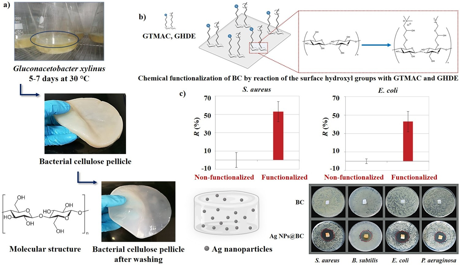
Collagen
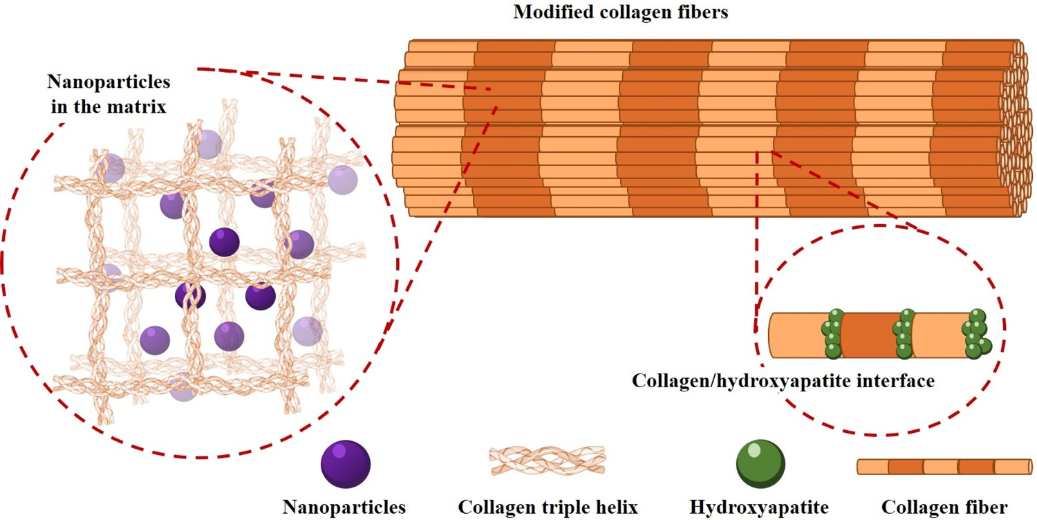
Bioengineering approaches
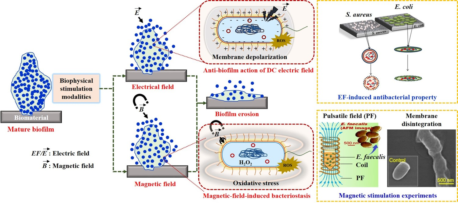
Electrical stimulation
Magnetic stimulation
Future perspectives
Smart, multifunctional and combinatorial approaches
Translational research
Regulatory aspects
The promise of artificial intelligence and machine learning
Footnotes
References