
Abstract
Calcium phosphate ceramics, owing to their resemblance to bone structure, are widely used for different biomedical applications. However, such a specific bone-structure pattern is making those materials susceptible to colonisation by pathogenic bacteria. This work presents results of the study on physical characterisation and microbiological assessment of biocide properties of modified man-made fluor- and hydroxyapatites. The structures and physical properties were characterised by a diffuse reflectance spectra, FTIR, Raman spectroscopy, TEM and Zeta-potential analyses. The bactericidal properties were assessed with Staphylococcus aureus, the main pathogenic species responsible for implant-associated infections. It was found that manufactured materials had structures typical for the biological (bone) apatites. Doping with silver has not changed their morphology. Results from the assays have confirmed that built-in silver substantially increased their biocidal properties, thus Ag-doped fluorapatite is a promising new resistant biomaterial with great bactericidal effect that potentially could be applied in tissue engineering, or dentistry.
Introduction
Nowadays, new biomaterials with distinctive chemical and physical properties and targeted biological functionalisation are of great interest in various medical applications. Consequently, the existing materials are being modified to obtain new features that could improve the efficiency of their applications. In recent years, the introduction of different bioceramics, like aluminium oxide, zirconium dioxide, or hydroxyapatite, has been revolutionising several biomedical fields [1-4]. Moreover, the improved ceramics doped with different metals are promising biomaterials that could be employed in new fields, as well. For example, the commonly known fluoride, an essential element in the skeletal system of bone and teeth, combined with various forms of silver [5,6], could be used to produce solution/gel/paste for impeding caries. In those applications, fluoride acts as a mineralisation component, while silver acts as an antimicrobial agent preventing formation of biofilms. On the other hand, it is well known that ionic or metallic silver could be toxic to the mammalian cells and cause cell's senescence or death [7,8]; therefore, doping processes have to be carefully monitored and possible release/action of silver or nanoparticles must be thoroughly evaluated for unwanted side effects. It was reported, that some types of biomaterial doped with silver may be not suitable for dentistry, because of staining teeth's enamel. Diamine fluoride (SDF) is such an example. SDF has been proved to be an effective treatment for anti-caries [9], but at the same time, it has stained teeth [6]. Nanopowders of silver-doped fluorapatite, tested for their antibacterial properties, have shown the bactericidal effects. It was demonstrated that the inhibition in cell proliferating was owing to the nanoparticles action combined with the release of the silver ions into the medium [10]. Interestingly, when powders of Al2O3 were incorporated along with silver nanoparticles, the final materials showed the inhibitory effect on the growth of different bacteria strains, but at the same time, they have not exhibited any cytotoxicity towards mammalian cells [11]. There are a number of published reports on the mechanisms of inhibitory effects of metals doped into apatites/ceramics, where the bactericidal actions were triggered either by the release of ions, or by the action of sole nanoparticles [1214].
The calcium phosphate, e.g. hydroxyapatite (HAP) and fluorapatite (FAP), the main mineral component of bone tissue, has been widely used in many medical fields, such as bone tissue engineering [15], drug delivery system [16], skeleton implants [17], bactericidal agents [12-14,1820] and in dentistry [21]. They have similar functions, but the fluorapatite has been recognised as being more chemically and structurally resistant when compared to hydroxyapatite [22]. In addition, in vivo studies have shown that fluorapatite materials have the osteoconductive properties, as well [23]. In the face of challenges of modern transplantation, it is very important to prevent infections caused by different pathogens introduced during surgery and/or post-surgery care. Hydroxyapatites are widely used in medical devices and health care products [24], thus, it is crucial to minimise the risk of infection by such implants. What is more, microorganisms responsible for implant-related infections are often resistant to classical antibiotics [25]. Those persistent infection/contamination problems may be reduced or eradicated by the replacement of typical implant's material with biomaterial, which has already built-in antimicrobial's capability.
Numerous well-known antibacterial agents, such as fluorine and biocidal metal ions, incorporated within the implant's material were found to be effective in the prophylaxis [26]. In this regard, nanoscale silver metallic inclusions (or ions) are promising antibacterial species that after combination with calcium apatite, have improved the overall biomaterial's disinfecting properties with absent, or low cytotoxicity towards mammalian cells [14,27]. Several other studies were concentrated on the development of calcium apatite with the sole silver inclusions, or incorporation of silver into complex structures based on calcium apatite. The examples include the synthesis of hydroxyapatite doped with silver [28,29], synthesis fluorapatite doped by silver by electrochemical deposition and co-precipitation methods [30-32].
Over the years, numerous methods have been developed for preparation of the fluorapatite, including neutralisation, deposition, sol–gel processing, mechanochemical, microwave and flame syntheses [22,33-38]. As expected, all these methods have several advantages and limitations. Current research is focusing on the development of safe for patients, inexpensive and suitable for industrial use, methods of synthesis of calcium apatite doped with silver. Understandably, the additional investigations are necessary for full evaluation of interactions between the modified HAP or FAP with mammal cells, crucial for the optimal design of new biological components with bactericidal activities.
This paper is presenting results from the comprehensive evaluation of properties (physical, structural and biocidal) of biomaterials, produced by the modified method of synthesis of calcium apatite doped with silver from saline melts at moderate temperature and with the addition of lutetium ions for increased stability of apatites during silver modification. The employed technique was based on the one previously developed by us, synthesis of calcium apatite [39]. Molecular structures of fluor- and hydroxyapatites doped with silver, obtained in this study, were fully characterised by diffuse reflectance spectra, FTIR, Raman spectroscopy, TEM and Zeta-potential analyses, while new embedded bactericidal effects were evaluated using Staphylococcus aureus.
Experimental methods
Materials, bacteria strain
All reagents (chemical grade) were purchased from Sigma Aldrich (U.S.A) and from POCH (Poland) companies. Nutrients, broth and agar, used for the preparation of suspensions of bacterial cultures or evaluation of the colony-forming unit (CFU), were purchased from BioCorp, Poland. The reagents employed in this study were as follows. For serial dilutions, the phosphate-buffered saline (1×PBS; 137 mM NaCl, 2.7 mM KCl, 10 mM Na2HPO4, 1.8 mM KH2PO4) was used. For the synthesis of tested apatites (HAP and FAP), a NaCl, KCl, AgNO3, Lu(NO3)3, CaCO3, CaO, Ca(OH)2 (for HAP), or CaF2 (for FAP), NaPO3 and hydrazine were used. A KBr was used as a diluent for the infrared spectroscopy measurements. Staphylococcus aureus ATTC 25923 (Microbiological Collection deposited in Biotechnology Centre for Applied and Fundamental Sciences, University of Rzeszow, Poland) was chosen for the study of antimicrobial properties of obtained biomaterials.
Synthesis and modification of apatites
The HAP, FAP, HAP/Ag and FAP/Ag were synthesised under the same experimental conditions to allow the no-bias comparison and assessment of their properties. Initial components for the synthesis of apatites in chloride melt were mixtures of CaCO3, CaO, Ca(OH)2 (for HAP), or CaF2 (for FAP), and NaPO3. Modification of apatites doped by Ag required an addition of 0.1 mol dm−3 AgNO3 solution supported by Lu(NO3)3. The details of the method of synthesis of apatites in the saline melt medium were provided in our earlier article [39]. The doping of synthesised apatites was conducted in AgNO3 solution (0.1 mol dm−3), for 1.0–1.5 h. To avoid undesirable processes, such as apatite's structure destruction, caused by the discrepancy of charges and the sizes of Ca2+ and Ag+ ions, the 0.018 mol dm−3 Lu(NO3)3 was added to AgNO3 solution. The process of Ag+ and Lu3+ ions sorption on HAP and FAP was carried out at 18–20°C. It was found that the concentration of Lu3+ ions in doping solution has changed only slightly, indicating better adsorption of Ag+ ions compared to Lu3+ ions. The overall content of Ag was found to be 9.5 and 13.0 wt-% for HAP and FAP, respectively. Finally, to avoid elution of silver from modified HAP and FAP samples, and possible transformation into nano-dispersed forms, a reduction of Ag+ ions to Ag with hydrazine was carried out according to the standard [39].
Characterisation of modified, silver-doped HAP and FAP
The full evaluation of physical properties of manufactured biomaterials was achieved by the TEM, Zeta potential and dynamic light scattering (DLS), FTIR, Raman spectroscopy and electronic spectroscopy of diffuse reflectance in the UV-range measurements and images, and their antimicrobial activity was confirmed through a series of assays.
Transmission electron microscopy measurements
Transmission electron microscopy (TEM) images were generated to characterise the size and shape of tested material. All measurements were performed on a JEOL-JEM-1011 microscope, operated at an accelerating voltage of 100 kV with a resolution of 0.2 nm. The powder samples of apatites were prepared by suspending in ultrapure water and ultrasonicating for 10 min in the Branson 3510 Ultrasonic Cleaner and; then few droplets of prepared slurry were placed on a carbon-coated copper grid, and dried in room temperature.
Zeta-potential measurements
To specify the differences in stability of the pure, and doped by Ag apatite's suspensions, the Zeta potentials of the HAP, FAP, HAP/Ag and FAP/Ag were measured and compared. Zeta potential was measured by the Zeta Sizer (Nano Series, Malvern Instrument Ltd, UK) equipped with disposable capillary cells. The tested samples (200 µg/ml concentration) were suspended in ultrapure water, vortexed and sonicated (10 min) before measurement. The pH was neutral for all tests; the volume of each suspension was 1 ml; and measurement's temperature was 25°C (±0.1°C). The instrument calculated electrophoretic mobility and Zeta potential according to Smoluchowski's equation [40]. These measurements were conducted three times (n = 3) for each sample.
DLS measurements
DLS measurements for characterisation of the size and the size distribution of particles in colloidal apatite's dispersions were also performed by a Zeta Sizer (Nano Series, Malvern Instrument Ltd, UK) equipped with a He–Ne laser (λ = 633 nm, max 5 mW). Sample's preparation was analogous as for the measurement of Zeta potential: particles were suspended in ultrapure water, vortexed and sonicated (10 min) before measurement. In all experiments, 2 ml of particle suspensions (200 µg/ml concentration) was placed in a 10 mm × 10 mm regular polystyrene cuvette.
The spectroscopic measurements
The pure and silver-doped HAP and FAP nanoparticles were characterised by the two optical methods: absorbance in the infrared region (FTIR) and Raman spectroscopy. The aim was to study the chemical characterisation of prepared materials by the identification of existing functional groups and chemical bonds, and further comparison of structures of pure and modified apatites. The FTIR spectroscopy measurements were performed by the Nicolet 6700 (Thermo Scientific) for powder specimens diluted with KBr powder of spectroscopic grade. The measurements were performed in the region of middle infrared (400–4000 cm−1) using 32 scans with a spectral resolution of 1 cm−1. The Raman spectra were obtained using the Smart Raman DXR (Thermo Scientific) spectrometer. The semiconductor laser of 14 mW power and 780 nm wavelength was used as a light source. The data were collected within the range of 100–2500 cm−1 with 2 cm−1 spectral resolution. The Raman scattering was measured in a backscattering geometry directly from the powder placed in the measuring cuvette. Diffuse reflectance spectra of the samples were recorded on a Perkin Elmer Lambda-9 spectrophotometer with smoked MgO as a standard.
Antimicrobial activity tests
A series of antimicrobial activities tests were designed to collect information about potential biocidal effects of all tested materials (HAP, FAP, HAP/Ag and FAP/Ag). The developed protocol was as follows. The apatite powders were sterilised for 30 min at 130°C, and all media and supplements were autoclaved for 20 min at 121°C. Quantitative analysis was performed for the determination of the percentage of microorganism's reduction on the powders according to methods of the Stanić et al. and Jastrzebska et al. [11,19] with some modification. Briefly, the 2.75 ml of sterile potassium hydrogen phosphate buffer solution was inoculated with 30 mg of each powder (HAP, HAP/Ag, FAP and FAP/Ag) and 250 µl of the prepared overnight culture of S. aureus. The incubated samples were shaken (150 rev min−1 at 37°C for 4 and 18 h of incubations. Then, 0.1 ml aliquots were serially diluted and plated onto agar plates for CFU counting. Control experiments were conducted in the absence of the apatites. The percentage of microorganism reduction (R) was calculated according to formula:
Results and discussion
The size and shape of pristine and Ag-doped FAP, HAP characterised by the transmission electron microscope technique are shown in Figure 1. The images of the synthesised nanocrystals revealed two different types of morphologies. Most of nanocrystals were spherical and polygonal and had rodlike structures, which closely resembled nanocrystals in bone. The crystals of fluorapatite were more elongated compared to those of hydroxyapatite (Figure 1(b,d)). It was reported in the literature that the type of morphology of the particles has an influence on the interaction of minerals with the organic materials and their physiochemical properties [41,42]. Thus, rodlike and needle-like man-made apatites closely resembled nanocrystals in bone, and have shown similarity to tooth enamel crystals [43]. It was concluded that manufactured FAP's morphology was successfully imitating the natural bone structures. In addition, it was found that there were no apparent changes in the morphology of the FAP/Ag nanoparticles after doping by silver.
TEM images of hydroxyapatite, HAP (a), silver-modified hydroxyapatite, HAP/Ag (b), fluorapatite, FAP (c) and silver-modified fluorapatite FAP/Ag (d).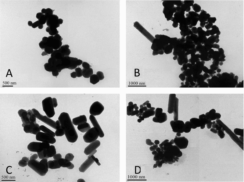
The results from the measurements of Zeta potential of HAP, FAP, HAP/Ag and FAP/Ag are shown in Table 1 and Figure 2. Zeta potential is described as the electrical potential difference between the dispersion medium and the stationary layer of fluid attached to the dispersed particle [44]. Negative Zeta potential of hydroxyapatite is usually reported, which could be a beneficial property for implantation in bone [45,46]. However, the positive Zeta potential does not preclude the usefulness of such materials. The value of Zeta potential is related to total calcium concentration [36,47] and indicates adsorption of protein on the material, which is associated with charge and distribution of adsorption sites on the surface of the substrate. The attachment of proteins, having different isoelectric point (pI), is depending on the Zeta potential of apatites [48]. Results from this part of the study have shown that values of Zeta potentials of HAP, HAP/Ag and FAP were negative; with only FAP/Ag Zeta potential positive at neutral pH (Table 1). In the first three measurements, the values in close proximity of −25 mV (which is known as threshold value) have confirmed the stability of tested materials. Increased value of Zeta potential observed for the FAP/Ag might suggest a tendency towards agglomeration and colloid instability [49].
Zeta potential (a) and DLS spectra (b) for HAP, HAP/Ag, FAP and FAP/Ag samples, and their distribution comprising a standard deviation for Zeta potential (c) and for DLS (d). The Zeta potential, electrophoretic mobility and DLS analysis (hydrodynamic diameter and polydispersity).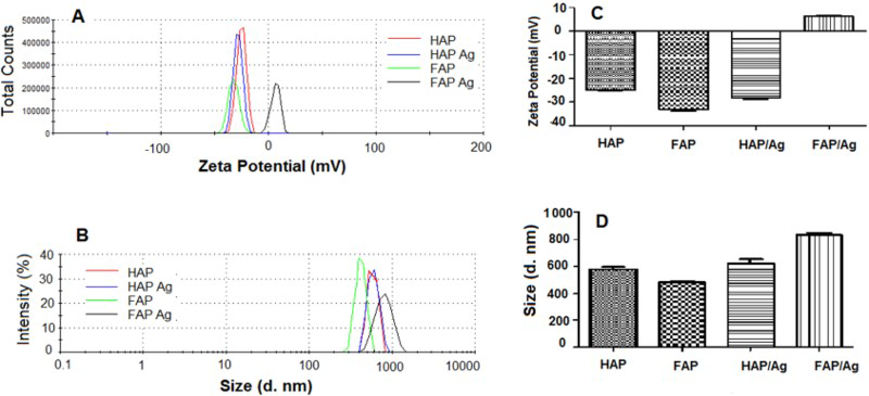
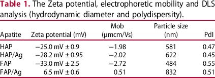
The size and distributions of particles were assessed with a laser DLS in water solution (Table 1). Similarly to Zeta potential, a significant change between pure and Ag-doped HAP particles was not detected. The hydrodynamic radius of the particle increased from about 581–622 nm (about 7%). When compared with pure or doped FAPs, the average sizes of particles were 484 and 832 nm, respectively. These results were consistent with those obtained during the Zeta-potential analysis, and confirmed that FAP/Ag nanoparticles were more unstable and could flocculate and/or aggregate.
Functional groups bonded to hydroxyapatite and fluorapatite molecular structures were identified by the FTIR spectroscopy (Figure 3(a)). The presence of stretching vibration around 3570, 3496 cm−1 and bending vibration at 633 cm−1 has confirmed the presence of a hydroxyl group only in HAP structure. The most intense lines attributed to phosphate groups were observed in HAP and FAP samples. There are PO43− ν3 mode at 1088/1093 cm−1, PO43− ν1 mode at 960/965 cm−1, PO43− ν4 mode at 603/604 and 568/569 cm−1 and PO43− ν2 mode at 470 cm−1 [50-53]. The weak band at 1457 cm−1 is corresponding with CO3 ν3 vibration [49,54]. The observed functional groups are presented in Figure 3(b).
FTIR spectra of the pure HAP (1) and FAP (2) NPs (a). The identification of the lines observed in HAP and FAP nanoparticles FTIR spectra (w: weak, s: strong line) (b).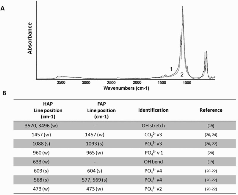
Apatite, containing carbonate groups, has been classified as type A or B, depending on the presence of CO32− groups. Carbonate ion can substitute for OH− (Type A), and for PO43− (Type B). The mixed type AB also can occur. All of the synthesised nanoparticles of HAP, FAP, HAP/Ag and FAP/Ag can be classified as apatites of type B, which was in agreement with other studies [51,55]. Predominantly, biological apatites are type B, and obtaining the same classification for the synthesised materials has confirmed their good biocompatibility. Apatites of this type give the FTIR bands associated with stretching vibrations of CO32− at about 1450–1460 cm−1, which is noticed in the presented spectra (Figures 3 and 4(a)) at 1457 cm−1. The main characteristic line of type A is ν3 band at around 1545 cm−1 [55-57].
We have found that manufactured HAP and FAP nanoparticles had good crystal quality, which indicated the stoichiometric composition between PO43− and Ca2+ ions [50]. After doping with Ag, the peak's position and crystal structure were not changed, which confirmed negligible influence of added silver on the crystal's structure. Figure 4 presents FTIR and Raman spectra of all studied apatites.
FTIR (a) and Raman (b) spectra of pure and silver-doped HAP and FAP NPs. (c) Identification of Raman lines observed mentioned nanoparticles (w: weak, m: medium, s: strong line).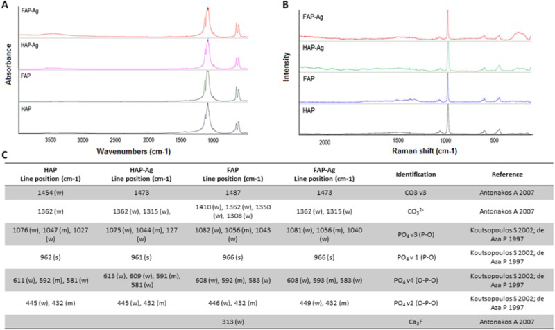
Complementary to FTIR, the Raman analysis has confirmed the development of HAP/Ag and FAP/Ag with correct crystal structure of apatites. As shown in Figure 4(b), the Raman spectra of studied samples were similar, and all the most intense bands have been assigned to internal vibrational modes of the phosphate groups [58-60]. They have been placed in correct wavenumber positions as shown in Figure 4(c). A very strong PO4 ν1 line has been noticed at 962 and 961 cm−1 in the HAP and HAP/Ag samples, respectively, which is assigned to a typical feature of HAP crystalline [58,60]. This line is shifted by 5–966 cm−1 in FAP and FAP/Ag samples. Other modes of PO4 vibration lines are situated around 1027–1082, 581–611, 432 and 446 cm−1 [5860]. In the 100–300 cm−1 region occurred some weak lines, which can originate from lattice modes or cation–oxygen modes [61].
Electronic diffuse reflectance spectra in the UV-range for samples after sorption are shown in Figure 5. The absorption bands of rather high (F (R)≈1 and more) intensity at 205–210 nm were observed. These bands belong to 4d–4d electronic transitions in Ag+ ions, thus confirming their presence in apatite structures. It worth noting that careful washing of samples with water resulted in gradual desorption of Ag+ ions, which was expressed through considerable reduction of intensity of adsorption bands.
Diffuse reflectance spectra of the samples of HAP (1) and FAP (2) after Ag+ sorption before (a) and after (b) washing.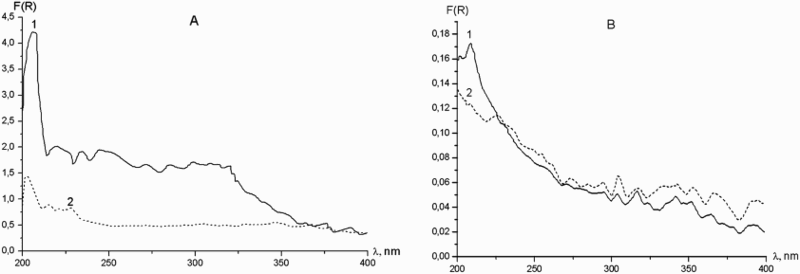
The recent studies on the characterisation of hydroxyapatite and fluorapatite have shown that fluorapatite has a higher thermal stability and better mechanical properties than pure hydroxyapatite [22]. Moreover, the presence of fluoride ions is important owing to the protection of teeth from dental caries; and it also enhances mineralisation and crystallisation [62]. Thus, our biomaterial that includes fluoride ions, in combination with silver, could be expected to have enhanced built-in antibacterial properties.
To confirm our assumptions, we compared the antimicrobial effect of the modified FAP to pure FAP or HAP, as well as to Ag-doped hydroxyapatite against the Staphylococcus aureus; the main pathogenic species among orthopaedic clinical isolates of implant-associated infections [25]. To examine the effects of apatite powders on S. aureus, a colony-forming capability tests were carried out. We chose this method according to the guidelines of the ASTM (International-Standards Worldwide) and CLSI (Clinical and Laboratory Standards Institute) [63,64] norm for the determination of antibacterial activity. Chosen assay was a quantitative assay, which generates more reliable results when compared with qualitative zone of inhibition (qualitative method) [65].
Figure 6 summarise the results from the study on the antimicrobial properties of HAP, HAP/Ag, FAP and FAP/Ag composites against Staphylococcus aureus, with an exposed time of 4 or 18 h. It could be seen that the modified apatites (HAP/Ag or FAP/Ag) strongly affected the viability of the bacteria, when compared to the pure apatites (pattern vs. clear bars in Figure 6). For the silver-doped hydroxyapatite (HAP/Ag), reduction ratio of viable cells was about 33 or 62% (p < 0.01) for 4 or 18 h of incubation, respectively. Interestingly, the 100% of inhibition of bacteria proliferation, (p < 0.001) was observed for silver-modified fluorapatite (FAP/Ag), regardless of the time of incubation. Generally, the results showed that the bactericidal activity was dependent on the type of apatite, and the antibacterial effect could be substantially increased by the doping FAP with silver. Moreover, the higher efficacy of the antibacterial activities of the FAP/Ag could be explained by the possible adhesion of bacteria to this type of the apatite. It is known that the physicochemical properties of the biomaterial may have a contributory effect on the bacterial adhesion [66]. In summary, the observed 100% inhibition of Staphylococcus aureus proliferation has suggested that addition of silver is crucial in obtaining built-in antimicrobial properties.
Bactericidal effect of biomaterials: HAP and HAP/Ag or FAP and FAP/Ag of S. aureus. Bars indicate SD, n = 3, **p < 0.01, ***p < 0.001 (ANOVA, Bonferroni's Multiple Comparison Test).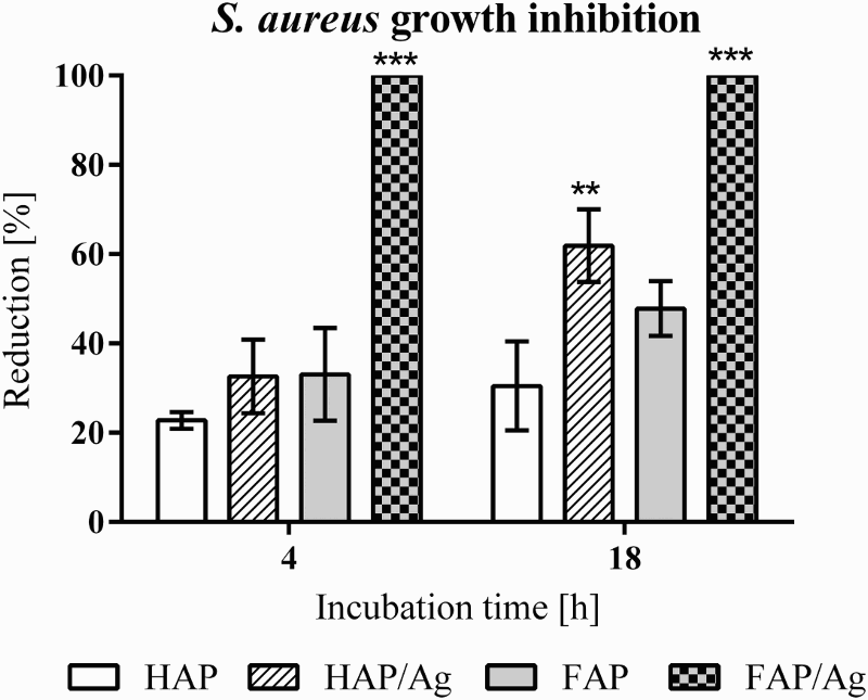
Conclusions
The presented work was focused on the physical and bactericidal characterisation of man-made pure and silver-modified apatites. It was found that manufactured HAP and FAP had structures typical for the biological (bone) apatites. It was confirmed that doping with silver has not changed the morphology of apatites (FAP/Ag and HAP/Ag), and lutetium ions added during doping process have not entered structures of developed material. This was an important observation, because final products with or without silver have kept their natural bone morphology. The aim of adding silver was to increase biocidal properties of new material. A series of assays with S. aureus, representative of bacteria responsible for infections during or after implanting procedures, have confirmed that the addition of silver substantially increased biocidal built-in properties. Interestingly, the inhibition growth rate of pure HAPs and FAPs was about 20 and 30%, respectively. After 4 and 18 h of incubation, the growth inhibition rate of the HAP/Ag has increased to 33 and 62%, respectively. Similar assays for FAP/Ag have shown that even after 4 h of incubation, the growth inhibition rate has reached 100%, which indicated complete eradication of bacteria. Our promising results have confirmed that FAP/Ag apatites successfully demonstrated biocidal effect against Staphylococcus aureus. Based on the obtained results, we can conclude that man-made, Ag-doped fluorapatite (FAP/Ag) is a promising new resistant biomaterial with great bactericidal effect that potentially could be applied in tissue engineering.
Footnotes
Disclosure statement
No potential conflict of interest was reported by the authors.