
Abstract
Aurivillius phase BaBi4Ti4O15 micro-sized powders were produced by solid-state reaction and their photocatalytic properties were reported for the first time. X-ray diffraction revealed the polar orthorhombic structure. BaBi4Ti4O15 ceramics exhibited diffuse phase transition at ∼ 410°C. The freezing temperature of 274°C was obtained by fitting the Vogel–Fulcher law. The distinct ferroelectric domain switching current peaks in current – electric field (I-E) loop and piezoelectric coefficient d33 value of 7.0 ± 0.1 pC/N at room temperature further demonstrated relaxor ferroelectric behaviour of BaBi4Ti4O15. UV-vis absorption spectra indicated that BaBi4Ti4O15 had a direct band gap of 3.2 eV. The photocatalytic study showed 15% degradation of Rhodamine B (RhB) solution by BaBi4Ti4O15 powders after 3.5 h UV-vis irradiation. The RhB degradation rate was further enhanced by depositing Ag nanoparticles on the BaBi4Ti4O15 powders surface. This work suggested that the relaxor ferroelectric BaBi4Ti4O15 is promising for photocatalytic applications.
Introduction
Semiconductor photocatalysis has attracted intensive attention due to their promising applications in environmental remediation and solar energy conversion. The hazardous organic pollutants from industrial effluent can be effectively decomposed through various chemical redox reactions induced by photoexcited charge carriers in an excited semiconductor [1]. One of the most important challenges for commercial applications of traditional semiconductor photocatalysts, such as TiO2, CdS, ZnO and WO3, is their low photocatalytic activity caused by fast recombination of photo-induced electrons and holes before they can initiate the photocatalytic processes [2]. The utilisation of ferroelectric materials as photocatalysts is considered to be a promising way to address this challenge in traditional semiconductor photocatalysts [3,4]. The polar structure of ferroelectrics induces an internal electric field, which facilitates the transport of photo-induced charge carriers, and thus enhances their separation. Photocatalytic activity of typical ferroelectrics with perovskite structure, such as BaTiO3 and BiFeO3, has been demonstrated [5-7]. In addition, perovskite materials can be modified with noble metals (such as Ag, Au, Pt, etc.) to optimise their plasmonic characteristics to design effective photocatalysts [810]. Although great efforts in the studies on the photocatalytic properties of ferroelectric materials with perovskite structures have been made, it is important to explore alternative ferroelectric materials with different crystal structures for potential photocatalytic applications.
The Aurivillius phases consist of intergrowth of [Bi2O2]2+ layers and pseudo-perovskite blocks (A n -1B n O3n+1)2-, where n is the number of perovskite-like layers. They have received increasing attention due to their unique properties. For example, their high Curie points make them attractive for high-temperature piezoelectric devices [11]. They are also promising for nonvolatile ferroelectric random-access memory applications due to their large polarisations and fatigue-free properties [12]. In addition to the aforementioned applications based on their ferroelectric and piezoelectric properties, efforts have also been made to explore their potential applications as photocatalysts. Kudo [13] and Tang et al. [14] firstly reported Bi2WO6 exhibited photocatalytic activity and could degrade organic compounds under visible-light irradiation. Afterwards more Aurivillius phase materials have been identified to exhibit photocatalytic behaviour, such as Bi3TiNbO9 [15], SrBi2Ta2O9 [16], Bi6Ti3WO18 [17] and Bi4Ti3O12 [18]. BaBi4Ti4O15 (BBT) is one of the Aurivillius oxides, which exhibits relaxor ferroelectric behaviour [19]. Although the crystal structure and electrical properties of BBT single crystal or ceramics have been investigated [20-25], to the best of our knowledge, there is no report about the photocatalytic properties of BBT.
In this paper, dielectric, ferroelectric and piezoelectrical properties of BBT ceramics were investigated to confirm the nature of their relaxor ferroelectric behaviour. Before the characterisation of photocatalytic properties, some of BBT powders were coated with Ag nanoparticles (hereafter named as BBT-Ag). Optical absorption property of the BBT powders was investigated to evaluate their energy band. The degradation rate of Rhodamine B (RhB) was used to compare the photocatalytic activity of BBT powders with and without Ag coating. By investigating photocatalytic properties of BBT, we report here for the first time that BBT is promising for photocatalytic applications.
Materials and methods
BBT powders were produced by solid-state reaction. BaCO3 (99.0% purity), Bi2O3 (99.975% purity) and TiO2 (99.6% purity) were used as starting materials. The stoichiometric mixtures of oxides were thoroughly milled for 96 h. The powder mixture was firstly heated at 600°C for 1 h and at 800°C for 2 h, then finally calcined at 950°C for 4 h. The pre-calcination step at 600°C and 800°C allowed time for Bi2O3 to react and form an intermediate phase with TiO2. This not only prevented an eutectic melt between Bi2O3 and one of the intermediate phases, Bi12TiO20, but also prevented the volatilisation of Bi2O3 which might occur before the formation of the desired compounds [26]. Calcination caused an agglomeration of BBT powders. In order to reduce agglomerate size to facilitate subsequent sintering and photocatalytic behaviour, the calcined BBT powders were re-milled for 168 h. To characterise the electrical properties, BBT ceramics were sintered at 1100°C for 1h. The density of sintered samples was 93%. Platinum paste (Gwent Electronic Materials Ltd, C2011004D5) was coated on the surfaces of disc BBT ceramic pellets as electrodes for electrical property measurements. The Pt electrodes were heated at 900°C for 30 min for strong adhesion on BBT ceramic surface.
The deposition of Ag nanoparticles on the surface of BBT powders was conducted through a photoreduction reaction [27]. 0.5 g BBT powders were added into a Petri dish with 50 ml of 0.01 M AgNO3 solution. The mixture was irradiated by a UV illumination source (UV Cube with a high pressure Hg lamp, Honle) for 10 s under constant stirring. The powders were separated from the solution by centrifuging, then washed with DI water, and dried at room temperature.
The phase of BBT powders after 950°C calcination and milling was characterised by using X-ray diffraction (XRD). The morphology of the above BBT powders and the polished ceramics were characterised by using a scanning electron microscope (SEM, FEI Inspect F). The particle size distribution of BBT powders was measured by laser diffraction (Zetasizer Nano, Malvern Instruments Ltd, UK). The changes in the dielectric constants and losses with temperature were tested at various frequencies using an LCR meter (Agilent 4284A). The coercive field of Aurivillius phase ferroelectric is usually very high at room temperature, which makes ferroelectric domains hard to be switched during P-E loop measurement at room temperature [28]. Therefore, in order to reduce coercive field of BaBi4Ti4O15 ceramics, P-E loops was tested at elevated temperature. The polarisation – electric field (P-E) and current – electric field (I-E) loops were measured by using a ferroelectric hysteresis measurement tester (NPL, UK) at 200°C and 100 Hz. Triangular voltage waveforms with two complete cycles were applied to the test samples during P-E and I-E loops measurement. The poling of BBT ceramics were conducted in silicone oil at 180°C with a DC electric field strength of 6 kV/mm for 15 min. A piezo d33 meter (ZJ-3B, Institute of Acoustics, Chinese Academic of Science, Beijing) was used to measure the piezoelectric constant, d33, of the poled BBT ceramics.
Composition and chemical states of BBT-Ag were detected by X-ray photoelectron spectroscopy (XPS, Thermo Scientific™ Nexsa™) with Al Kα X-ray source. The optical absorption spectra were obtained from a UV-vis spectrophotometer (PerkinElmer LAMBDA 950). The photocatalytic activity of both BBT and BBT-Ag powders was assessed by degradation of Rhodamine B (RhB, Sigma, 99.99%) dye solution [3]. 0.15 g catalyst powders and 50 ml of 10 ppm dye solution were mixed in a glass Petri dish and stirred in the dark for 30 min before exposure to a solar simulator (Newport, class ABB). The irradiation intensity was kept at 1 sun (100 mW/cm2) with a silicon reference cell. The degradation reaction was checked by taking 2 ml of the solution for measurement of the absorbance at the wavelength (554 nm) of maximum absorbance of RhB using the UV-vis spectrophotometer. According to the Lambert−Beer law [29], the absorbance is proportional to the RhB concentration:

Results and discussion
Figure 1 is XRD pattern of BBT powders after 950°C calcination and milling. The powders show a single phase, which can be well indexed to orthorhombic structure with space group A21am (JCPDS no 43-0973) [30,31]. SEM image [Figure 2(a)] shows the BBT powders were extensively agglomerated with an average agglomerate size of 2.5 ± 0.1 μm. Figure 2(b) shows the grain morphology of BBT ceramic. The sample is composed of randomly oriented plate-like grains. The pores are visible in BBT ceramic. Similar microstructure in conventionally sintered BBT was also observed [32]. Although the Aurivillius phase ceramics are difficult to be densified by the conventional sintering due to a low packing density caused by plate-like grains [33], the current ceramic samples are dense enough for electrical property characterisation. Figure 3 presents the particle size distribution of BBT powders after 950°C calcination and milling. A broadened unimodal distribution peak at around 1.46 µm was observed.
XRD pattern of BBT powders after 950°C calcination and milling. SEM micrographs of (a) BBT powders after 950°C calcination and milling, and (b) BBT ceramic. Size distributions of BBT powders after 950°C calcination and milling.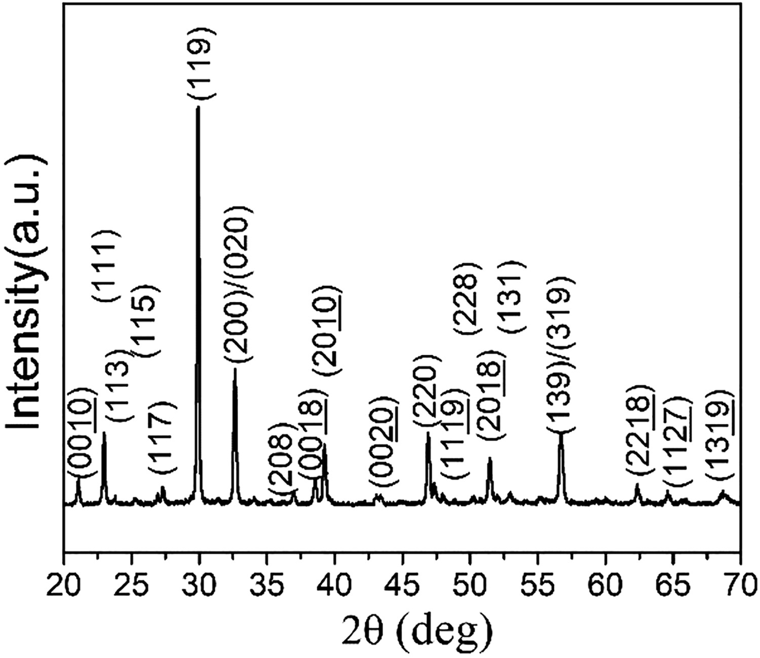
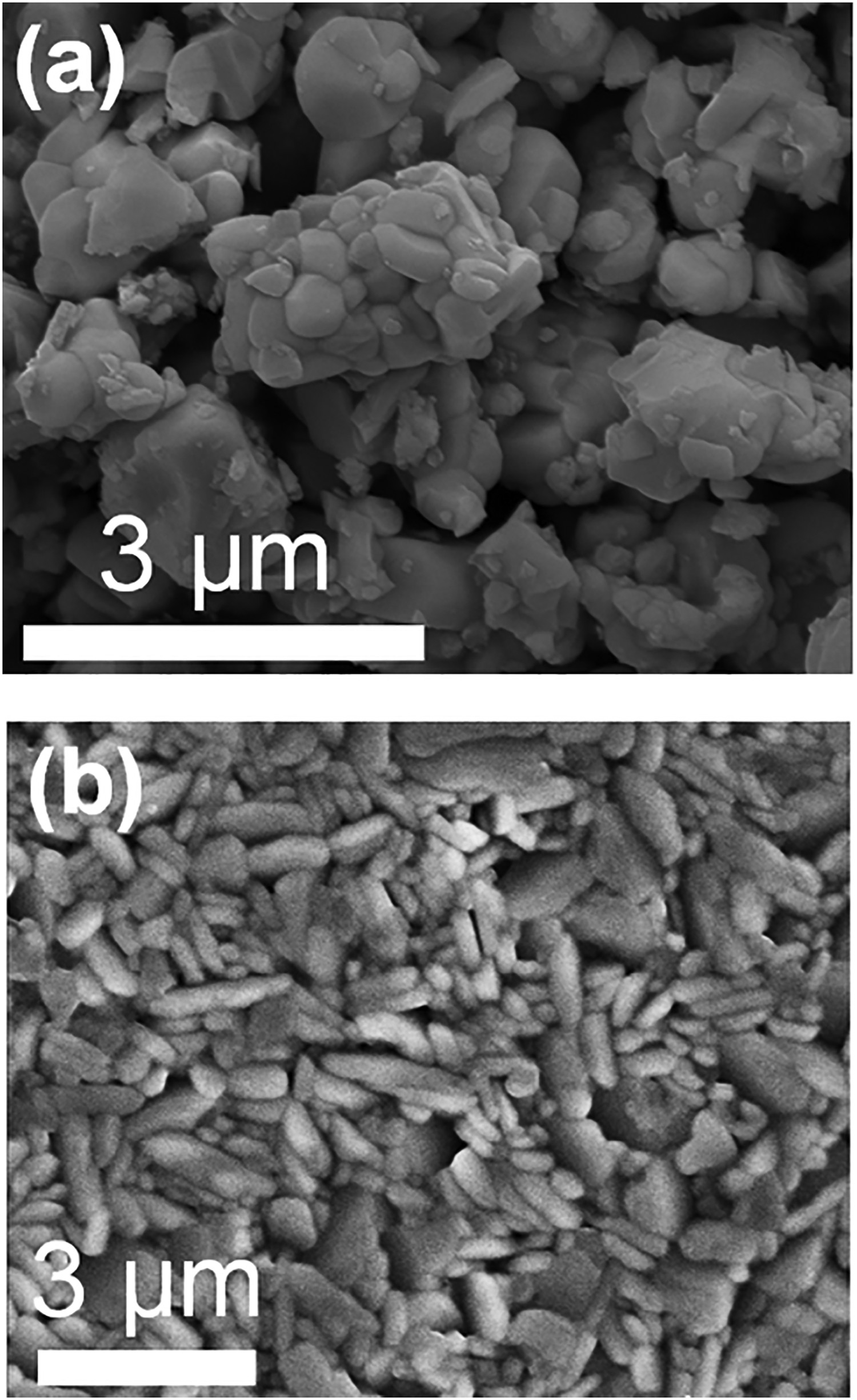
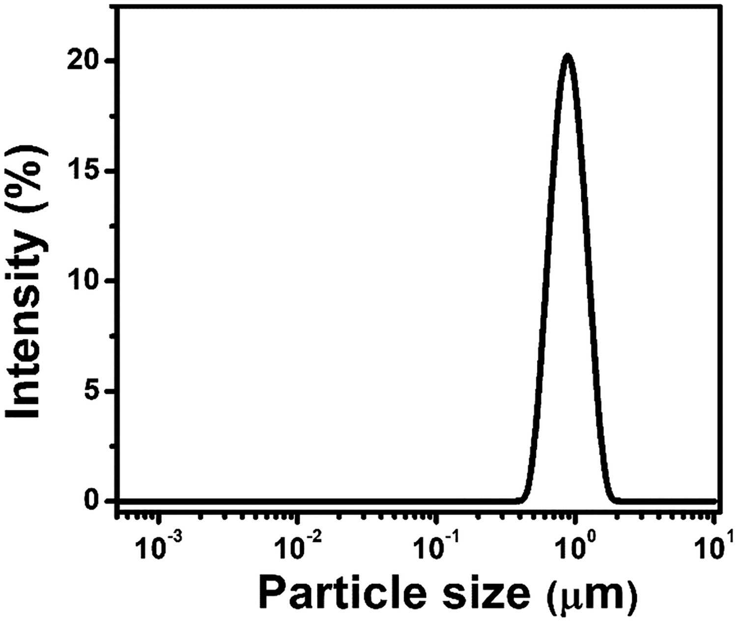
Figure 4 illustrates the dielectric constants and losses of BBT ceramics as a function of temperature at different frequencies up to 500°C. There is diffuse dielectric anomaly at around 410°C. In addition, the temperature (Tm) of the dielectric constant maximum increased with increasing frequency, which is the typical relaxor ferroelectric behaviour. The loss peaks are observed at all frequencies below Tm. Above Tm the dramatically increased losses are assumed to be caused by an increase in conductivity with the increasing temperature. Our results are consistent with the results previously reported for BBT [34]. It is well known that the relaxor ferroelectric behaviour can be described by Vogel–Fulcher law [35]:
Temperature dependence of the dielectric constants and losses at different frequencies. Inset: the Vogel-Fulcher fit of the frequency-dependent transition temperature Tm.
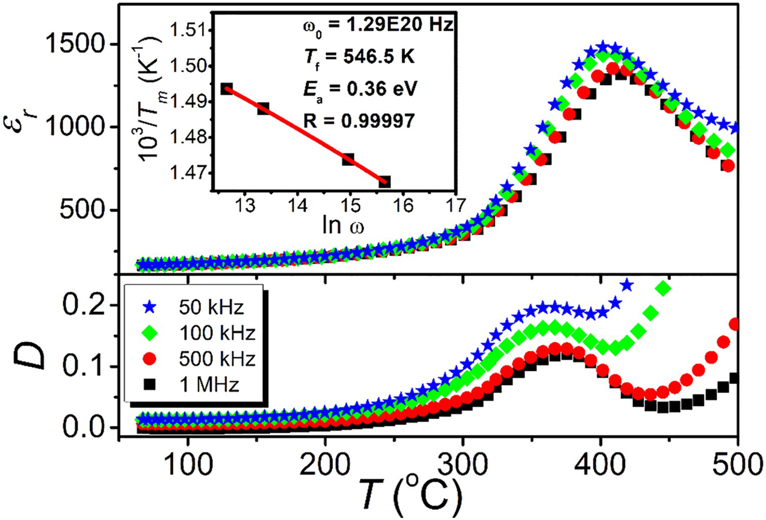
Figure 5 shows the I-E and P-E loops of BBT ceramics tested at 200°C and 100 Hz. The ferroelectric domain switching is evidenced by the current peaks at the first and third quadrant in I-E loop [28]. After poling at 180°C, BBT ceramics showed a d33 value of 7.0 ± 0.1 pC/N at room temperature. When the external electric field is absent, relaxor ferroelectrics contain randomly oriented polar nanoregions with strong thermal fluctuation above Tf temperature [35]. The number, size and the interaction of polar nanoregions increase during cooling. When the temperature reduces to around Tf temperature, the thermal fluctuation is frozen and micro-domains appear. When the temperature is further reduced below Tf, the micro-domains are able to be oriented along the direction of electric field and the macro-domains appear [35]. The temperatures for both poling (180°C) and P-E loop measurement (200°C) are significantly lower than Tf value (274°C). Consequently, distinct current peaks in I-E loop and d33 value is consistent with micro- to macro-domain transition in relaxor ferroelectric BBT ceramics. Obviously, the spontaneous polarisation in relaxor ferroelectric BBT represents an attractive property for photocatalytic activity, which will be demonstrated in the following context.
I-E and P-E loops of BBT ceramics measured at 200°C and 100 Hz.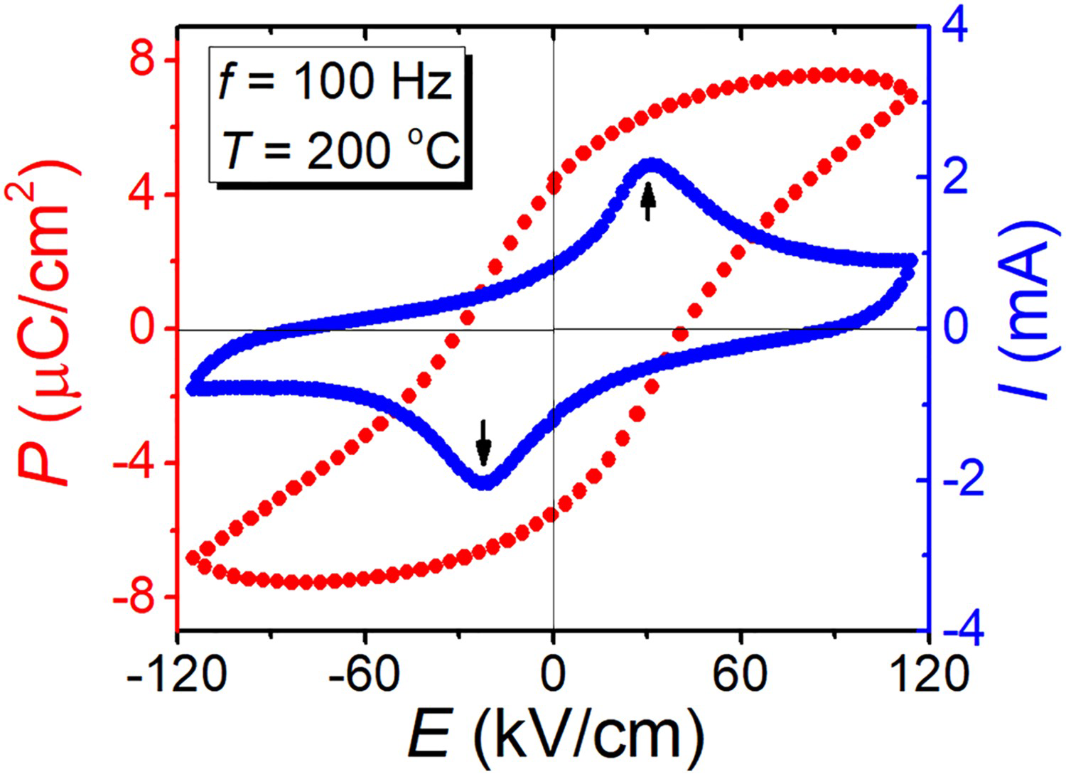
XPS was conducted to confirm the existence and chemical state of BBT-Ag powders. Figure 6(a) is the XPS survey spectrum of BBT-Ag, which shows the existence of Ag on BBT surface. Figure 6(b) is the XPS spectrum of Ag 3d. Two peaks at 368.28 and 374.28 eV are characteristic of Ag 3d5/2 and Ag 3d3/2 and consistent with the XPS spectrum of metallic Ag [36]. Accordingly, the XPS results confirm that Ag has been successfully deposited on the BBT powder surface.
XPS spectrum of BBT-Ag (a) Survey spectrum; (b) Ag 3d. The spectrum of Ag 3d is characteristic of metallic Ag.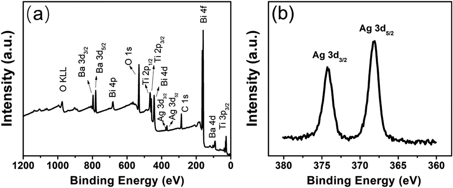
Figure 7(a) shows UV-vis absorption spectrum of both BBT and BBT-Ag powders. The vertical dash line is the boundary between UV and visible light. The BBT powders had a small visible-light absorption at the wavelength range of 400–550 nm, and a sharp increase in light absorption at the wavelength of < 400 nm [Figure 7(a)]. Incorporation of Ag nanoparticles on the surface of BBT powders increased light absorption of the photocatalysts in the visible-light region. This can be attributed to the effect of surface plasmon resonance (SPR) [37]. The derived Tauc plot of BBT powders is shown in Figure 7(b). The optical band gap (Eg) of BBT powders could be calculated using the Tauc relationship [38]:
(a) UV-vis absorption spectrum of BBT and BBT-Ag powders, where vertical dash line is the boundary between UV and visible light; (b) the derived Tauc plot of BBT, where the dot line is tangent of the linear part.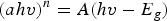

The photocatalytic activity of both BBT and BBT-Ag powders was evaluated by photodegradation of RhB. Figure 8 shows the photodegradation profiles of RhB with BBT and BBT-Ag powders, in which the photocatalytic degradation ratio (C/C0) was presented as a function of the degradation time t. After the BBT powders were irradiated by UV-vis light for 3.5 h, 15% RhB was degraded, which demonstrates photocatalytic activity of BBT powders. As shown previously, BBT is a relaxor ferroelectric. Therefore, BBT can effectively separate photo-induced charge carriers due to the existence of internal electric field. When BBT powders were deposited with Ag nanoparticles, the degradation rate significantly increased and a complete degradation of 100% was achieved within 3.5 h. The enhanced photodegradation behaviour can be attributed to the catalytic function of the Ag nanoparticles, which can provide chemically active sites for relevant chemical reactions to start with lower activation barriers [37]. Furthermore, the SPR of Ag nanoparticles can potentially provide charge carriers that take part in photocatalytic reactions by electrons injection from surface plasmon states of Ag to the conduction band of the BBT photocatalyst [37]. Clearly, BBT is very promising for photocatalytic applications, which has not been reported previously. Since the catalytic activity of a catalyst greatly relies on its specific surface area, the photocatalytic properties of BBT is expected to be further improved when the powder particle size is reduced from current micro-size to nano-size.
Degradation profiles of RhB with BBT and BBT-Ag under solar simulator.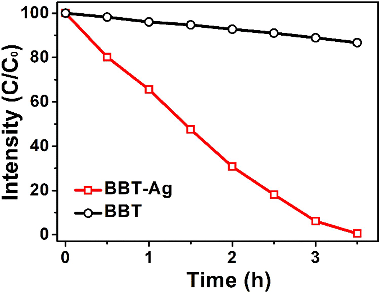
Conclusion
BBT powders with micrometre size were produced by conventional solid-state reaction. The XRD pattern indicated that the BBT powders were single phase with orthorhombic structure. BBT ceramics with a density of 93% were produced for electrical property characterisation. The dielectric measurement showed diffuse dielectric anomaly at around 410°C. The freezing temperature Tf obtained by fitting of Vogel–Fulcher law was 274°C. The distinct current peaks in I-E loop due to ferroelectric domain switching were observed during P-E loop testing at 200°C and 100 Hz. BBT ceramics showed a d33 value of 7.0 ± 0.1 pC/N after poling at 180°C and cooling down to room temperature. The electric properties confirmed the relaxor ferroelectric behaviour of BBT. UV-vis measurement suggested that the direct band gap of BBT was 3.2 eV. The BBT powders demonstrated the photocatalytic activity with a RhB degradation of 15% under UV-vis light irradiation for 3.5 h. A significantly increased RhB degradation up to 100% was achieved when the surface of BBT powders were deposited with Ag nanoparticles.
Footnotes
Disclosure statement
No potential conflict of interest was reported by the authors.