
Abstract
Coral reefs provide habitat for most oceanic life forms, however, they are declining worldwide due to pollution, increased ocean temperature, diseases and other factors. In this paper, structural and mechanical properties of endangered coral (Acropora cervicornis) skeletons cleaned by chemical bleaching and biological processes were studied. The structure of CaCO3 aragonite skeleton is porous and very complex and affects the deformation behaviour of coral skeletons. The average compressive strength was equal to 9.5 ± 2.3 MPa and 14.5 ± 6.4 MPa, while Vickers hardness was measured to be equal to 3.67 ± 0.33 GPa and 3.65 ± 0.24 GPa for the chemically bleached and biologically cleaned skeletons, respectively, thus showing similar values regardless of the cleaning techniques. The results are important for understanding the fundamental relationship between the coral skeleton's microstructure and mechanical behaviour.
Introduction
Coral reefs are very diverse and occur worldwide in warm, clear and clean ocean waters. However, in the last half-century many environmental factors, including elevated ocean temperatures and other pollution, caused catastrophic declines of many coral populations [1]. Acropora cervicornis (Lamarck, 1816) is a weedy coral that once formed extensive thickets in shallow water (to ∼25 m deep) with low to moderate wave exposure throughout the Caribbean Sea. At present, most of these thickets have disappeared or are greatly reduced in size, and A. cervicornis is listed as threatened under the US Endangered Species Act. Several coral nurseries are propagating A. cervicornis asexually from fragmented pieces of live colonies. The mechanical properties of skeletons of nursery-reared A. cervicornis are of considerable interest because premature fragmenting may cause colonies to decline.
Acropora cervicornis is a reef-building coral with a branching, deer antler-like skeleton comprised of needle-like aragonite, which is a form of CaCO3. This skeleton bears the cyclic mechanical load constantly imposed on this branching coral by ocean waves, and thus the mechanical behaviour of coral skeletons is important for understanding their structural integrity and time to failure (i.e. fragmentation, which can be a form of asexual reproduction).
To study the mechanical behaviour of coral skeletons in the laboratory, skeletons are cleaned of organic matter: mostly proteins from the living polyps, which reside in the skeleton's corallites [2]. Different cleaning techniques can be used to remove the proteins: two typical processes are chemical bleaching and biological decomposition. Bleaching is performed by soaking corals in a 2 × 10⁻¹ M aqueous solution of sodium hydroxide (NaOH), which decreases coral pigmentation because it chemically digests protein from the skeleton [3]. In contrast, biological decomposition uses natural processes to remove protein; dead coral branches are simply buried on the sandy ocean floor and proteins are removed by microorganisms residing in the sand [4]. This paper reports, for the first time, the effect of these alternate protein removal techniques on mechanical properties of the staghorn coral skeleton.
Experimental
Skeletons of A. cervicornis were retrieved from Nova Southeastern University's coral nursery off Broward County, FL, USA, and proteins were removed using either bleaching or biological decomposition. During the bleaching process, the skeletons were soaked overnight in a bleach solution. In the biological decomposition process, the skeletons were buried for several days underneath sand at the nearshore coral nursery. Both processes produced a clean coral skeleton but chemically bleached skeletons were noticeably brighter. After cleaning, skeletons were cut into cylindrical samples with 1:2–2:1 height-to-diameter ratios. Top and bottom surfaces of these cylindrical samples were then polished and ground with P1000-3000 SiC sandpaper to prepare them for hardness and compression tests.
X-ray computed tomography (GE Phoenix Nanotom-MTM; GE Sensing & Inspection Technologies GmbH, Hamburg, Germany) was used to analyse both chemically bleached and biologically cleaned coral skeletons at 8 μm voxel size. Phoenix datos software (Baker Hughes; a GE company, Texas, USA) was used to reconstruct the 2D images into 3D volume renderings for the skeletons. Analysis and image production of the 3D volumes was performed with VGStudioMax (v 2.1) and myVGL software (v 3.2.3; Volume Graphics GmbH, Heidelberg, Germany).
Hardness measurements were performed using a Vickers hardness tester (Tukon 2100B; Wilson Instruments, Illinois, USA) with 10 indentations under a 100 gf load and 15 s dwell time under the load. A universal testing machine (Critertion® 43; MTS, Minnesota, USA) was used for uniaxial compression tests on the cylindrical samples, with a strain rate of 0.003 mm s−1. Weibull statistics were used to calculate the probability of failure and Weibull parameters [5].
Results and discussion
Computed tomography scans
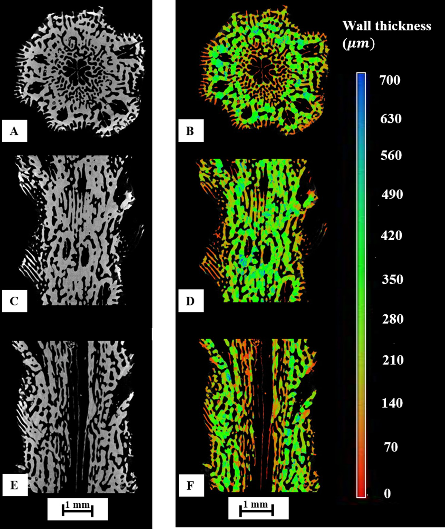
As shown by CT scans, the CaCO3 aragonite skeleton structure is highly porous with complex non-homogenously distributed cavities (Figures 1 and 2). The wall thickness of both chemically bleached (Figure 1) and biologically cleaned (Figure 2) skeletons decreased from their outer wall toward the centre of samples in a longitudinal direction, and was coupled with increased porosity in the same direction (Figure 1D and F) and (Figure 2D and F). No differences were apparent in the structure of chemically bleached and biologically cleaned skeletons except for overall wall thickness, which is explained by the sample of chemically-bleached coral skeleton being from the bottom of the coral; thus, it was older than the skeleton that was cleaned by biological processes.
(A, C, E) CT images and (B, D, F) wall thickness distribution of chemically bleached Acropora cervicornis CaCO3 aragonite coral skeleton. (A, C, E) CT images and (B, D, F) wall thickness distribution of biologically cleaned Acropora cervicornis CaCO3 aragonite coral skeleton.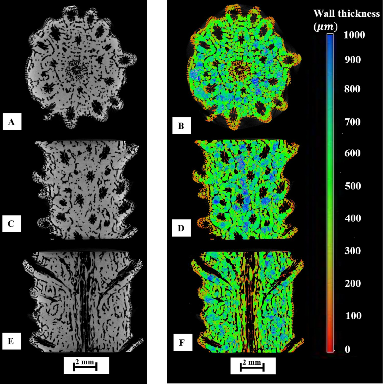
Vickers hardness
Vickers hardness values of CaCO3 aragonite skeletons measured on polished sample surfaces were 3.67 ± 0.33 GPa and 3.65 ± 0.24 GPa for the chemically bleached and the biologically cleaned samples, respectively, which is typical for aragonite materials [6,7]. Vickers hardness impressions of chemically bleached and biologically cleaned coral skeletons are shown in Figure 3. The highly porous structure of the skeleton prevented crack propagation, which resulted in very few radial cracks that grew from the corners of impressions (Figure 3A and B). No difference was found in the hardness values of chemically bleached and biological cleaned skeletons of A. cervicornis.
Optical micrographs of Vickers hardness impressions of (A) chemically bleached and (B) biologically cleaned Acropora cervicornis CaCO3 aragonite skeletons.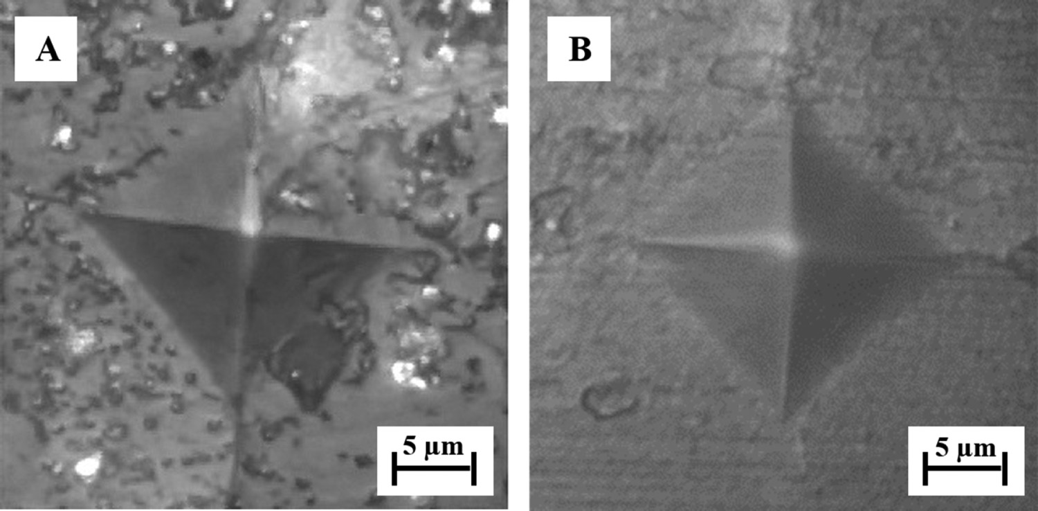
Uniaxial compressive strength
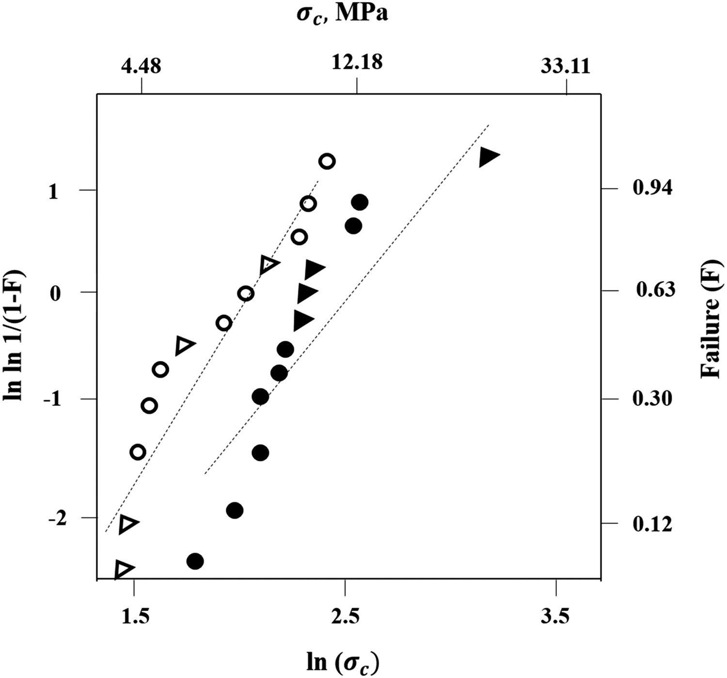
Stress–strain plots of the chemically bleached and biologically cleaned coral skeletons are shown in Figure 4. The response of the skeletons under compressive load was linearly elastic with many pop-ins occurring upon loading. Each pop-in represented a crack propagation event, where the crack propagated until it reached the next closest pore. This pore then causes the crack to stop growing because it reduces the stress concentration at the crack tip and the applied load must increase again until another cracking occurs. Crack accumulation eventually causes the specimens to fracture completely. Mean compressive failure stress σavg for chemically bleached samples was 9.5 ± 2.3 MPa, while for the biologically cleaned samples σavg was 14.5 ± 6.4 MPa. The calculated strength values are considered to be nominal due to no uniform shape of the coral skeletons. These values measured in the current study are similar to those reported by Chamberlain [8]. Young's modulus values, measured from the linear portion of the stress–strain deformation plots, were 0.19 ± 0.16 GPa and 0.29 ± 0.13 GPa for the chemically bleached sample and the biologically cleaned sample, respectively. Strength measurements at the point of failure and at the appearance of first pop-in were analysed using Weibull statistics (Figure 5). From the distribution, the scale parameter σo at first pop-in was equal to 7.31 MPa and its shape parameter m was equal to 3.45. At skeletal failure, the scale parameter σo was equal to 12.63 MPa and the shape parameter m was equal to 3.10, which are quite similar. No difference in deformation behaviour was apparent between chemically bleached and biologically cleaned coral skeletons.
Stress–strain deformation plots of chemically bleached and biologically cleaned Acropora cervicornis CaCO3 aragonite skeletons upon uniaxial compression. Weibull plots of chemically bleached and biologically cleaned Acropora cervicornis CaCO3 aragonite skeletons, ○ first pop-in event stress, chemically bleached. • Failure stress, chemically bleached. ▵ First pop-in event stress, biologically cleaned. ▴ Failure stress, biologically cleaned.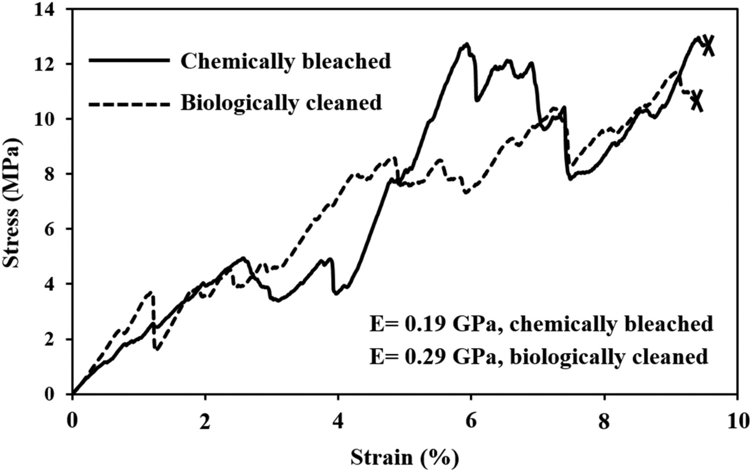
Conclusion
It was established for the first time that two different protein removal techniques – chemical bleaching and biological decomposition – had no appreciable effect on mechanical properties of staghorn coral (A. cervicornis) skeletons. Coral skeletons cleaned using either process had very similar values for Vickers hardness, Young's modulus and uniaxial compressive strength. The results are important to better understand the mechanical behaviour of coral (Acropora cervicornis) CaCO3 aragonite skeletons for further testing of their time dependent mechanical behaviour or their applications as bone substitutes in medicine. Ultimately, the study allows insights into the complex evolutionary designs of these coral structures from mechanics and materials perspectives.
Footnotes
Bridget Masa's and Zachary Shepard's help with performing mechanical tests of CaCO3 aragonite coral skeletons is greatly appreciated. This research was supported in part by NSF MRI award #133775. The GE Phoenix Nanotom-M™ X-ray CT was acquired through NSF Award # 0959511. Initial harvest of staghorn coral fragments to establish the NSU coral nursery was authorised under Florida Fish and Wildlife Conservation Commission Special Activity License # SAL-10-1086-SCRP.
Disclosure statement
No potential conflict of interest was reported by the author(s).