
Abstract
Bare and functionalised haematite nanoparticles (NPs) are synthesised through the co-precipitation method and characterised using XRD, SEM, TEM, EDX, FTIR, and VSM. XRD confirmed the rhombohedral-based hexagonal structure of Fe2O3, while, TEM and SEM showed the formation of lamellar-shaped NPs with the grain size varied from 22 to 80 nm. VSM presented soft ferromagnetic behaviour. From in vivo evaluations, changes in serum levels are analysed at different days of study after administration of bare and coated NPs through the intraperitoneal route of administration in albino rats. Toxicity parameters revealed a transient increase at day 1 but a gradual decrease thereafter. For biodegradation studies, NPs exposure in lysosomal-like medium showed insignificant changes in the absorbance spectrum for a short interval of time, however, dynamical degradation effects were observed on Day 8. Apoferritin studies disclose the growth of absorbance near 260 nm, which reveals progressive and partially filled proteins with iron ions.
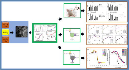
GRAPHICAL ABSTRACT
Introduction
Understanding the relationship between reactivity and structure of nanomaterials (NMs) in living bodies is an essential step towards the safe and efficient biomedical applications. The exploitation of iron oxide nanoparticles (IONPs) in nanomedicine is considered as the most important field among different areas of therapeutics. IONPs are potential treatment candidates for many in vivo applications, including magnetofection [1], cellular therapy [2], tissue repair [3], magnetic resonance imaging [4], controlled drug delivery [5], and hyperthermia therapy for cancer treatment [6]. In stem cell research and cell biology, IONPs can be used for cell separation [7], cell sorting [8], cell labelling [9], and purification processes. IONPs can be coated with suitable ligands for making them more biocompatible to enhance suitable targeting abilities. Increasing applications of IONPs also raised many concerns among scientific communities, public, and regulatory authorities due to their long-term toxicological effects on living organisms [10-13].
Haematite is considered as one of the most important polymorphs of iron. Haematite particles with micrometer size possess low toxicity at cellular levels, but with the size reduction, toxic effects have been witnessed because of an increase in structural defects. Hence, toxicity studies must be first conducted on animals to evaluate the effect of these nanoparticles (NPs) on living organisms because studies based on cell cultures are not reliable for longer exposure time. Additionally, NPs uptake and distribution are much complicated in biological systems as compared to cell culture. Besides the above-mentioned issues, the hydrodynamic size can cause in vivo changes owing to multiple factors such as opsonisation with biological macromolecules, supplementary salt deposition [14], conglomerates formation of NPs clustering due to surface charge of functionalised moiety. From a toxicity point of view, the main concern associated with NPs is excessive exposure, which requires complete elimination of these accumulated particles from different body organs. Many researchers tried to promote the NPs excretion from the body by reducing the tissue inflammatory effects through different anti-inflammatory drugs. In vivo studies also revealed that submicron and nano-sized haematite NPs usually induce pulmonary injury and neurotoxicity in rats [15].
Synthesis methods also play important role in the determination of toxicity associated with these NPs due to the presence of cationic distribution, physicochemical properties, and residual reagents. Different preparation methods are used to synthesise IONPs such as microemulsion [16], sol–gel [17], co-precipitation [18], conventional hydrothermal [19], laser ablation [20] and polyol methods [21]. However, co-precipitation is considered as the most common method due to simplicity, the use of less hazardous chemicals, and the formation of more uniformly sized NPs for their use in nanomedicines.
When NPs are injected into living organisms, after cellular uptake, they start enclosing in lysosomes and undergo the effect of various proteins, lipids, acidic environment, and hydrolytic enzymes that are responsible for the metabolism of NPs [22]. At the surface of metal/oxides, the protective layer is removed as the chemical etching leads towards the metallic ion release and crystal disintegration which form metal complexes [23]. Therefore, intracellular recycling of IONPs [24], silver NPs [25], zinc oxide NPs [26], and quantum dots [27] produce highly reactive metallic ions that alter the physical properties and functionalise the NPs as they interact with the microenvironment. As a result of the intracellular degradation of IONPs, harmful iron ions are produced that are usually channelised through ferritin. Ferritin possesses cytoprotective properties that control iron overloading as it can stimulate lysosomal stress [28].
The aim of this study is to provide a complete cycle from synthesis to the recycling of haematite NPs for in vivo applications. This study consists of four phases. In phase I, bare and coated haematite NPs are synthesised through the co-precipitation method. Characterisation of the prepared NPs are performed through X-ray diffraction (XRD), transmission electron microscope (TEM), scanning electron microscope (SEM), dynamic light scattering (DLS), energy dispersive spectroscopy (EDX), and Fourier transform infrared spectroscopy (FTIR). Phase II is based on acute toxicity studies. Phase III details the biotransformation studies of NPs to observe the change in pH, electrical conductivity, and optical properties through Ultraviolet–Visible (UV-Vis) spectroscopy results. In Phase IV, degraded ions transferred into ferritin proteins. By monitoring colloidal behaviour and storage ability of IONPs in ferritin presence, we investigate the role of protein in aggregation and erosion of IONPs. In ferritin cages, accommodation and storing ability of degraded products of IONPs are studied through in solution approach.
Materials and methods
Chemicals
Iron (III) chloride anhydrous (FeCl3), iron (II) chloride (FeCl2.4H2O), citric acid, and polyethylene glycol (PEG-6000) and Ammonia water 32% (NH3) were supplied by Daejung Korea. All solvents and chemicals were of analytical grade and used without any further purification. Apoferritin (ApoF) extracted from equine spleen was supplied by Sigma-Aldrich.
Synthesis protocol for iron oxide NPs
Haematite NPs were prepared by the co-precipitation method. To synthesise iron oxide NPs, FeCl2·4H2O and FeCl3 were dissolved with a 1:2 molar ratio in deionised water. The brown colour solution was obtained after mixing both the solutions. Ammonia (32%) solution was added dropwise in the prepared brown solution. Subsequently, the solution colour changed from brown to dark brown after a few minutes of stirring. Solution colour further changed from dark brown to black after 40 min of continuous reaction, which indicated the formation of haematite NPs. Precipitates were filtered after 24 h of aging and washed many times with deionised water to remove residual salts. Obtained precipitates were dried overnight in an oven at 60°C. After drying, the sample was shifted into the furnace for calcination under 600°C for 3 h. The same method was followed for the preparation of the other two samples. The coating was done after furnace treatment. For coating, the ligand exchange method was used to functionalise IONPs and the purpose was to make them stable in the suspension form. The ligand exchange method is very effective that can associate and substitute ligand on the metal surface. For the second sample, the prepared powder was stirred in deionised water. After 10 min of stirring, 1gram PEG (6000 kDa) was added to the haematite suspension and stirred for 24 h. Filtration and washing were performed again to remove impurities and residual PEG. Prepared powder was dried in an oven under 60°C for 4 h. The same method was followed for the third sample by adding 1 g citric acid in the NPs suspension. Prepared samples were characterised through XRD, SEM, TEM, EDX, DLS, VSM, and FTIR.
Characterisation tools
The characterisation of prepared samples was performed through respective tools. For the analysis of phase purity, Philips Xpert X-ray diffractometer was used, where Cu Kα radiations with wavelength 0.15406 nm were used for the analysis of prepared powder in the angle ranges from 20° to 70°. TEM and SEM of (JEOL-JEM-1010) were used for the morphological analysis. Bruker Alpha spectrophotometer was used to obtain the FTIR spectrum. VSM (Quantum Design, PPMS, Inc.) was used for the magnetic study. Zeta sizer (ZEN5600 Malvern) was used for analysing the zeta size and zeta potential. Optical properties were analysed by using T80 UV-Vis spectrophotometer (PG instruments LT9).
Toxicity evaluation
Acute toxicity testing was performed according to the rules of the Organisation for Economic Co-operation and Development. Studies were performed on four groups of albino rats. A total of 32 rats with an average weight in the range of 112–137 grams were bought. These animals were placed in separate cages and retained in a 12-h dark and light cycle. Full access to drinking water and ad libitum was provided.
Experimental design
For treatment allocation to rats, the following experimental design was adopted.
Group 1 received the water, routine diet and refers to the control group.
Group 2 received dose of 100 mg Kg−1 of bare haematite NPs administrated intraperitoneally.
Group 3 received dose of 100 mg Kg−1 of PEG-coated haematite NPs administrated intraperitoneally.
Group 4 received dose of 100 mg Kg−1 of citrate-coated haematite NPs administrated intraperitoneally.
Albino rats were kept under observation to note any change in mortality and routine intake of food/water. Any variation in physical signs e.g. mucous membranes, diaphoresis, variations in skin texture, diaphoresis, comma, sleep disturbances, and excessive salivation was observed for 8 days on a daily base. 50% of animals were sacrificed on day 1 and remaining on day 8. Day 1 was selected to observe adverse effects following the acute toxicity of NPs. While day 8th was chosen to study histopathological variations due to self-healing capacity of vital organs. Blood samples were collected for haematology and biochemical analysis. For histopathological analysis, vital organs were collected and preserved in 10% formalin solution.
Bio-transformations of NMs in the model biological media
To mimic the lysosome-like medium, NMs were dispersed in the buffer solution by maintaining the potential hydrogen (pH) at 4.7. By taking the same concentration of 0.01 M citric acid and sodium citrate tribasic in 250 ml of deionised water, a buffer solution was prepared [23,29]. A specific amount of haematite NPs was added in the 20 ml of prepared solution. The concentration of all nine solutions was kept the same during the analysis. All the solutions were kept under controlled conditions in the incubator by maintaining the temperature at 37°C [30]. To study the change in properties at a different time interval, UV-Vis spectroscopy, electrical conductivity meter, and highly precise pH meter were used. UV-Vis measurements were taken at a small interval of time till Day 8. Variations in pH and electrical conductivity in lysosomal media were also recorded.
Transfer of NMs into ferritin proteins
In one step complex, lysosomal media was prepared by the addition of ApoF in buffer solution used for transformation studies [31]. In Eppendorf's, 1 ml buffer solution, 1 mg NPs/salt, and 10 µL ApoF were mixed and kept at 37°C. Haematite NPs and iron salt (FeCl2) were added separately in two solutions. Transfer studies were conducted using UV-Vis spectra were taken after 2, 4 h, 1, 2, 3, 7, and 8 d.
Results and discussion
Characterisation of NMs
Structural analysis of bare iron oxide NPs is performed through XRD (Figure 1), which confirmed the formation of the single-crystal haematite phase matched with the JCPDS card no. 00-002-0919. Diffraction peaks appeared at angles 24.18°, 33.21°, 35.90°, 41.06°, 49.46°, 54.15°, 62.78°, and 64.13° correspond to the planes (012), (104), (110), (113), (024), (122), (214) and (300), respectively. No other phase has appeared which confirms the purity of the prepared sample. Prepared haematite NPs possess empirical and chemical formula (Fe2O3) with the rhombohedral crystal structure.
XRD pattern of synthesised haematite NPs.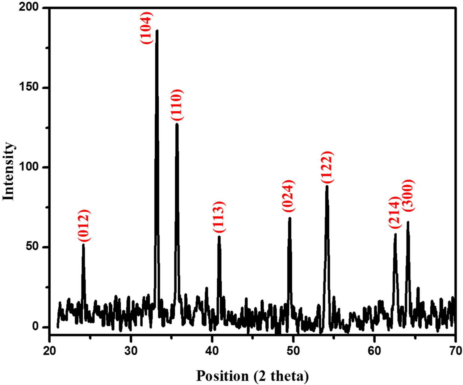
SEM image of synthesised haematite NPs is shown in Figure 2(a) that reveals irregular agglomerates of NPs with lamellar morphology. The average grain size of bare NPs is 70.86 nm with skew-symmetric distribution (Figure 2(a) (inset)). TEM images also confirm the lamellar morphology of the NPs. Representative bright-field TEM micrographs of bare and coated haematite NPs are shown in Figure 2(b–e) confirming the size range identical to SEM images. Bare haematite NPs have size variation having width from 22 to 80 nm. PEG and citrate-coated NPs have shown higher size and aggregation of the NPs can also be observed. The aggregation of the NPs is observed due to functionalisation resulting in the increased size of citrate-coated NPs. EDX provides the compositional analysis of elements present in the material. Spectrum in Figure 2(f) shows the elemental composition of haematite NPs and indicates the presence of iron and oxygen peaks. The emergence of copper signals in the EDX spectrum belongs to the Cu grid used for sample support. No other signal is observed within the detection limit which confirms the formation of purified haematite NPs.
(a) SEM image of bare haematite NPs (Inset: size distribution histogram), TEM images of (b–c) bare haematite NPs, (d) PEG-coated, (e) citrate-coated haematite NPs, (f) EDX spectrum of haematite NPs.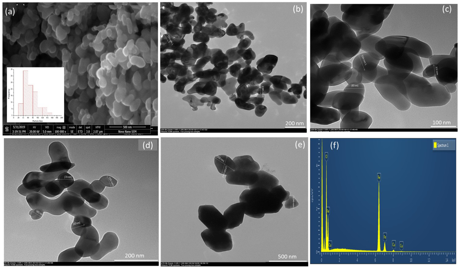
Surface charge and hydrodynamic size are important parameters as these criteria decide the behaviour of the materials in the organism. Zeta potential and hydrodynamic size variations of bare and functionalised haematite NPs are shown in Figure 3(a,b), respectively. The typical hydrodynamic size for bare and coated NPs are 68.08, 190.14, and 141.77 nm, whereas, the surface charge of −9.34 mV, −11.49 mV, and −26.67 mV are observed, respectively. Negative zeta potential values confirm the presence of hydroxyl groups at NPs surface and moderate stability of prepared haematite NPs [32]. Smaller zeta potential value of citrate-coated NPs shows their enhanced stability in the moderate colloidal solution owing to steric repulsion of the carboxyl group [33,34]. PEG-coated particles show the larger hydrodynamic size as compared to citrate and bare NPs, which indicates the thicker PEG ions layer presence around the prepared NPs.
DLS analysis showing zeta potential (a), size variations (b), zeta potential. (c), hysteresis loop (d), FTIR spectrum.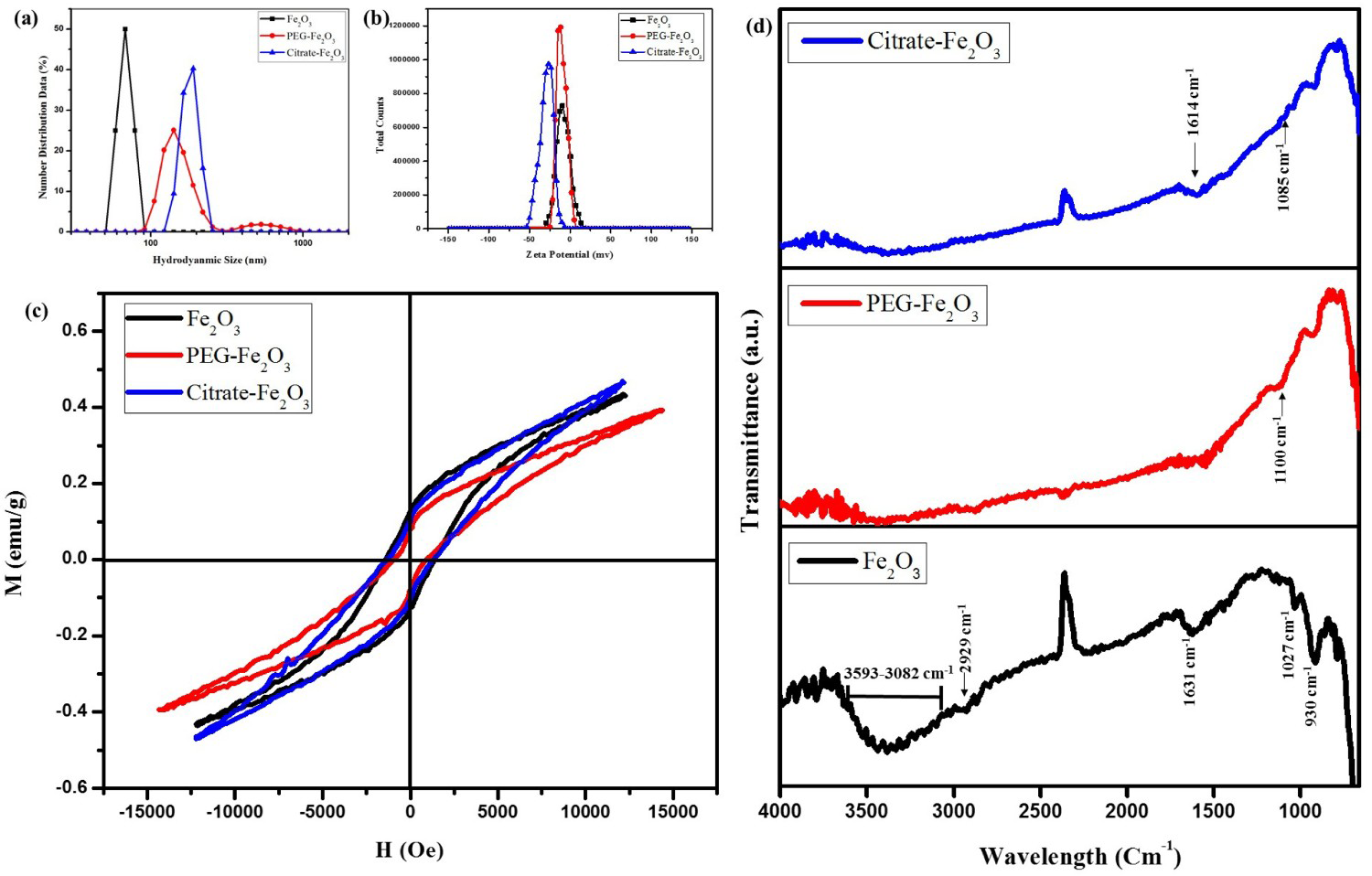
VSM is a useful tool to measure the magnetic properties of the NMs [35-37]. Magnetic properties of bare and functionalised haematite NPs are measured and the hysteresis loop between −12500 and 12500 Oe is determined (Figure 3(c)). Magnetic properties show the soft magnetic behaviour of the haematite NPs. Different magnetic parameters of bare, PEG, and citrate-coated NPs with saturation magnetisation 0.43, 0.001 and 0.001 emu g−1, retentivity 0.13, 0.0002 and 0.0003 emu g−1, and coercivity 1448.28, 687.92 and 1256.14 Oe values are observed, respectively. The squareness ratio is 0.29 for bare NPs that changes up to 0.23 for coated NPs. Squareness is a quantity of the progression to which the hysteresis loop shape is estimated by a rectangle. It shows how adjacent the bottom and top stupors of the loop by the horizontal. The estimated value of the squareness ratio is representing the particle growth and presence of particles with multi-domain nature. The hysteresis loop of coated NPs possesses small saturation magnetisation, retentivity, and coercivity values. Literature supports the significant decrease in retentivity and saturation magnetisation for coated NPs when compared with the uncoated NPs, which can be related to the thickness of the synthesised samples. High coercivity values of coated NPs primarily originate from magnetoelastic anisotropy, usually generated due to fine particles by internal strains while in large crystals because of crystal defects. Reduction in saturation magnetisation may be linked to the quenching of surface moments [38,39]. Henceforth, these results show that magnetic characteristics of iron oxide NPs change to some extent after coatings.
FTIR spectra are shown in Figure 3(d) for bare and coated NPs. In case of bare haematite NPs, the peak appeared at 930 cm−1 attributed to Fe2O3 precipitated in aqueous media, whereas, at 1631 cm−1 attributed to the asymmetric stretching vibrations of C–O. Bands appeared in the range from 1600 to 1670 cm−1 related to the bending vibrations of O–H bonds. The broad band appeared at wavelength range 3082–3593 cm−1 is instigated by stretching vibrations of the OH group present in water molecules, adsorbed at the surface of NPs. FTIR spectrum of PEG-coated haematite NPs shows that extra peak appearing at 1100 cm−1 is the major characteristic absorption peak of PEG because of C–O–C bending vibrations, which suggest successful coating of PEG at the surface of haematite NPs [40,41]. Citrate-coated haematite NPs show vibrational modes at 1085 cm−1 ascribe to symmetric stretching of C–O, COO– and OH group of citric acid. Hence, it is proposed that citric acid binds at the surface of haematite NPs by chemisorption of carboxylate as citrate ions. The peak appearing at 1631 cm−1 slightly shifts toward 1614 cm−1, which is assigned to the C = O vibrations arises from the COOH group of citric acid and reveals the successful binding of citric acid on the haematite NPs surface. All previously discussed characterisations confirm haematite NPs and their functionalisation with PEG and citrate ions. Haematite NPs are recently considered for biomedical applications due to higher oxidation stability, low cost, and non-toxic nature. Therefore, after characterisations, the toxicity of these NPs has been evaluated on albino rats and then biotransformation in the model lysosomal medium has been studied [42].
Toxicity studies
In liver function tests (LFTs), kidney function tests, and complete blood count (CBC) analysis, data is analysed through two-way analysis of variance (ANOVA) trailed by Tukey's multiple comparison test. Results are statistically significant (p < 0.05). **** specifies highly significant results with p < 0.0001, while, ** shows significant results with p < 0.0012.
Liver function tests
Regarding Figure 4, parameters that elucidate the effect of bare and coated haematite NPs effect on liver functions are resoluted. To investigate the toxic effects of bare and coated NPs on the liver of adult albino rats, key clinical indices related to liver function including bilirubin (Figure 4(a)), ALT (Figure 4(b)), AST (Figure 4(c)), and ALP (Figure 4(d)) have been evaluated. Serum shows a transient increase in all the indicators for 24 h, but these levels decline slowly thereafter. As regard liver function markers, the results of the current study reveal that bare and coated NPs cause a significant increase (p ≤ 0.05) in serum levels of ALP, AST, ALT, and bilirubin than control group after 24 h of intraperitoneal administration. This reveals that bare and coated NPs significantly increase the liver's metabolic burden. The increase in levels of LFTs might be due to some underlying inflammatory processes and hepatocytes degeneration. After 8 days, bilirubin, ASP, ALT, and AST levels of all three treatment groups are reduced. This might be referred to as the regenerative capability of the liver. The liver uses macrophages to get rid of the externally administrated particles. In the case of PEG-coated NPs, content enhancement after day 8 is close to the normal values when compared with citrate-coated or bare NPs. Contents of liver functions (ALT, ALP, bilirubin, and AST) of all treatment groups specify that PEG-coated haematite NPs eradicate more effectively without prompting any liver damage. More biocompatibility is evidenced in PEG-coated NPs. PEG coatings prevent the active uptake of NPs and as a result delay in the degradation process can be observed along with the release of (Fe3+/Fe2+) ions. The liver is among the most important target organs that provides a major site for NPs accumulation. Our studies reveal that due to acute dosages of bare and coated haematite NPs, ALT, AST, ALP, and bilirubin target enzymes significantly increase in the liver caused by hepatocytes degeneration and inflammatory process after 24 h of administration, while these enzymes decrease because of the regenerative capability of liver till day 8.
Liver function tests of haematite NPs: (a) bilirubin (b) ALP, (c) AST, (d) ALT. Results: Bilirubin, ALT, ALP and AST values of control, bare, PEG and citrate-coated haematite NPs at day 1 are (0.72, 1.29, 0.84 and 1.06 mg dL−1), (32.02, 135.8, 81.48 and 115.43 IU L−1), (114.46, 220.19, 170.7 and 205.62 IU L−1) and (44.99, 185.27, 145.5, 164.9 IU L−1), respectively. While, these values change to (0.74, 1.02, 0.8 and 0.94 mg dL−1), (34.92, 96.03, 43.65 and 64.02 IU L−1), (124.74, 156.17, 125.17 and 145.5 IU L−1) and (45.55, 106.7, 67.9 and 88.27 IU L−1), respectively.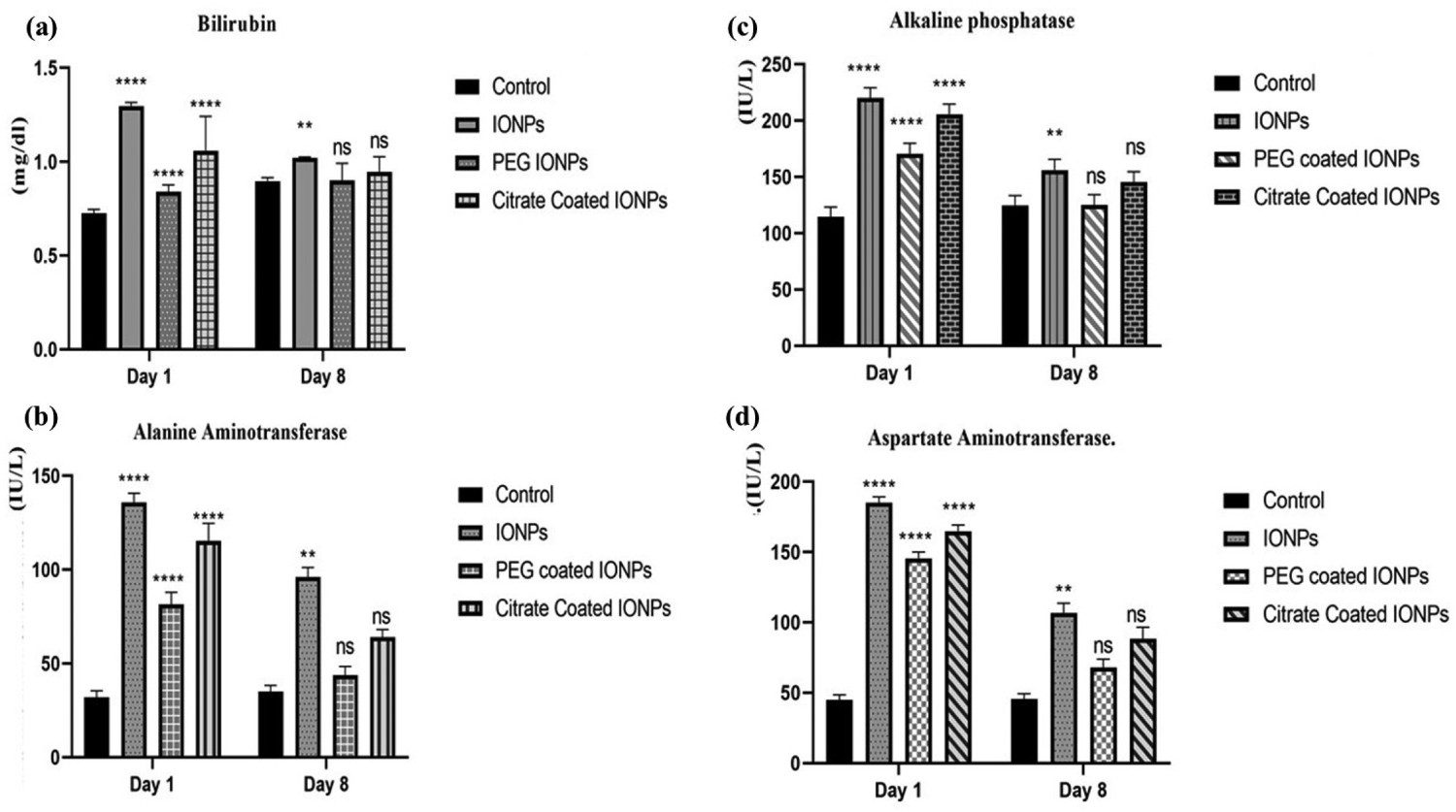
Kidney function tests
Changes in the BUN, creatinine, and urea levels are used as biological markers for kidney function as shown in Figure 5. Preliminary studies were carried out to determine the enzymatic activities followed by the NP injection on day 1 and day 8. In the first part of the experiment, the effect of administrated NPs on serum enzymes are compared with the animals of control group. Renal functional analysis reveals a significant increase with p < 0.05 in the concentration of BUN (Figure 5(a)), creatinine (Figure 5(b)), urea (Figure 5(c)) in coated and bare NPs treated groups. Transient increase is observed in all three groups after 24 h of intraperitoneal administration. However, urea, BUN, and creatinine levels were observed to be reduced after 8 days. After the administration of doses, NPs might practice chemical degradation which leads towards the release of iron ions due to the transformation of NPs. Intracellular distribution of NPs may accompany due to the biodegradation progression, this results in a possible increase in cell death and a defensive process as observed in macrophages when foreigner elements come in contact. But as a whole, kidney function analysis shows neither significant alterations nor the cell membrane damages.
Kidney function tests of haematite NPs: (a) Urea (b) Creatinine (c) BUN Results: BUN, creatinine and urea values of control, bare, PEG and citrate-coated haematite NPs at day 1 are (15.08, 45.66, 33.20 and 35.28 mg dL−1), (0.71, 0.92, 0.82 and 0.86 mg dL−1) and (32.3, 97.75, 71.1 and 75.56 mg dL−1), respectively. While, these values change to (14.52, 33.20, 24.91 and 29.08 mg dL−1), (0.70, 0.81, 0.73 and 0.88 mg dL−1) and (31.1, 71.1, 53.32 and 62.22 mg dL−1), respectively.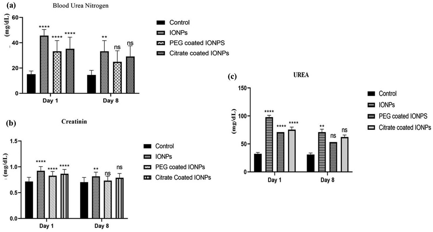
Complete blood count
Regarding Figure 6, haematological parameters including haemoglobin, white blood cells, red blood cells, and platelet count are studied. Representative haematological results with the administration of bare and coated haematite NPs (Figure 6(a)), which shows high Hb levels on both days of study. Significant changes with p < 0.05 are detected after the administration of coated NPs, which cause a transient increase in the Hb values as compared to the control group on day 1. Figure 6(b,c) reveals the transient increase in the platelets and WBCs on day 1, but recovery after day 8. A significant increase with p < 0.05 in platelets count is seen when citrate and bare haematite NPs are injected. Reduce enzyme levels for PEG-coated haematite NPs on day 1st and day 8th are observed when linked with the control group [43-45]. Haematite NPs encounter immune cells and plasma proteins when they enter the bloodstream. Uptake of these particles may occur through different pathways like complement activation, thrombogenicity, and haemolysis. Numerous activities take place when they entered through different pathways including simulation of oxidative stress, anti-mitotic, reduction in blood cells/cellular antioxidants, anti-mitotic properties and increase in resistant cells. The current study tells that haematite NPs may persuade variations in blood cell count, increase white blood cells with the administration of haematite NPs.
Complete blood count of haematite NPs: (a) haemoglobin (b) platelets (c) red blood cells (d) white blood cells. Results: Hb, WBCs, PLT and RBCs values of control, bare, PEG and citrate-coated haematite NPs at day 1 are (11.3, 11.5, 13 and 13.8 g dL−1), (9, 13.6, 17 and 10 103 µL−1) and (725, 829, 634 and 759 103 µL−1) and (6.39, 6.39, 7 and 7.65 106 µL−1), respectively. While, these values change to (11, 11.6, 11 and 10.6 g dL−1 mg dL−1), (8.9, 15, 13.7 and 12.3 103 µL), (734, 715, 936 and 921 103 µL−1) and (5.74, 6.39, 6.25 and 5.79 106 µL−1), respectively.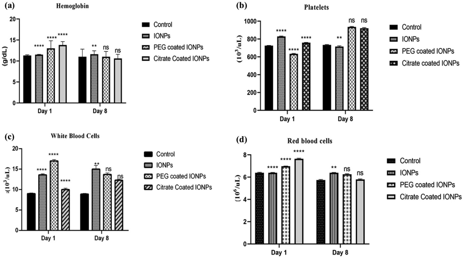
Histopathological analysis
Figure 7 shows histopathological investigations of different organs including the spleen, kidney, and liver of albino rats following the parenteral administration of bare and functionalised haematite NPs. In the kidneys of rats belong to the control group, normal parenchyma tissues can be observed. Kidney micrographs of NPs treated rats reveal cellular infiltration of polymorphonuclear cells resulting in the accumulation of some extracellular fluid which becomes the cause of edema. In the case of bare NPs, this infiltration is higher but is significantly reduced when treated with coated NPs on day 1. Inflammation reduces greatly on day 8. After the interaction of NPs with xenobiotics, the kidney's defensive mechanism activates. In all groups, Bowman's capsule remained intact indicating no degenerative changes in the nephrons of the kidney. Noticeable reduction in inflammatory changes can be observed in PEG-coated haematite NPs. Minor to moderate variations are observed in liver parenchyma tissues in NPs treated groups when compared to the rats of the normal group. Owing to the accretion of extracellular fluid, slight caseation can be observed, which is the major sign of inflammation after treatment with the NPs on day 1, while no signs of hepatocyte necrosis are observed in the control group. The hepatic duct and hepatic triad can be visualised normally, whereas, on day 8, a slight increase in the hepatic duct is seen which goes normal in PEG-coated NPs. The liver has a greater regenerative capacity, which provides more support to the defensive system of the body to respond against external intoxication/stimuli too. In iron recycling, immune responses, and blood filtration, the spleen actively plays a major role. The fibrous capsule covered the spleen. The main splenic artery is divided into many small arteries, which is normal in texture even on day 1st and day 8th. In experimental animals, PEG-coated NPs are showed safer than bare and citrate-coated NPs.
Histopathological images captured of the spleen, kidney, and liver samples collected from control, bare and functionalised treated rats at day (1st and 8th) after administration.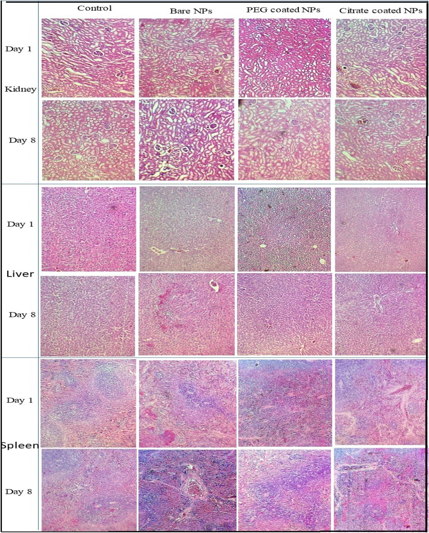
Phase III: In solution bio-transformations
After administration of NPs inside the body, NPs pass through different physio-chemical processes and finally confine into the lysosomes. The lysosome is organelles consisting of degradative enzymes in the cytoplasm enclosed by a membrane and present in all types of eukaryotic cells. Processing of NPs undergoes in these compartments. Hence, to determine the degradation process, this lysosome-like environment is reproduced, and the transformation of NPs is studied. This medium consists of pH 4.7 and citrate ions play the role of the chelating agent. Arbab et al. proposed the lysosomal environment for the first time [46] and many studies validate this lysosome-like medium. NPs transformations are investigated by recording the possible changes in pH, electrical conductivity, and optical properties for the different intervals of time.
UV-Vis spectroscopy studies
Haematite NPs usually exhibit strong absorption in the visible region and redshift with the increase in size and aggregation. Absorption spectra from 450 to 900 nm is recorded at different time intervals. Figure 8(a) is showing the characteristic UV-Vis spectra of haematite NPs for 2 h. The absorption peak of haematite NPs can be observed at 681.87 nm. The broad hump-like shoulder that appears around 681.87 nm is mainly due to agglomeration of spherical-shaped α-Fe2O3 NPs to form large size aggregates that result in scattering the visible light overlays on the absorption of synthesised particles (Figure 8(a–f)) [47]. Results reveal that citrate-coated NPs show loss in absorption signal more rapidly than bare and PEG NPs during the first 2 h of observations. However, at longer incubation time, bare NPs lose their optical properties whereas functionalised NPs stabilise in the lysosomal-like medium. In the optical spectra of Fe3+ based species, three major types of electronic transitions usually arise: (a) transitions based on the ligand to transfer metal charge (b) Fe3+ ligand-based transitions (c) simultaneous excitations of Fe3+ cations due to pair excitations that are coupled magnetically. Absorption edge that appeared from 210 to 400 nm mainly occur due to the transition of metal charge transfer, where the partial contributions of Fe3+ transitions associated with ligand field are observed to be 6A1 → 4E (4G) at 390 nm, 6A1 → 4T2 (4D), and 6A1 → 4E (4D) at 360–380 nm, 6A1 → 4T1 (4P) at 290–310 nm. The absorption band that appears near 430 nm is due to 6A1 → 4E, 4A1 (4G) ligand field transition that usually corresponds to Fe3+. Additionally, wavelength region ranging between 600 and 750 nm can be assigned as ligand field transition 6A1 → 4T2 (4G) of Fe3+ [48]. Transition associated with ligand field at the wavelength range between 600 and 750 nm are dominated by the spherical-shaped particles. The size and shape of NPs possess an excessive influence on the optical properties as well. These findings agree well with the results reported in the literature.
UV-Vis spectra of haematite NPs, when added in the lysosomal media (a–c–e) short duration, (b–d–f) long duration.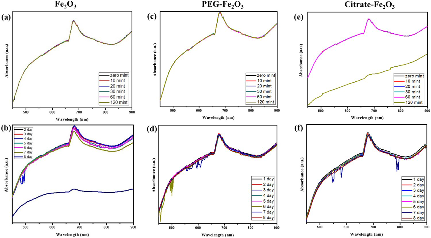
Insignificant alteration in absorbance spectrum for a short interval of time is observed except for citrate-coated NPs (Figure 8(c)), however, absorbance decreases gradually from day 4 to day 8 that indicate dynamical degradation of haematite NPs. Delay in the degradation of haematite NPs indicates the nonreactive nature of the material to sustain in the biological microenvironment (Figure 8(d–f)). The monitoring of the particle degradation process reveals the stochastic corrosion procedure, which depends on the distribution and nature of NPs. In the transformation process, the organisation of particles in a prepared sample plays the main role. Aggregation protects the degradation process although segregation enhances the iron release process from the nanocrystals. Crucial issues that regulate the degradation kinetics involve their access to the core of NPs and chelating agents’ availability.
Electrical conductivity and pH variations
After the addition of haematite NPs in the lysosome-like environment, the pH of the medium is measured at different time intervals and this study is continued to observe all possible changes in pH for 11 days (Figure 9(a,b)). There is some reduction in pH initially, whereas overall pH remains the same with some variation after 11 days. pH is a logarithmic scale, 0.1 increment in pH indicates that the initial solution becomes one time acidic. pH variation exposed the chemical nature of the prepared lysosomal environment by revealing any addition of ion/dopant in it. Loss of optical properties may be linked with biodegradation, but the transformation in morphology is because of the detachment of metallic/iron ions from the haematite NPs that are present in buffer solution and result in varying the environment pH. Electrical conductivity is also considered an important aspect to understand the interaction between biological tissues and fluid. This can distress the pumping ability of fluid in the body. Variation of electrical conductivity for the short interval of time till day 11 is noted (Figure 9(c,d)). Rapid escalation can influence numerous body functions such as damage to the nervous system and heating of tissues. A short interval of time, electrical conductivity values increase initially, which is indicating the increasing number of metal ions inside the mimicked environment and may change the inclusive conductivity of the biological environment.
Short and long follow up of (a–b) pH and (c–d) electrical conductivity changes of bare and functionalised haematite NPs.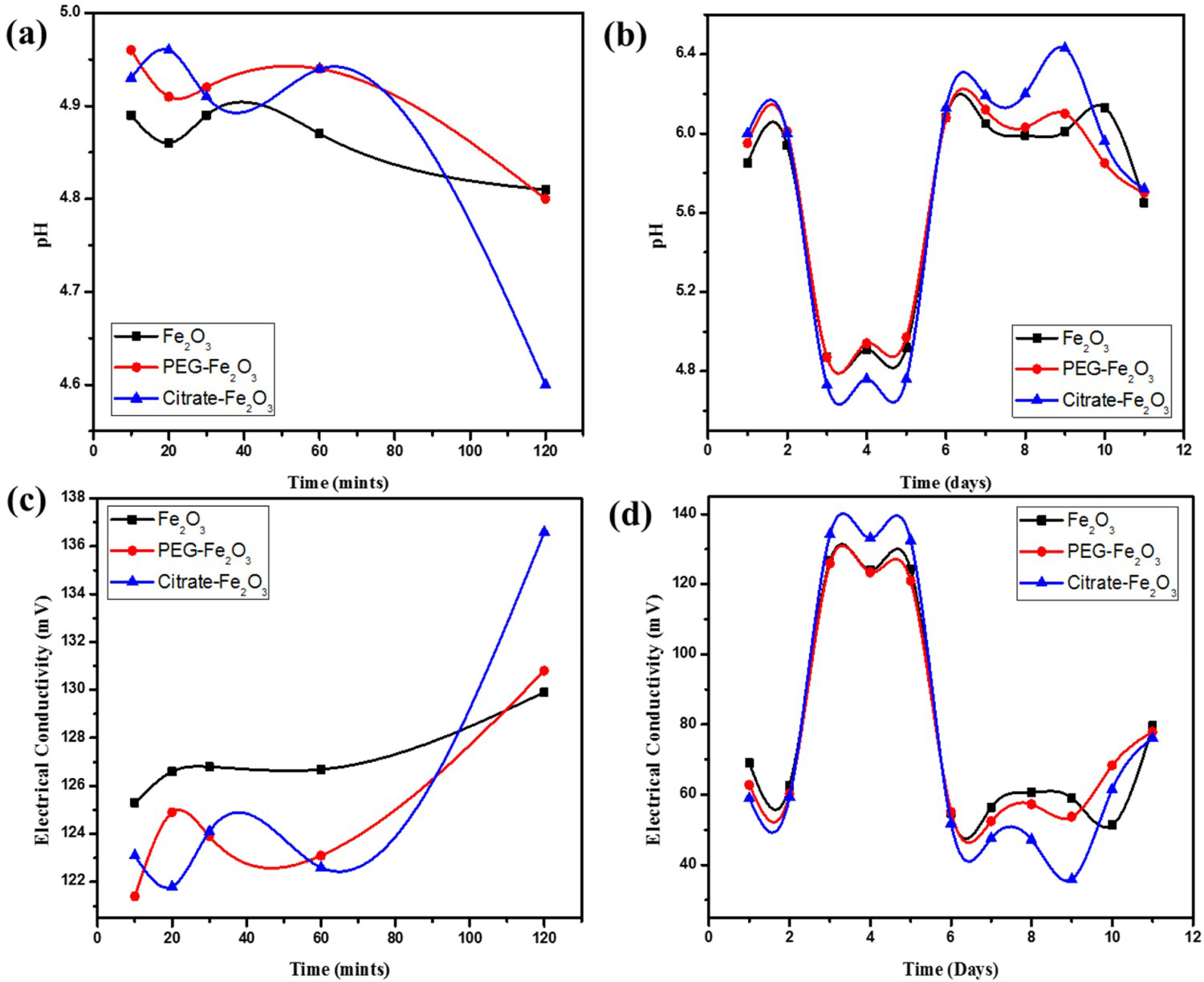
Phase IV: Transfer Studies of Degraded NMs into Ferritins
In vivo studies in solution investigations are performed to examine the possible degradability when ApoF is incubated in a simplified lysosomal medium to evaluate its ability to integrate the haematite NPs. ApoF dispersion in the citrate buffer leads to the phase separation and protein aggregation. ApoF can reutilise the byproducts of iron-based NMs. ApoF possesses the intrinsic ability to sequester iron and shifted the direction of NPs dissolution. For this study, ApoF is added in the lysosome-like medium along with haematite NPs. Changes in optical properties and transfer of degraded ions in the ApoF are analysed by UV-Vis spectroscopy. In parallel, the ability of ApoF core to accommodate the iron ions from iron salt in lysosomal-like media is also investigated. The evolution of the ApoF absorption signal in UV-Vis spectroscopy with haematite NPs and iron salts at different time intervals is shown in Figure 10(a,b). During 2 h of incubation at 37°C, the absorbance signal appeared near 260 nm, an increase in absorbance over time reveals incremental filling of iron ions in ApoF protein. Growth of absorbance near 260 nm is indicating progressive and partial filling of the protein. Till day 8, absorption signals confirm the ApoF ability to simultaneously sequester iron ions resulted from the degradation of haematite NPs and iron salts in the lysosomal-like medium.
UV-Vis spectra NMs taken when ApoF is added in the mimicked lysosomal medium at different periods (a) bare haematite NPs (b) iron salt.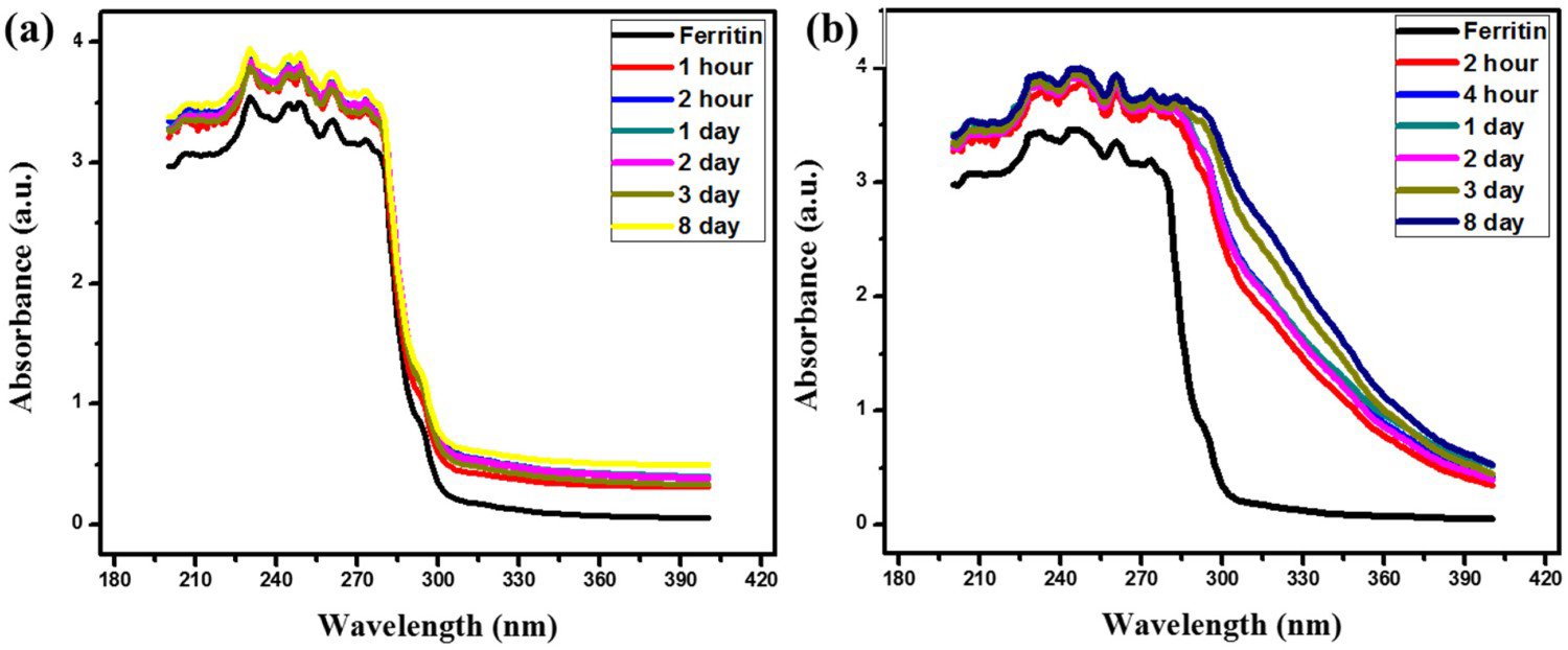
Conclusion
This study provides new aspects of the degradation of iron oxide-based NM. Bare and coated IONPs are synthesised by the co-precipitation method and characterised by different analytical tools such as SEM, XRD TEM, DLS EDX, VSM, FTIR, and UV-Vis spectroscopy. Acute toxicity study is performed by administrating bare and coated NPs through the intraperitoneal route on a group of albino rats. Increased levels of kidney and liver enzymes are evidenced at day 1 that reinstate after day 8. Slight inflammatory changes are evidenced in normal parenchyma tissues of the kidney, spleen, and liver through histopathological investigations, while no severe damage is observed. PEG functionalised NPs showed better biocompatibility than bare and citrate-coated NPs. Degradation of NPs depends on various factors such as the nature and distribution of the coating layer and shape of NPs. The experiments further showed that iron ions released from haematite NPs and iron salt can be stored in ferritin proteins. In situ transformations are studied with the transfer of iron ions in ApoF, which offer insights into IONPs fate in organisms.
Footnotes
Disclosure statement
No potential conflict of interest was reported by the author(s).