
Abstract
Objective:
This research was conducted to increase precision of point location and standardize acupoint targets in relation to the facial nerve (CN VII).
Materials and Methods:
A literature review, cadaver dissection, and electrostimulation of the CN VII were performed, focusing on the anatomical locations of the acupoints along the facial-nerve trajectory. The results were contrasted against established acupoint locations described in the 4th edition of Chinese Acupuncture and Moxibustion.
Results:
Triple Energizer (TE)–17 targets the facial nerve at its cranial exit; Qian Zheng at the facial nerve trunk; Tai Yang at the frontotemporal branch; Gallbladder (GB)–3 at the zygomatic branch; Small Intestine (SI)–18 at the buccal branch; Stomach (ST)–5 at the marginal mandibular branch; SI-17 at the cervical branch; TE-18 at the posterior auricular branch; TE-19 at the posterior auricular nerve; TE-20 at the superior auricular nerve; and TE-22 at the anterior auricular nerve.
Conclusions:
This study demonstrated the specificity with which acupuncture points are located in relation to the facial nerve. Standardization of facial acupuncture points to distinct branches of the facial nerve will facilitate reproducible research designs and interpractitioner reproducibility.
INTRODUCTION
Although typically self-limited, the facial paralysis that occurs in Bell's palsy may lead to potential eye injuries and poor long-term outcomes that can be devastating to patients. 2 Currently, treatments are generally designed to improve facial function and facilitate recovery. There are many treatment options for Bell's palsy, including medications (steroids and antivirals), surgical decompression, and physiotherapy. However, some controversy exists regarding the effectiveness of several of these options. 2 In this context, acupuncture has emerged as an alternative therapy for Bell's palsy in both adults and children. Yet, the use of acupuncture as a modality treatment for facial paralysis remains controversial. 3
Although early Chinese Medicine practitioners knew about branching and functioning of the facial nerve, as detailed in A Revised Neuromyofascial Understanding for the Neck, Head and Facial Channel Sinews based on the Ling Shu, 4 at present, the practice of acupuncture focuses on specific and overall accepted therapeutic effects of the acupoints, while modern anatomy is generally not described or emphasized during acupuncture education. This lack of anatomical specificity in acupuncture practice creates difficulty in developing consistent and reliable results in practice and for clinical research trials. 5
As a potential treatment for facial nerve paralysis, it is important to standardize, increase the precise locations—and confirm the anatomical targets—of acupoints in relation to the facial nerve. A deeper understanding of the acupoints' anatomical targets is needed to enhance clinical research and treatment outcomes.
MATERIALS AND METHODS
Literature Review
Acupoint locations and their associated neuroanatomy were sourced from the 4th edition of Chinese Acupuncture and Moxibustion, an official translation of a standardized textbook from the People's Republic of China. 6 The CN VII neuroanatomy described for each acupoint on the face were crossreferenced with peer-reviewed medical and surgical articles, with a focus on anesthesia literature. When possible, nerve targets were crossreferenced further with 2 versions of Gray's Anatomy.7,8 To compare facial neuroanatomy to the acupoints, an established cun measurement system was utilized. A cun is a proportional measurement that is scaled to any body size but can also be described as approximately the width of a person's thumb (∼ 1").
Dissection
A single cadaver fixed with vascular perfusion, using a mixture of liquid phenol, 95% ethanol, glycerin, and water, was used. The specimen was the cadaver of an 82-year-old male that was acquired for anatomical dissection via the Division of Anatomy, Department of Surgery, of the University of Toronto, Ontario, Canada. Bioethics approval was obtained through the Human Research Ethics Program (#32018). Facial-nerve dissection was performed between 2015 and 2016.
Electrostimulation
When there was a gap in the literature, or when detailed nerve ramification was not dissected successfully, electrostimulation via inserted needles was conducted on 10 healthy volunteers. All volunteers were licensed acupuncturists who gave their written consents prior to professional continuing-education events offered by the current authors between 2019 and 2020. A Pointer Plus device (a 10-Hz monopolar electrostimulation device manufactured by Mayfair Medical Supplies Ltd., Hong Kong) was used for the electrostimulation. To reveal the trajectory of nerve branching, electrostimulation was applied to inserted needles for 1–2 seconds to record the pattern of muscle recruitment and activation.
RESULTS
The present research found Triple Energizer (TE)–17 targets the facial nerve at its cranial exit; Qian Zheng at the facial nerve trunk; Tai Yang at the frontotemporal branch; Gallbladder (GB)–3 at the zygomatic branch; Small Intestine (SI)–18 at the buccal branch; Stomach (ST)–5 at the marginal mandibular branch; SI-17 at the cervical branch; TE-18 at the posterior auricular branch; TE-19 at the posterior auricular nerve; TE-20 at the superior auricular nerve; and TE-22 at the anterior auricular nerve.
The reinterpreted acupuncture target and how it compares to the Systematic Classic 9 location and the modern location—including innervation and vasculature descriptions from the Chinese Acupuncture and Moxibustion, 4th edition, text-book, 6 —are shown in Table 1.
Reference 9.
Cheng XN, ed. Chinese Acupuncture and Moxibustion, 4th ed. Beijing: Foreign Languages Press; 2019. m., musculus.
Figure 1 shows the re-interpreted point locations superposed over facial-nerve anatomy, over the main branches, revealing the specificity with which acupuncture points are located in relation to the facial nerve. The specific acupuncture points used for electrical stimulation of the facial nerve and its branches, as well as the muscle-stimulated and expected muscle action, are shown in Table 2.
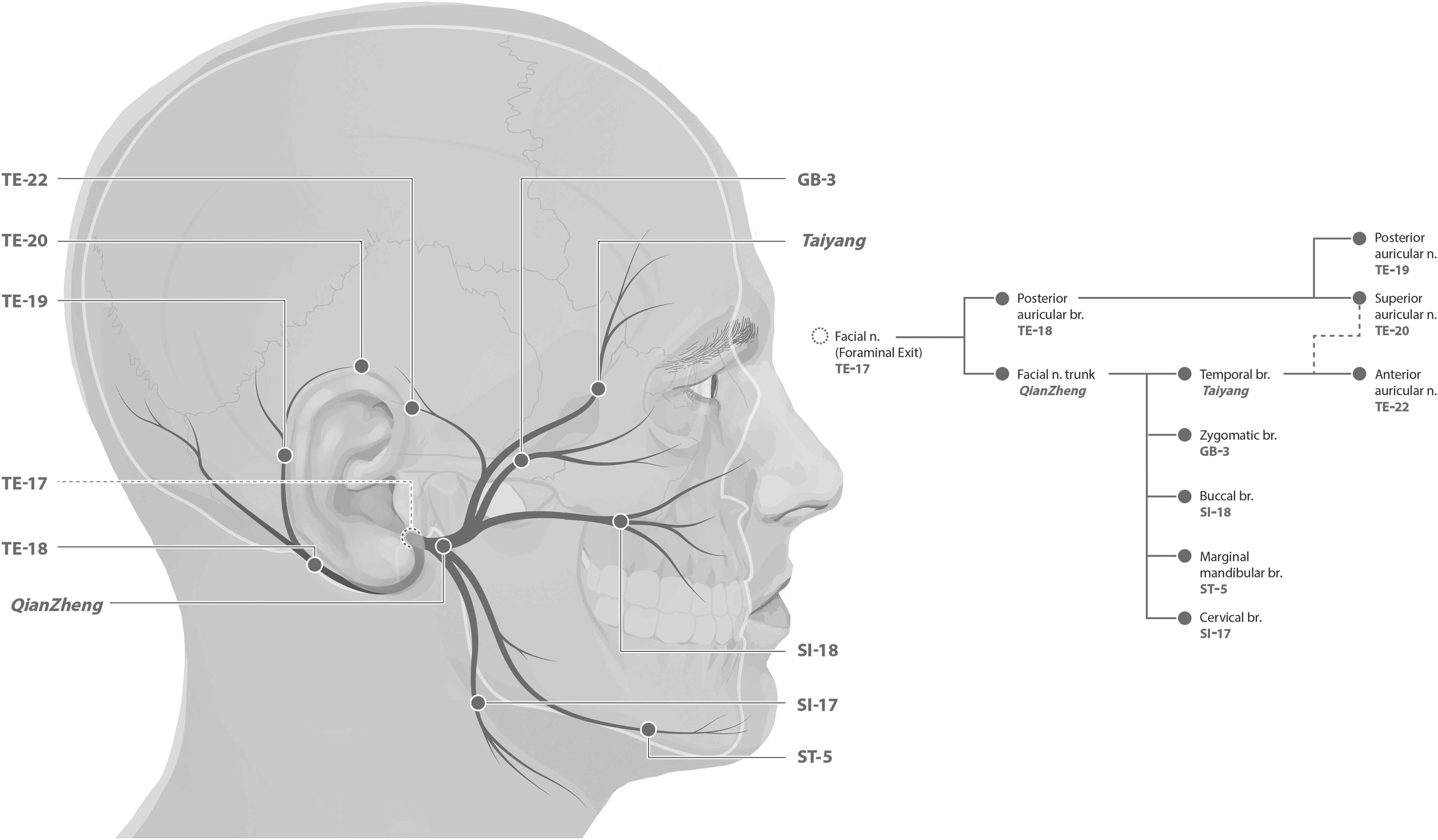
Reinterpreted acupuncture point locations superimposed over the facial nerve (n; CN VII) main branches (brs).
Acupuncture Points Used for Electrical Stimulation, Branch of the Facial Nerve Stimulated, Corresponding Muscle Stimulation and Expected Muscle Action
N/A, not applicable.
Figure 2 shows the dissection anatomy of the buccal branch and corresponding area of the acupoint SI-18, and Figure 3 shows the marginal mandibular branch of the facial nerve and corresponding area of acupoint ST-5. Finally, the Supplementary Videos S1–S10 demonstrate the electrical stimulations of acupuncture points and respective branches of the facial nerve: TE17 (facial nerve foraminal exit), Qian Zheng (facial nerve trunk), TE18 (posterior auricular branch), TE19 (posterior auricular nerve), Taiyang (temporal branch), TE22 (anterior auricular nerve), GB3 (zygomatic branch), SI18 (buccal branch), ST5 (marginal mandibular branch), SI17 (cervical branch). (Supplementary data are available online at www.liebertonline.com/ACU).
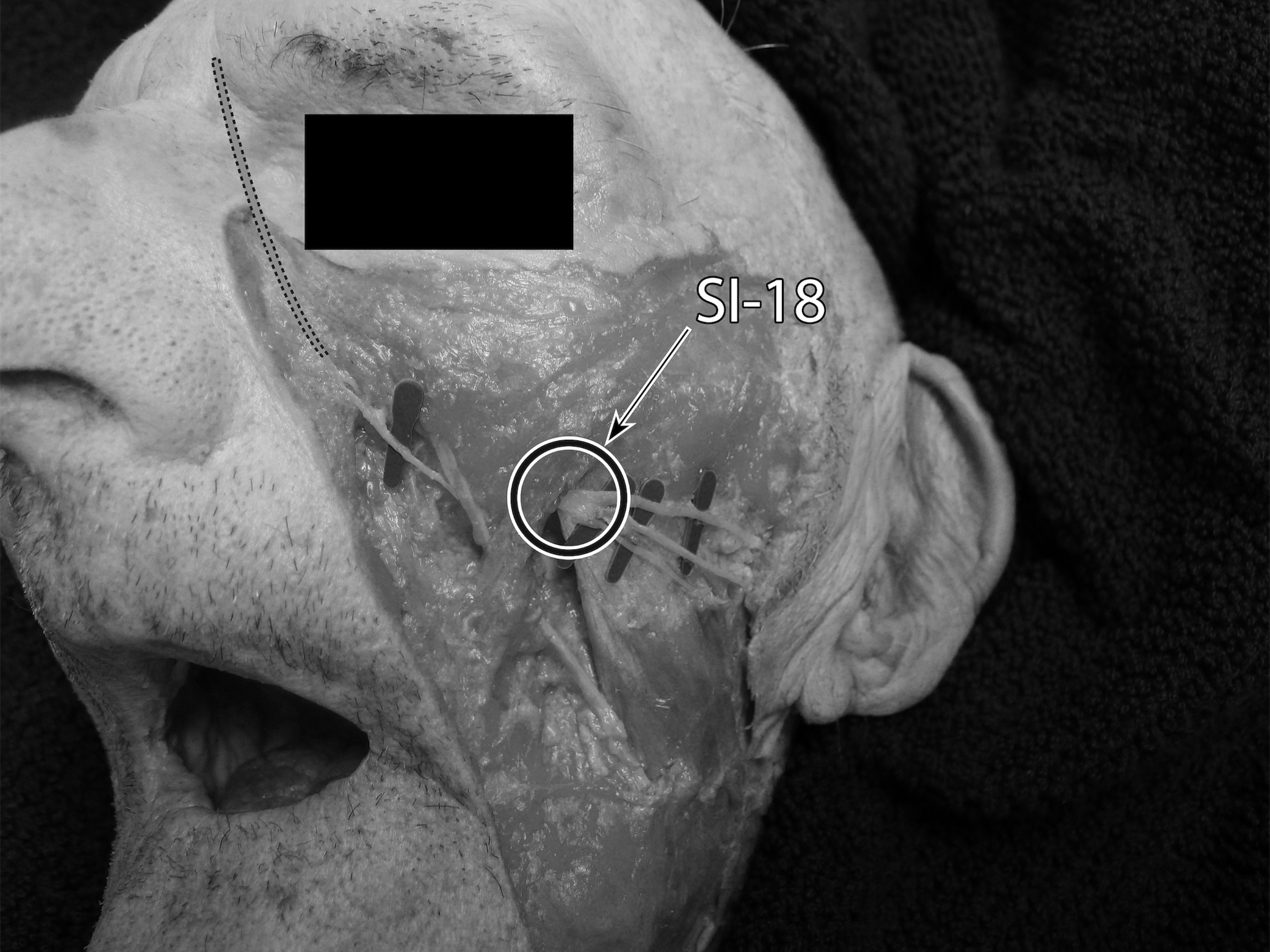
Acupuncture point SI-18, buccal branch of facial nerve (yellow) and facial artery (red).
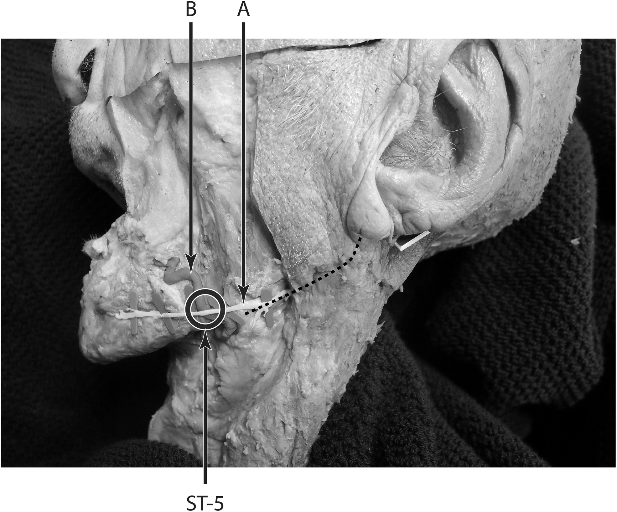
Acupuncture point ST-5, marginal mandibular branch of the facial nerve
DISCUSSION
Acupuncture has been increasing in popularity. In the West, this can be attributed, in part, to this modality's effectiveness for pain relief and the fact that scientific studies have begun to prove acupuncture's efficacy. 10 Despite acupuncture's rising in popularity, education has not been updated and, generally, modern anatomy is not emphasized. The existing anatomical information from standardized textbooks is incomplete and requires further investigation. Using one of the most-popular textbooks (Chinese Acupuncture and Moxibustion 6 ) as a reference, this study contrasted the lack of specificity and accuracy with which modern anatomy is described to the new reinterpreted anatomical targets, with the goal of updating anatomical knowledge of acupuncture points regarding their specificity in relation to the facial nerve. The main results are summarized in Tables 1 and 2 and are represented in Figure 1. Some selected points discussed further below
The facial nerve is the motor nerve to the majority of muscles of the face, with the exception of the mastication muscles. This nerve has a complex anatomy and, according to the nerve's topographic localization, can be divided into intracranial, intratemporal, and extratemporal components. The intratemporal component of the facial nerve exits the canal through the stylomastoid foramen behind the ear, where the extratemporal facial nerve begins. After leaving the stylomastoid foramen, the facial nerve extends to suprahyoid motor branches to the posterior belly of the digastric muscle and stylohyoid muscle, and the posterior auricular division produces branches to the superior and posterior auricular muscles and Occipitalis muscle. Then, the facial nerve courses anterior to the posterior belly of the digastric muscle to penetrate the posterior edge of the parotid gland. Here, the nerve divides into superior and inferior divisions, also referred to as temporofacial and cervicofacial divisions. A trifurcation or quadfurcation of the facial-nerve trunk is also possible.
The typical peripheral branching of the facial nerve into 5 terminal branches is in the substance of the parotid gland. The 5 main branches represented in Figure 1 include the (1) temporal branch, (2) the zygomatic branch, (3) the buccal branch, the (4) marginal mandibular branch, and (5) the cervical branch. 11
Qian Zheng
Qian Zheng is located right before the bifurcation into the temporofacial and cervicofacial divisions of the facial nerve. The meaning of its name, “pull upright,” 9 suggests traditional insight into the potential to correct facial drooping associated with CN VII palsy.
TE-18, TE-19, TE-20, and TE-22
In humans, 3 extrinsic auricular muscles—the auricularis superior muscle, auricularis anterior muscle, and auricularis posterior muscle—arise from the temporal aspect of the cranium and are inserted into the auricular cartilage. They hold the auricles in place and are responsible for reinforcement, positioning, and angle of the auricle. These muscles are innervated by the temporal (auricularis anterior) muscles) and posterior auricular division of the facial nerve (auricularis posterior and superior muscle). 12 There is a lack of agreement in the literature about whether or not the temporal branch also contributes to the superior auricular nerve. The extrinsic auricular muscles are considered to be vestigial muscles in humans. Unlike other mammals, such as dogs or horses, humans have lost the ability to control these extrinsic auricular muscles voluntarily. The TE points TE-18, TE-19, TE-20, and TE-22 are located in the trajectory of the posterior auricular division, posterior auricular nerve, superior auricular nerve, and anterior auricular nerve, respectively, and their stimulation has the ability to activate these muscles.
Despite the vestigial nature of these nerves and their associated muscles, there are some clinical applications for them, such as for treating spontaneous periauricular and occipitalis muscle spasms. Although these muscle spasms are not as readily perceptible as spasms associated with other facial-nerve branches, these spasms, nevertheless, can be highly distracting to the extent that one's ability to work, socialize, and sleep are affected. In addition, for cases of submandibular gland dysfunction—which is modulated via parasympathetic innervation by the facial nerve—electromyographic testing of the auricularis posterior muscle helps localize the site of the facial-nerve lesion. Needle electromyography to the auricularis posterior muscle is expected to be abnormal if the facial-nerve lesion is located at the motor nucleus or proximal to the stylomastoid foramen. In contrast, for lesions distal to the stylomastoid foramen, the electromyographic testing result is expected to be normal for the auricularis posterior muscle. 13
Tai Yang
Tai Yang is located in the trajectory of the temporal branch of the facial nerve. The temporal branch innervates the frontalis, superior aspects of the orbicularis oculi, and corrugator supercilli muscles. The facial nerve has a highly variable and complicated branching pattern and forms communications with several other cranial nerves, especially with the branches of the trigeminal nerve (cranial nerve V). 14 Communications between the temporal branch of the facial nerve and the zygomaticotemporal branch of the trigeminal nerve have been described as being in the same anatomical region as Tai Yang. 15 These communications between a motor nerve and a sensory nerve raise a question about the possibility of using this point to treat both sensory and motor conditions, or, to use motor stimulation to treat sensory conditions and vice versa. Further studies are necessary to elucidate the nature of these fibers.
SI-18
The SI point SI-18 is located in the trajectory of the buccal branch of the facial nerve (Fig. 2). The buccal branch of the facial nerve is responsible for innervation of the levator labi alaque nasi, levator labi superioris, and orbicularis oris, whose actions are noted in Table 2. The buccal branch has also been demonstrated to innervate the procerus muscle. 16 It is interesting to note that this innervation was recorded in Chapter 10 of the Ling Shu classical text, as a separating branch that arrives at “the inner angle of the eye.” 4
ST-5
The ST point ST-5, on the anterior border of the attached portion of the masseter, is located in the trajectory of the marginal mandibular branch of facial nerve. Point location instruction from the Systematic Classic, as shown in Table 1, describes locating this point with the aid of a pulsating vessel, which is likely to be reference to the facial artery. The facial artery intersects the marginal mandibular nerve. Therefore, palpating the pulsation provides an easy way to locate the marginal mandibular nerve branch. This anatomical peculiarity is shown in Figure 3.
CONCLUSIONS
The present study demonstrated the precise correspondence between distinct acupoints and unique branches of the facial nerve. These findings are congruent with the growing body of evidence that support acupoints, as transmitted from classical texts, are precise coordinates for stimulating the peripheral nervous system.5,17 These results offer acupuncturists greater clarity for targeting acupoints on the face. Refining point location with verified neuroanatomy is the first step in designing clinical studies for facial motor dysfunction. Without standardizing the neuroanatomical target of facial acupoints, it would not be acceptable to compare the results between any facial acupuncture studies. The significance of the present research is that it will raise the consistency of therapeutic results and the reproducibility of scientific research on facial acupuncture. Future facial acupuncture research is not limited to Bell's palsy, but can expand to areas such as stroke rehabilitation, facial tics, and cosmetology.
Footnotes
ACKNOWLEDGMENTS
AUTHORs' CONTRIBUTIONS
Dr. Chiang was responsible for conceptualization, methodology, project administration, resources, and supervision. Dr. Martins was responsible for visualization; writing the original draft of this article, and writing, reviewing, and editing the final version.
AUTHOR DISCLOSURE STATEMENT
No financial conflicts of interest exist.
FUNDING INFORMATION
No funding was received for this work.
References
Supplementary Material
Please find the following supplemental material available below.
For Open Access articles published under a Creative Commons License, all supplemental material carries the same license as the article it is associated with.
For non-Open Access articles published, all supplemental material carries a non-exclusive license, and permission requests for re-use of supplemental material or any part of supplemental material shall be sent directly to the copyright owner as specified in the copyright notice associated with the article.