
Abstract
Interleukin-6 (IL-6) family of cytokines are multifunctional proteins that play an important role in host defenses, acute phase reactions, immune responses, hematopoiesis, and tumorigenesis. The cytokines are produced by various lymphoid and nonlymphoid cells and mediate their biological activity through initial low-affinity binding to cell surface receptors, which are specific for their respective ligands. Ligand-specific receptor binding results in the receptor heterodimerization with ubiquitously expressed signal-transducing transmembrane component gp130 followed by activation of the gp130-associated Janus kinase, which, in turn, phosphorylates signal transducer and activator of transcription 3 (STAT3). Phosphorylated STAT3 (pSTAT3) dimerizes and translocates to the nucleus, where it activates gene transcription. Activation of STAT3 is essential to IL-6 family-associated physiological effects. Therefore, the ability to assess STAT3 phosphorylation is important for drug discovery efforts targeting IL-6 family cytokines. Various reagents and technologies are available to detect the effect of IL-6 type cytokines in treated cells. The present study describes the development of two pSTAT3 detection assays: the high-throughput screening assay based on Meso-Scale Discovery technology, which utilizes electrochemoluminescent signal measurements for the detection of pSTAT3 in treated cell extracts, and the secondary characterization assay based on fluorescent imaging analysis, which monitors pSTAT3 nuclear translocation in cells after activation. We have successfully utilized these assays to screen a small library of secreted proteins and identified inducers of STAT3 phosphorylation. The results obtained in this study demonstrate that both assays are robust, reliable, and amenable to high-throughput screening applications.
Introduction
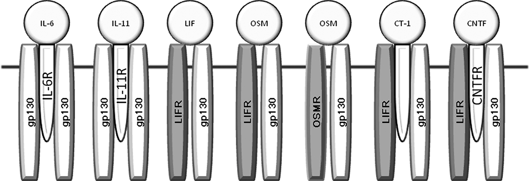
IL-6 type cytokines and their receptors. IL-6 family cytokines share the common receptor subunit gp130 for signal transduction and, therefore, have similar physiological responses that can sometimes overlap. Signal-transducing subunit gp130 is shown in white or light gray (LIFR and OSMR). Alpha receptor subunits (IL-6R, IL-11R, CNTFR) initially bind to IL-6, IL-11, and CNTF but are not involved in the intracellular signal-transducing cascade. IL, interleukin; LIFR, leukemia inhibitory factor receptor; OSMR, oncostatin M receptor; CNTF, ciliary neurotrophic factor receptor; CT-1, cardiotropin.
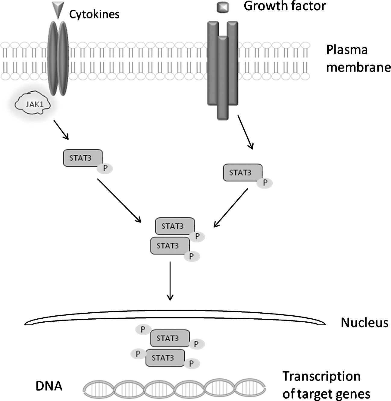
Activation of STAT3. Binding of IL-6 type cytokine to its receptor causes gp130 subunit to form a homo- or heterodimer, which then initiated intracellular signaling by activating members of Jak. Activated Jak tyrosine phosphorylates itself, the cytoplasmic tail of gp130, and cellular substrates. Jak are the main activators of the STAT pathway and phosphorylation of inactive cytoplasmic STAT1 and STAT3 causes their activation, which in turn leads to STAT dimerization and translocation to the nucleus, where they regulate transcription of target genes. Jak, Janus kinases; pSTAT3, phosphorylated signal transducer and activator of transcription 3.
The secondary assay described in this article employed a fluorescent-based cellular imaging approach for the detection of activated STAT3 nuclear translocation. This assay allows the detection of STAT3 protein with a specific primary antibody and a secondary antibody conjugated to a fluorophore. STAT3 nuclear translocation was monitored using Acumen eX3 laser scanning cytometer and quantified as the difference in cytoplasmic-to-nuclear intensity of the labeled transcription factor.
ECL and imaging-based activated STAT3 detection assays were developed and optimized using the A375.S2 cell line previously shown to be responsive to IL-6 and OSM treatments (Centocor internal data). Both assays were partially automated using Biomek FX and performed in a 96-well format. A small library of secreted proteins was tested in the ECL assay for the detection of pSTAT3 inducers in A375.S2 cells. Proteins identified as activators of STAT3 during the initial screening were tested in the imaging-based nuclear translocation assay. The results obtained from the two assay formats showed good agreement. Taken together, the results presented here demonstrate that the ECL pSTAT3 detection and STAT3 nuclear translocation assays both generate robust, reproducible data and are suitable for detection of activated STAT3 in cell lysates in high-throughput applications.
Materials and Methods
Cell Culture and Reagents
Human A375.S2 melanoma cells were purchased from the American Type Culture Collection (Manassas, VA). Cells were grown in Dulbecco's modified Eagle's medium with GlutaMAX™-I (catalog no. 10569; Invitrogen, Carlsbad, CA) supplemented with 10% fetal bovine serum (catalog no. SH30071; Thermo Fisher, Pittsburgh, PA), 1% of nonessential amino acids (catalog no. 11140; Invitrogen), and 1% sodium pyruvate (catalog no. 11360; Invitrogen) at 37°C in a humidified atmosphere of 95% air and 5% CO2. Cells were split twice a week using Accumax™ (catalog no. AM105; Innovative Cell Technologies, San Diego, CA) and utilized in the assay between the second and 10th split. Experiments were performed on cells plated in 96–well, black, clear-bottom tissue culture-treated plates (catalog no. 655090; Greiner, Monroe, NC).
Recombinant human IL-6 (amino acids 29–212; catalog no. 06-IL/CF), recombinant human tumor necrosis factor alpha (TNF-α; 210-TA), neutralizing anti-human IL-6 monoclonal antibody (MAB206), anti-human gp130 neutralizing monoclonal antibody(MAB228), and mouse IgG1 isotype controls (MAB002) were purchased from R&D Systems (Minneapolis, MN). A whole-cell lysate phospho-STAT3 (Tyr 705) assay kit (K110DID) was purchased from MSD and was used for the ECL pSTAT3 detection assay. A STAT3 activation kit (K01-0008-1) for monitoring STAT3 nuclear translocation was purchased from Thermo Fisher.
To generate the secreted protein library, the entire Invitrogen Orfeome collection (approximately 10,000 clones) was scanned to identify secreted proteins. The selected open reading frames were obtained in a gateway cloning vector and were subsequently subcloned into the pTT5-based mammalian expression vector. 7 The DNA for the transient transfection was prepared using QIAprep 96 Turbo Miniprep Kit (catalog no. 16191; Qiagen, Valencia, CA). A lipid-based transfection of human embryonic kidney cell line 293 (HEK293) cells was done in a 96-well format into the 293 Freestyle serum-free media (catalog no. 12338; Invitrogen), and the cells were incubated at 37°C with 5% CO2. Supernatants with expressed proteins were harvested after 72 or 96 h and were used directly for the assays. For positive hit confirmation, the DNA was prepared using Qiagen maxiprep kit (catalog no. 12663) and the transfection for each sample was performed in multiple wells in the 96-well plates.
MSD Technology
MSD sector 6000 from MSD is a high-throughput ECL reader that detects emitted light from a nonradioactive label when it is electrochemically stimulated, producing minimal background signal. The MSD assay principle is based on binding of phospho-STAT3 (Tyr 705) to a precoated capture antibody (total STAT3) that is immobilized on the working electrode surface of a plate and detection with an anti-phospho-STAT3 sulfo-tag–labeled antibody. The addition of the read buffer provides the appropriate chemical environment for ECL, and when a plate is loaded into the instrument, a voltage applied to the plate electrodes causes the label bound to the electrode surface to emit light. The instrument measures the intensity of the emitted light to provide a quantitative measure of the amount of pSTAT3 present in the sample.
Acumen eX3 Technology
Acumen eX3 from TTP LabTech (Melbourn Royston, United Kingdom) is a high-throughput screening fluorescent cytometer equipped with triple laser enabling simultaneous detection of multiple cellular markers. Acumen eX3 is applicable for live or fixed cell assays in both adherent and nonadherent cell lines. Acumen eX3 is capable of scanning the entire well and provides multiparametric analysis of acquired data. The developed Acumen eX3-based assay is a fixed end-point immunofluorescence assay, wherein activated STAT3 is measured by its translocation from cytoplasm to the nucleus.
ECL pSTAT3 Detection Assay
In this assay, A375.S2 cells were plated in 96-well tissue culture-treated plates at 25,000 cells per well and incubated overnight at 37°C with 5% CO2. The next day, the cells were treated with IL-6 for 20 min at room temperature. Then, the treatment reagent was removed simultaneously from all wells using the Biomek® FX liquid handling system from Beckman Coulter (Brea, CA) followed by the addition of a lysis buffer containing phosphatase and protease inhibitors. After 10 min of incubation, the obtained cell lysate was transferred using Biomek FX to an MSD plate that was previously blocked and washed. The cell lysate was incubated for 1 h on the MSD plate at room temperature on a shaker. Then the plate was washed with Tris buffer, followed by addition of an anti-phospho-STAT3 detection antibody. After 1 h of incubation at room temperature, the plates were washed and a surfactant-based read buffer was added to the plate before it was read on the MSD instrument.
STAT3 Nuclear Translocation Assay
A375.S2 cells were plated in 96-well, black, clear-bottom plates at 2,500 cells per well and incubated overnight at 37°C with 5% CO2. The next day, the cells were treated with IL-6 for 15 min at room temperature. After treatment, IL-6 was simultaneously removed from all wells using Biomek FX. The treated cells were fixed and permeabilized, followed by staining with anti-phospho-STAT3 antibody (primary) and secondary antibody conjugated to DyLight 488 Fluor. The plates were read using Acumen eX3 instrument. For each assay point, data for the entire well were collected and analyzed using Acumen 3.4.25 software. For data analysis, we used a fluorescent half-width intensity algorithm that was specifically developed by the instrument manufacturer to analyze protein translocation from cytoplasm to the nucleus (Fig. 3). Half width intensity is calculated as the peak intensity of the object divided by its half width. Half width is the width of the object at the point where the intensity drops to half of the peak.
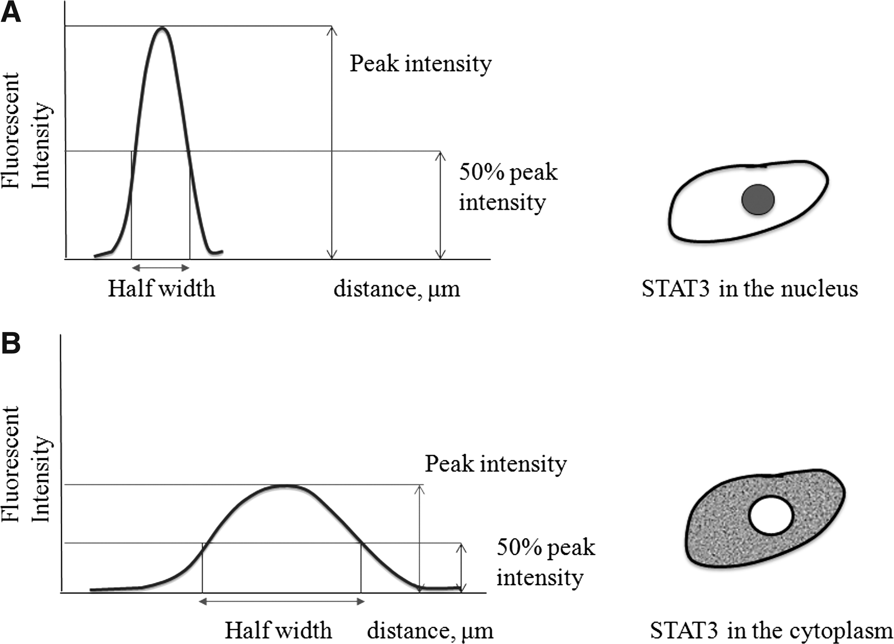
Cell fluorescent half-width intensity used as the measure of STAT3 nuclear translocation. Acumen eX3 laser progressively scans the bottom of the assay plate, followed by collecting cell fluorescent intensity readings at regular intervals using a photomultiplier tube detector. Thresholding algorithms identify all fluorescent intensities above the solution background for automatic object identification. The cartoon represents fluorescent intensity distribution within the objects/cells with nuclear (
Data Analysis
Data generated from both assays were analyzed using GraphPad Prism software, version 5.0 (La Jolla, CA). To demonstrate assay robustness, Z′ factor was calculated according to the following equation: Z′ = 1 − [3 × (SDtreated + SDuntreated)/(Meantreated − Meanuntreated)], where SD is standard deviation and Mean is average of data. 8
Results And Discussion
Development of ECL pSTAT3 Detection Assay
We intended to develop a screening assay suitable for the identification of various extracellular acute STAT3 activators that potentially could be utilized for the development of large-molecule therapeutics. We used IL-6 cytokine as a model activator of STAT3 signaling. However, we realized that various activators might have STAT3 activating potency and kinetics that are different from IL-6.
Based on our previous experience with A375.S2 cell response to different IL-6 cytokine family members, a cell density of 25,000 cells per well was selected for the assay. To optimize the ECL pSTAT3 detection assay, A375.S2 cells were treated for different time intervals with an IL-6 titration starting from 100 ng/mL followed by threefold serial dilutions. We found that within the time points tested, 20 min of incubation with IL-6 results in the most potent induction of pSTAT3 (Fig. 4A). In subsequent experiments, a wide range of IL-6 concentrations were tested to obtain full dose–response curves at a 20 min induction time (Fig. 4B). Phosphorylation of STAT3 in A375.S2 cells was found to be IL-6 concentration dependent, with a calculated EC50 of 11 ng/mL. We did not test additional IL-6 induction time points in this assay as it was developed for the potential detection of a variety of pSTAT3 inducers. As a full IL-6 dose–response experiment revealed robust induction of pSTAT3 in response to 20 min of IL-6 stimulation, we felt that other inducers of this signaling pathway should also give detectable responses in the same time frame. We demonstrated specificity of STAT3 phosphorylation by IL-6 using antibodies against IL-6 and cell surface gp130 receptor. Various antibody concentrations were preincubated for 10 min before addition to the cells with an IL-6 concentration that caused submaximal activation of STAT3. The cells were stimulated for 20 min and STAT3 phosphorylation was measured. Both anti-IL-6 and anti-gp130 antibodies inhibited the stimulatory effect of IL-6 in a dose-dependent manner, indicating that STAT3 phosphorylation is induced through IL-6/gp130 signaling pathway (Fig. 5A). IC50 parameters were found to be 0.1 and 0.7 μg/mL for anti-IL-6 and anti-gp130 antibodies, respectively. The treatment with isotype-control antibodies did not cause inhibition of IL-6–induced STAT3 phosphorylation (data not shown). During the development of the assay, we encountered some day-to-day variability in raw ECL signal. Potentially, this variability could be caused by assay plate or assay reagents lot-to-lot variability. To minimize this effect, all the experiments were performed with internal plate controls, and single assay plate and reagent lots were used for screening and hit characterization.
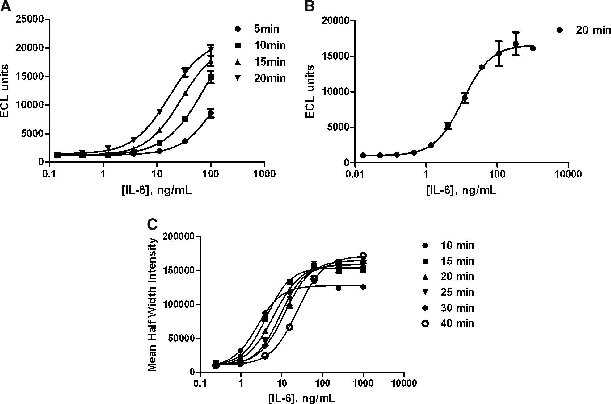
Assay optimization. Comparison of stimulation times with various concentrations of recombinant human IL-6. A375.S2 cells were treated with recombinant human IL-6 for different time periods, ranging from 5 to 20 min for the ECL pSTAT3 detection assay (
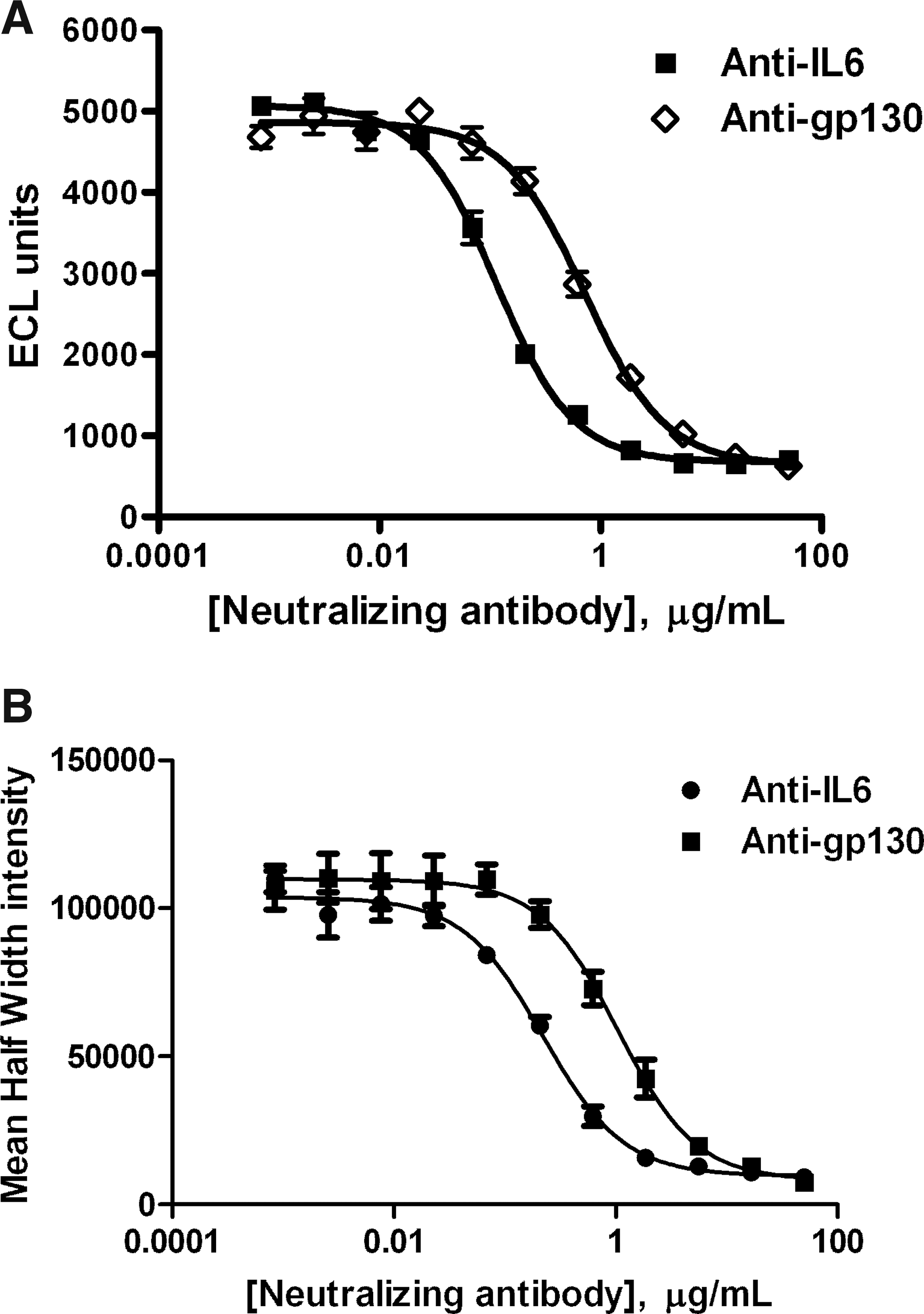
Inhibition of STAT3 activation with anti-IL-6 and anti-gp-130 antibodies. Various concentrations of anti-human IL-6 and anti-human gp130 antibodies were preincubated with IL-6 for 10 min and then added to A375.S2 cells. IL-6 (33 ng/mL) was used for the ECL pSTAT3 detection assay (
Development of STAT3 Nuclear Translocation Assay
In general, subconfluent cell densities are used in imaging assays that require single-cell identification and measuring of cellular translocation events. We have measured STAT3 nuclear translocation in response to a range of IL-6 concentrations in A375.S2 cells seeded at 2,500 and 5,000 cells per well in 96-well plates. The response was found to be similar at both densities tested. A density of 2,500 cells per well was selected for further experiments. To identify optimal stimulation conditions, we treated A375.S2 cells with titrations of IL-6 cytokine for 10–40-min time intervals. Titrations started from 100 ng/mL followed by fourfold serial dilutions. We found that 15–30 min of treatment of A375.S2 cells with IL-6 resulted in the most optimal induction of STAT3 translocation to the nucleus (Fig. 4C). EC50 parameters for IL-6–induced STAT3 nuclear translocation were found to be 4 ng/mL at 15 min of induction. Based on EC50 comparisons, we found that the STAT3 nuclear translocation assay is approximately twofold more sensitive than the ECL pSTAT3 detection assay. Similar to the pSTAT3 detection assay, we confirmed the specificity of the activation using anti-IL-6 and anti-gp130 neutralizing antibodies. Antibodies were preincubated with IL-6 for 10 min and then added to A375.S2 cells for 15 min of stimulation. The experiment showed that the antibodies inhibited IL-6–induced STAT3 nuclear translocation in a dose-dependent manner (Fig. 5B). IC50 parameters were found to be 0.2 and 1 μg/mL for anti-IL-6 and anti-gp130 antibodies, respectively. Treatment with isotype control antibodies did not cause inhibition of IL-6–induced STAT3 nuclear translocation (data not shown). Potentially, both the ECL pSTAT3 detection and STAT3 nuclear translocation assays could be used for the detection of Jak/STAT pathway inhibitors. In this case, additional modulators such as commercially available Jak inhibitors have to be evaluated for activity. Overall, both the ECL pSTAT3 detection and STAT3 nuclear translocation assays showed comparable response to IL-6 stimulation and similar neutralization pattern by anti-IL-6 and anti-gp130 antibodies.
Secreted Protein Library Screening
To evaluate whether the assays are amenable for high-throughput screening, we performed a Z′-score test, wherein the majority of wells of a 96-well assay plate were treated with IL-6 at 20 and 33 ng/mL for the STAT3 nuclear translocation and ECL pSTAT3 detection assays, respectively (Fig. 6).
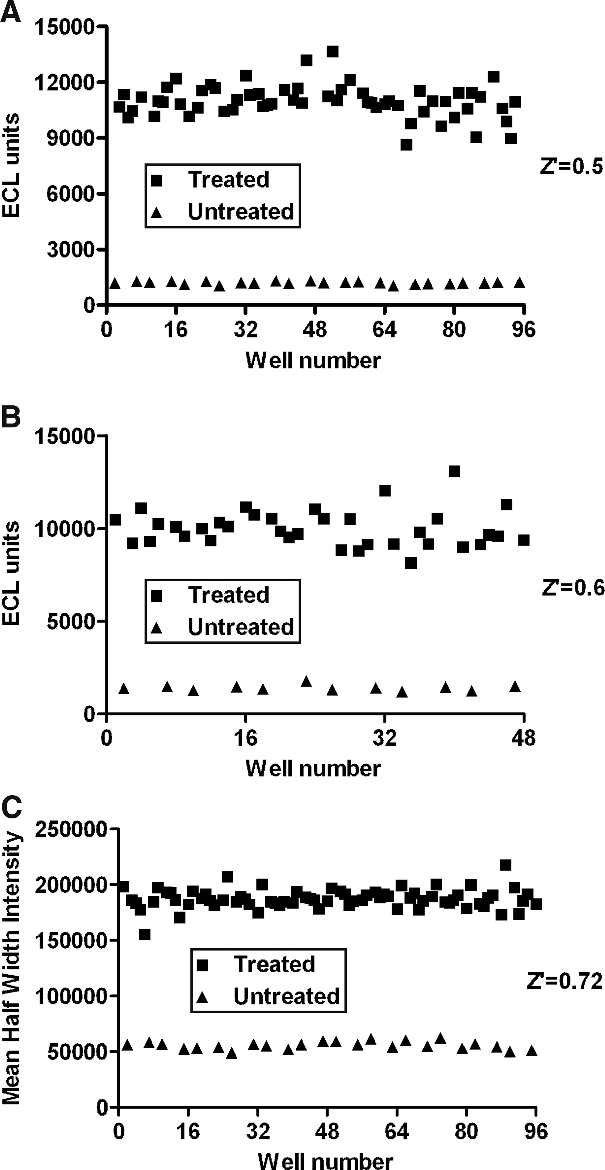
Assay reproducibility. To assess assay reproducibility for high-throughput screening, we conducted Z′-score tests, in which certain wells were treated with IL-6 at 33 and 20 ng/mL for the ECL pSTAT3 detection (
The Z′ test for the ECL pSTAT3 detection assay was repeated twice and resulted in values of 0.5 and 0.6 (Fig. 6A, B). A Z′ value of 0.72 was obtained in one experiment performed for the STAT3 nuclear translocation assay (Fig. 6C). According to industry standards, Z′ test scores between 0.5 and 1 indicate an excellent and robust screening assay. 8 Thus, both the nuclear translocation and ECL pSTAT3 detection assays are robust and reproducible and can be applied to high-throughput screening.
We used the ECL pSTAT3 detection assay to perform the initial screening of the secreted protein library, which is available in the form of cell supernatants from HEK293 cells transiently transfected with plasmids expressing the individual proteins. Prior to that, we evaluated the effect of cell supernatants containing selected secreted proteins on assay performance. As TNF-α does not signal through the Jak/STAT pathway, it was used as negative control, whereas IL-6 was used as positive control. To ensure that the transfection plasmid, transfection reagents, and cellular metabolites found in cell supernatants have no stimulatory or inhibitory effect, we performed the following experiment: Cells were treated with commercially available recombinant human IL-6 and TNF-α that were spiked into HEK293-conditioned medium and were used as positive and negative controls, respectively. In addition, supernatants from HEK293 cells transfected with vectors encoding human IL-6 (positive control) and TNF-α, along with empty vector and spent conditioned medium containing transfection reagents (negative controls), were also used to treat A375.S2 cells. (Fig. 7). The results of this experiment showed that empty vectors and transfection reagents do not have any nonspecific effect on STAT3 phosphorylation. Spent HEK293 medium also had no stimulatory or inhibitory effect. Recombinant IL-6 spiked into spent HEK293 medium at 60 ng/mL (positive controls) had a 10-fold signal over background (just spent HEK293 medium) and was comparable to the signal from secreted IL-6 from transfected cells. Both commercial and library-secreted TNF-α negative control worked as expected, with no stimulation of STAT3 pathway. As the secreted protein library was previously tested for protein expression by gel electrophoresis and Coomassie staining, this test indicted that the library can express bioactive protein and is suitable for cell-based screening.
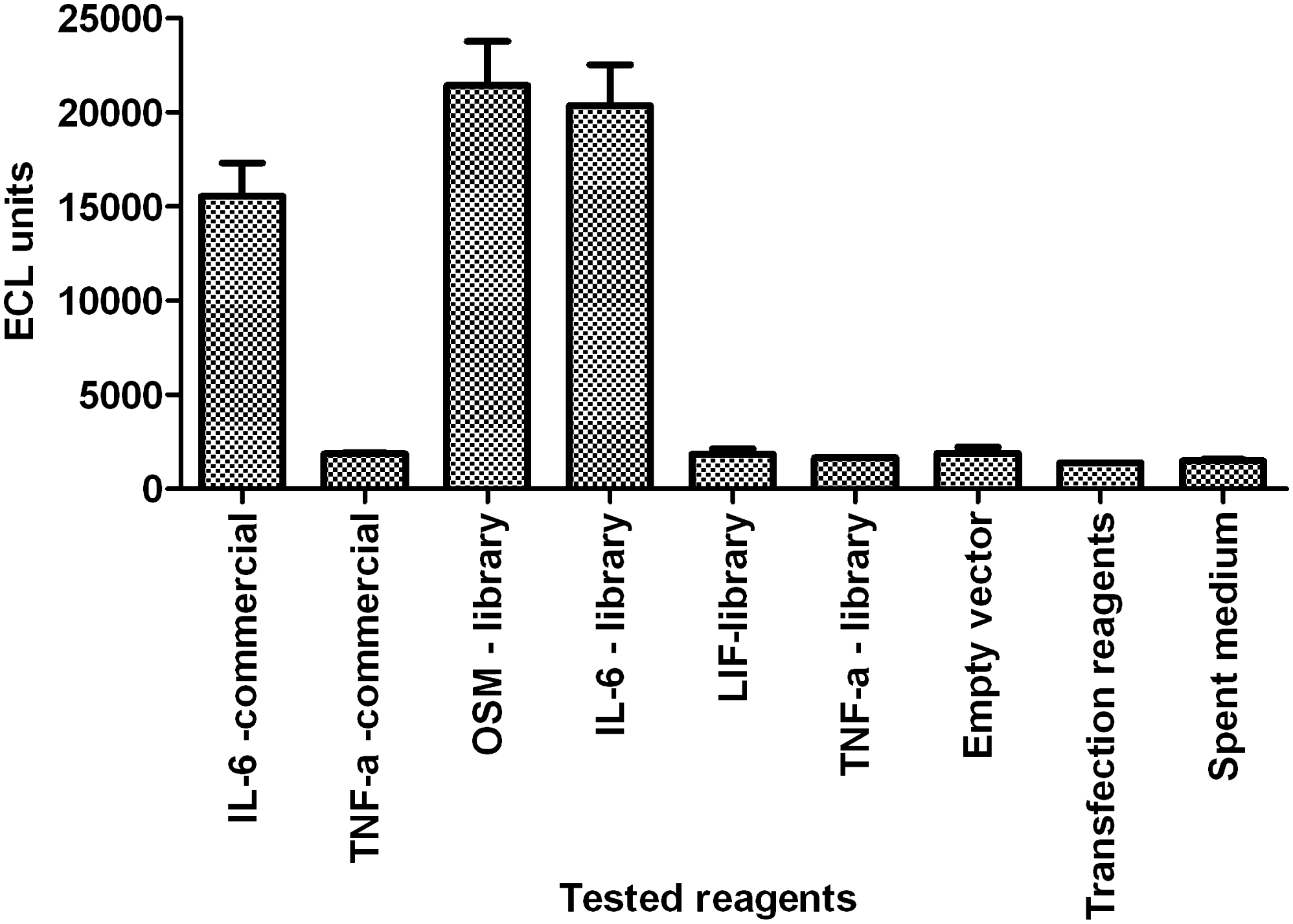
Evaluation of the effect of the transfection reaction components on the ECL pSTAT3 detection assay performance. HEK cells spent supernatants with or without cDNA library or empty vector or transfection reagents were tested for stimulation or inhibition of STAT3 pathway. Commercially available recombinant human IL-6 and TNF-α spiked into empty vector-transfected HEK293 cell supernatants were used as positive and negative controls, respectively. Data are presented as average from duplicate wells (n = 2), with error bars presenting variability. HEK, human embryonic kidney cell line; TNF-α, tumor necrosis factor.
For this case study, we screened a sublibrary of 480 samples from 1,440 proteins available in the Centocor-secreted protein library using the ECL pSTAT3 detection assay. The library consists of proteins that are involved in transcription, enzyme regulation, signal transduction, chemoattraction, catalysis, cellular transport, and more. The expression of these proteins was assayed by gel and western blot analysis of the transfected cells supernatants (data not shown). The expression levels were found to be dependent upon the specific proteins analyzed. A selected set of candidates were gel analyzed for each experiment to ensure efficient cell transfection and protein secretion. IL-6 and TNF-α were selected as assay controls and tested on each screening plate in the form of library-expressed or recombinant proteins spiked into HEK spent medium. Protein samples were tested in duplicate on replicate plates. Initial screening was performed using the ECL pSTAT3 detection assay and identified hits were titrated and tested in both the developed assays (ECL pSTAT3 detection and nuclear translocation). During the initial screening, we identified OSM, IL-11, tolloid-like protein 1 (TLL1), pancreatic lipase-related protein 1 (PNLIPRP1), and regenerating islet-derived protein 3 alpha (REG3A) as STAT3 activators (Fig. 8). As described earlier, OSM and IL-11 are known members of the IL-6 cytokine family that signal through the Jak/STAT pathway. 9,10 LIF is another member of the IL-6 cytokine family that was tested among library samples. LIF did not cause STAT3 activation because of the absence of LIF receptor in A375.S2 cells. 11 Assay controls, including spiked recombinant and library-expressed proteins, were easily distinguished, having a signal 4–5 times higher than background or negative controls (TNF-α, HEK spent medium). Identification of OSM and IL-11 as hits confirmed that the secreted protein library contains bioactive proteins at concentrations detectable by the assay. Three other hits (PNLIPRP1, TLL1, REG3A) identified during screening are not known to signal through the Jak/STAT pathway, but they were confirmed as positive hits during rescreening in the ECL pSTAT3 detection and nuclear translocation assays (Fig. 9). Those hits were tested at different dilutions, producing dose-dependent responses that agreed between the two assays.
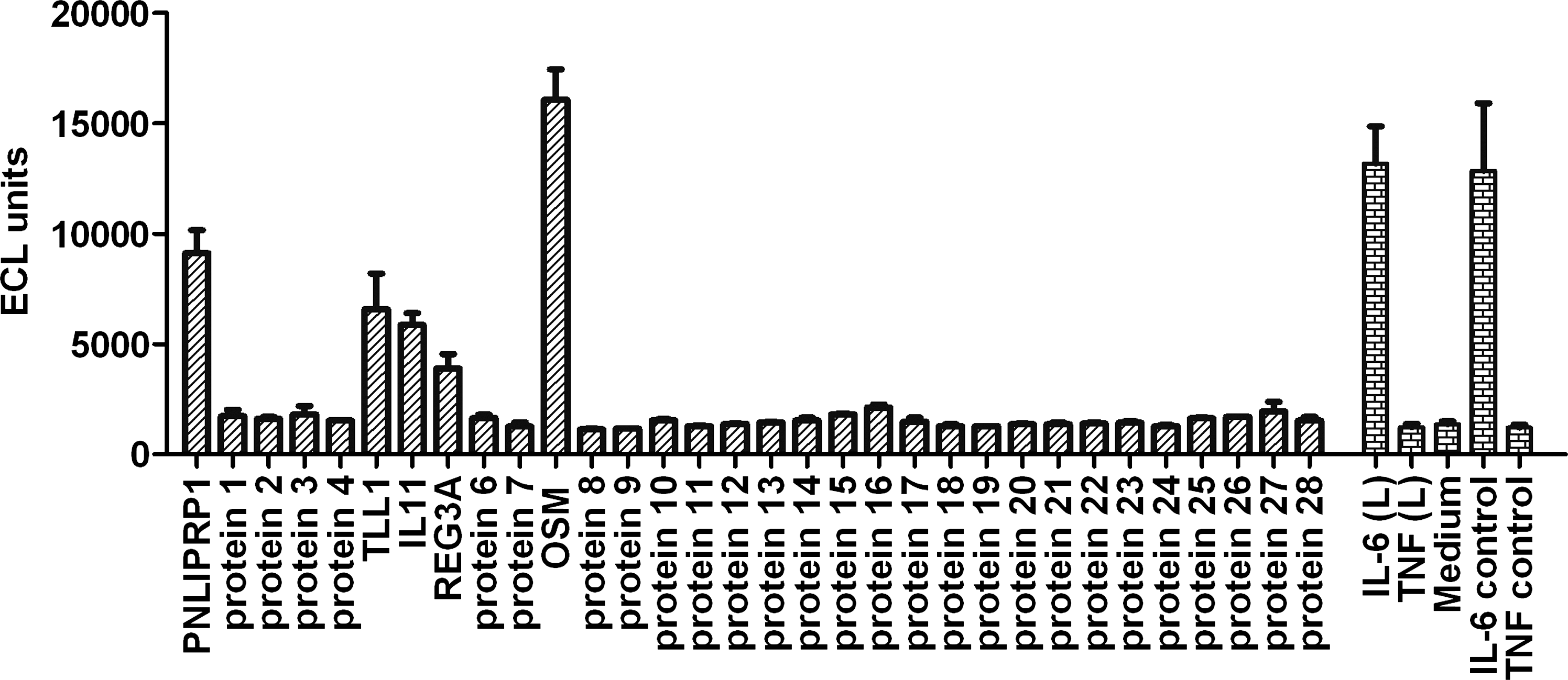
Centocor-secreted protein library screening results (representative data). Four hundred eighty proteins were screened using the ECL pSTAT3 detection assay. As a result, six proteins were identified as hits, including proteins known to signal through Jak/STAT pathway (OSM, IL-6, IL-11). In addition to the predicted hits, three new proteins were identified (PNLIPRP1, TLL1, REG3A) that were not previously reported to signal through Jak/STAT pathway. Commercially available recombinant TNF-α and IL-6 were used as negative and positive controls, respectively. Library-secreted IL-6 (L) and TNF-α (L) were used on each plate to confirm successful transfection. Data are presented as average from duplicate wells (n = 2), with error bars presenting variability. PNLIPRP1, pancreatic lipase-related protein 1; TLL1, tolloid-like protein 1; REG3A, regenerating islet-derived protein 3 alpha.
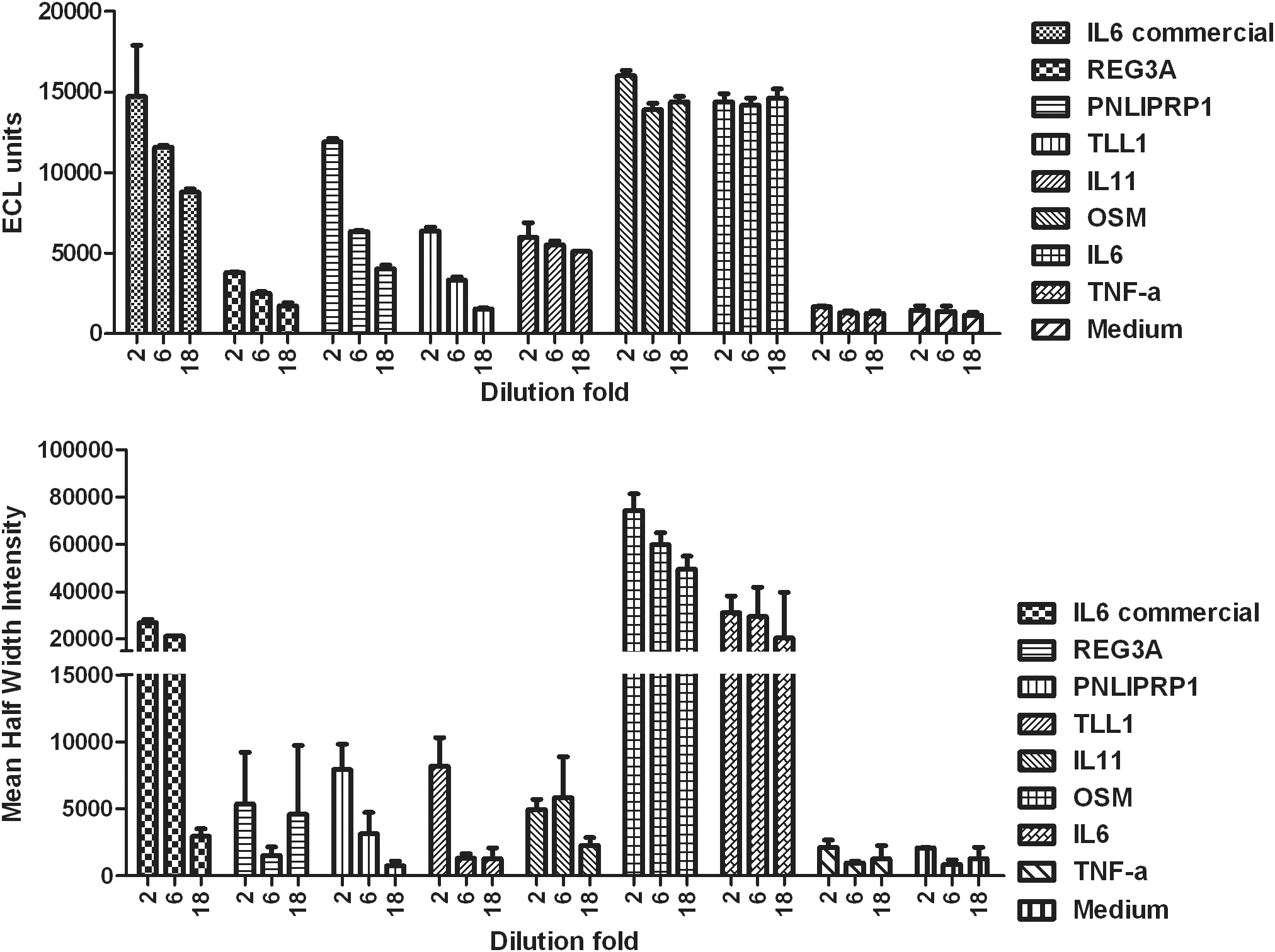
Confirmatory screening of previously identified hits. Protein hits identified during initial screening were retested in the ECL pSTAT3 detection and nuclear translocation assays and were confirmed as functional. Assay controls, commercial and library-transfected, worked as expected. Data are presented as average from duplicate wells (n = 2), with error bars presenting variability.
To prove that Jak/STAT signaling is indeed triggered by REG3A, PNLIPRP1, and TLL-1, we set up an experiment to test whether STAT3 activation by these proteins is blocked by anti-gp-130 and protein-specific antibodies. For this experiment, we needed a larger volume of secreted protein hits, and therefore, library transfection was done on a larger scale. For that reason, we prepared cDNA-containing plasmids encoding the aforementioned proteins and IL-6 as a positive control, using a Qiagen Maxiprep Kit, instead of the QIAprep 96 Turbo Miniprep Kit, which was originally used for screening. Using this methodology, the Maxiprep-expressed and commercial IL-6 triggered STAT3 activation and were neutralized by anti-gp130 and anti-IL-6 antibodies in a dose-dependent manner. PNLIPRP1, REG3A, and TLL-1 samples were produced by cDNA transfections with expression confirmed as protein bands of expected sizes by gel electrophoresis and Coomassie staining. However, these samples did not trigger pSTAT3 activity in two experiments. A potential explanation for these results is that contamination in the DNA preparation that was originally used for screening could account for some false positives in the initial screen. cDNA transfection samples used for the initial screen were made using an automated small-scale DNA extraction kit. The purity of the DNA samples isolated with the kit depends on the size of bacterial pellet used for the isolation. The false positives could be caused by Jak/STAT activating proteins present in the DNA transfection samples prepared by inefficient plasmid purification from overgrown bacterial cultures. Another potential source of false positives in the initial screen could be DNA cross-contamination, which occasionally happens during the handling of multiple samples.
The contaminating factors in the small-scale DNA preparations that generated false positives were eliminated using the large-scale DNA preparations for rescreening of selected hits. Large-scale DNA is less prone to contaminations because of the different resins used and the purification performed by gravity flow as opposed to the vacuum filtration applied in a small-scale preparation. Nevertheless, the developed screening assays proved to be efficient and suitable for high-throughput screening.
In this article, we described the development of pSTAT3 detection assays using two different technologies: ECL and immunofluorescence imaging. The developed assays proved to be robust and produced very comparable results, thus allowing them to be used interchangeably. The ECL pSTAT3 detection assay employs only one washing step that makes it more amenable for screening purposes compared with the imaging assay, which requires a number of washing steps. The advantage of using the STAT3 nuclear translocation imaging assay lies in the opportunity to monitor multiple cellular parameters and also detect pSTAT3. This makes the imaging assay suitable for confirmatory screens and compound mechanism-of-action studies. The developed assays were used in a Centocor-secreted protein library feasibility study in which we tested the utility of the library for identification of signaling pathway modulators. We used the ECL pSTAT3 detection assay for screening of 480 proteins that were expressed and secreted from cDNA-transfected HEK293 cells. The STAT3 nuclear translocation assay was used for hit confirmation testing. During the initial and confirmatory screenings, we identified IL-6, OSM, and IL-11 proteins that were previously known to activate STAT3. Three additional hits found during primary screening that were not described as STAT3 activators were later shown to be false positives.
Overall, the results provide evidence that the developed assays are robust, reproducible, and suitable for identification of STAT3 activators. Sublibrary screening results confirm that the library contains functional proteins and can be used for screening purposes.
Disclosure Statement
No competing financial interests exist.