
Abstract
We have developed a multiplex assay to measure the expression of anti-apoptotic proteins and caspase 3 activation using the Luminex platform. In this report, we show three applications for this assay. First, we used this assay to identify biomarkers for BCL2 inhibitors to obtain a quantitative measure of expression of anti-apoptotic proteins (BCL2, BCLxL, and MCL1) in a panel of cell lines and correlated their response to BCL2/BCLxL inhibitor, ABT-263 (navitoclax). Second, we used this assay to monitor the change of MCL1 protein expression and induction of active caspase 3 after treatment with cyclin-dependent kinase inhibitor flavopiridol. Finally, we used this assay to screen for small molecules that decrease MCL1 protein and identified new combinations with ABT-263. This method provides a quick and convenient way to measure basal expression of the anti-apoptotic proteins and monitor expression change upon drug treatment. It is also applicable for high-throughput screening for compounds that decrease the expression of these anti-apoptotic proteins.
Introduction
A hallmark of cancer cells is defects in the apoptotic cell death program. 1 Apoptosis is a highly regulated cell suicide process that functions to remove unwanted cells. One means of inhibiting the apoptotic pathway in cancer cells is through up-regulation of the anti-apoptotic BCL2 family members, including BCL2, BCLxL, BCLw, Bfl-1/A1, and MCL1. 2 To restore the ability of cells to undergo apoptosis, small molecules have been designed to inhibit these proteins.
ABT-263 (navitoclax) is an orally bioavailable BCL2 family protein inhibitor that inhibits BCL2, BCLxL, and BCLw. 3 It exhibits potent activity as a single agent against several tumor types. However, most solid tumors are resistant to ABT-263 due to high expression of MCL1, to which the drug has a low affinity. 3,4 Multiple reports have shown that the expression of BCL2, BCLxL, and MCL1 determines sensitivity to ABT-263. 4 –12 In addition, compounds that modulate expression of MCL1 would sensitize resistant cells to ABT-263. 6,11,13 Our goal was to develop an assay that can measure expression of these proteins simultaneously in a single sample. A multiplex protein assay would facilitate the evaluation of basal expression of these proteins to predict response to BCL2 inhibitors. In addition, this assay could monitor the modulation of these proteins upon drug treatment. Therefore, we developed a custom triplex assay for BCL2, BCLxL, and MCL1 based on the Luminex platform. The assay can be viewed as a combination of flow-cytometry and enzyme-linked immunosorbent assay (ELISA) technology. This technology utilizes Luminex fluorescent color-coded microspheres that can be coated with specific antigens or antibodies for the capture and detection of specific analytes from a sample. To identify the microsphere particle, a laser excites the internal dyes of the microsphere and allows the determination of the fluorescent dyes, which acts as an address marker for the analyte, allowing for multiplexing. Then, a second laser excites the reporter molecule streptavidin-phycoerythrin (SAPE) that readily binds to the biotin-conjugated detection antibodies, allowing for quantification of the amount of bound analyte. The mean fluorescent intensity (MFI) of the sample is calculated based on the SAPE signal. This technology allows multiplexing of up to 500 unique bioassays within a single sample. However, the actual number of feasible bioassays is dependent on the ability of the multiple selected antibody pairs to retain sensitivity/specificity in multiplex. The advantages of the Luminex platform over standard protein detection platforms, such as ELISA and Western blot, include the ability to multiplex, which conserves precious samples due to lower protein input; quantitative measurement of the proteins; and ease of use. In addition, this assay is amendable for high-throughput screening purposes in a 96-well platform. In this article, we describe three applications of this assay that could facilitate drug discovery and identify rational drug therapy combinations.
Materials and Methods
Reagents
ABT-263, SNS-032, flavopiridol, and PF-562271 were synthesized at AbbVie (North Chicago, IL). The C-terminal domain (CTD) of RNA polymerase II and phospho-CTD (p-CTD) antibody were purchased from Covance (Princeton, NJ) and Bethyl (Montgomery, TX), respectively. Anti-MCL1 and anti-tubulin for Western analysis were purchased from Epitomics (Burlingame, CA; cat. no. 1239-1) and Santa Cruz Biotechnology, Inc. (Santa Cruz, CA), respectively. BCL2 and BCLxL antibodies for developing the Luminex assay were purchased as DuoSetIC kits (DYC827B and DYC894, respectively) from R&D Systems (Minneapolis, MN) and MCL1 antibodies (Sc-12756 for capture recognizes whole MCL1 protein and Sc-819 for detection recognizes MCL1 residues 100–150) were purchased from Santa Cruz Biotechnology, Inc. Standard BCL2 and BCLxL proteins were included in the DuoSetIC kits from R&D Systems.
Cell Culture and Cell-Based Assays
Non–small cell lung cancer (NSCLC) cell lines A549, H23, Hcc827, H1993, H838, and H1838 were purchased from ATCC and were cultured in RPMI supplemented with 10% fetal bovine serum, 1% sodium pyruvate, 4.5 g/L glucose, and antibiotic-antimycotic (cat. no 15240; Invitrogen Corp., Grand Island, NY). U2932 cells were cultured in IMDM (Invitrogen Corp.) supplemented with 10% human serum (Sigma, St Louis, MI). All cell lines were maintained in a humidified chamber at 37°C containing 5% CO2. Cells were treated with indicated agents in 96-well tissue culture plates for 1 day before assaying for viability using CellTiter Glo Luminescent cell viability assay according to the manufacturer's protocol (Promega, Madison, WI).
Western Blot Analysis
Cell lysates were prepared in RIPA buffer (Sigma, St. Louis, MI) plus protease inhibitor cocktail (Roche, Indianapolis, IN). Twenty micrograms of total protein was resolved on a 12% SDS polyacrylamide gel and probed with anti-CTD, anti-p-CTD, anti-MCL1, and anti-tubulin.
Protein Assays
BCL2, BCLxL, MCL1, and activated caspase 3 protein expression were measured using an assay developed based on the Luminex technology. In brief, MCL1, BCL2, and BCLxL capture antibodies were custom conjugated to Luminex carboxyl beads (bead region 9, 33, and 64, respectively) by Millipore (St. Charles, MO). MCL1 detection antibody was also conjugated to biotin through custom service provided by Millipore. BCL2 and BCLxL detection antibodies conjugated to biotin were included in the DuoSetIC kits from R&D Systems. Activated caspase 3 assay was purchased from Millipore (cat. no. 48-670). The bead region for this assay is 6.
Cells were harvested by centrifuging cells at 335 × g. Cells were lysed in MILLIPLEX MAP lysis buffer 1 (Millipore cat. no. 43-040, Danvers, MA) plus protease inhibitor cocktail (Sigma), and diluted with equal volume of MILLIPLEX MAP cell assay buffer 1 (Millipore, cat. no. 43-010). BCL2, BCLxL, MCL1, and activated caspase 3 capture antibody beads were diluted in 25 μL of MILLIPLEX MAP cell assay buffer 1 and added to a Beadlyte filter plate (Millipore, cat. no. 40-008). Then, 25 μL of the diluted cell lysate was transferred to each well of the filter plate and incubated overnight at 4°C with shaking. After the overnight incubation, beads were washed twice with cell assay buffer 1, and a combination of biotinylated BCL2, BCLxL, MCL1, and activated caspase 3 detection antibodies diluted in 25 μL cell assay buffer 1 was added and incubated for 1 h at room temperature with shaking. After filtering, 25 μL of MILLIPLEX MAP SAPE (Millipore, cat. no. 45-001d) was added and incubated for 30 min at room temperature with shaking. Finally, 150 μL cell assay buffer 1 was added after filtering and incubated for 5 min at room temperature. The signal was read using a Luminex FlexMap 3D system (Luminex, Austin, TX) using standard PMT settings. Data is presented as mean fluorescent intensity (MFI).
Small Molecule Library Screen
The AbbVie bioactive small molecule library consisted of 120 compounds that target multiple cancer pathways. Multiple chemotherapeutic agents were included. All agents were dissolved in dimethyl sulfoxide (DMSO) and dispensed in 96-well plates in four concentrations (0.5, 1.67, 5, and 15 μM) with equal volume of DMSO. The control wells contain the same volume of DMSO as the tested wells. Adherent cells were incubated with these agents for 4 h. After removing the medium, lysis buffer was added to the plate and incubated for 20 min on ice (Table 1). Cell lysates were used immediately for assay or stored at −80°C until the assay was performed.
Multiplex Luminex Assay Protocol
SAPE, streptavidin-phycoerythrin; RT, room temperature; PBS, phosphate-buffered saline.
Results and Discussion
Conjugation of BCL2 and BCLxL to Microbeads Allows Multiplex Capability
The Luminex system provides an advantage to multiplex assays for quantitative high-throughput screening. Previously, we have developed a Luminex quantitative assay to detect the protein expression of MCL1 in a 96-well format. 8 Since the levels of BCL2 family proteins is crucial for determining whether cells undergo apoptosis, 12 we set out to develop a bead-based assay for quantitating BCL2 and BCLxL together with MCL1 and active caspase 3 in this work (Fig. 1A).
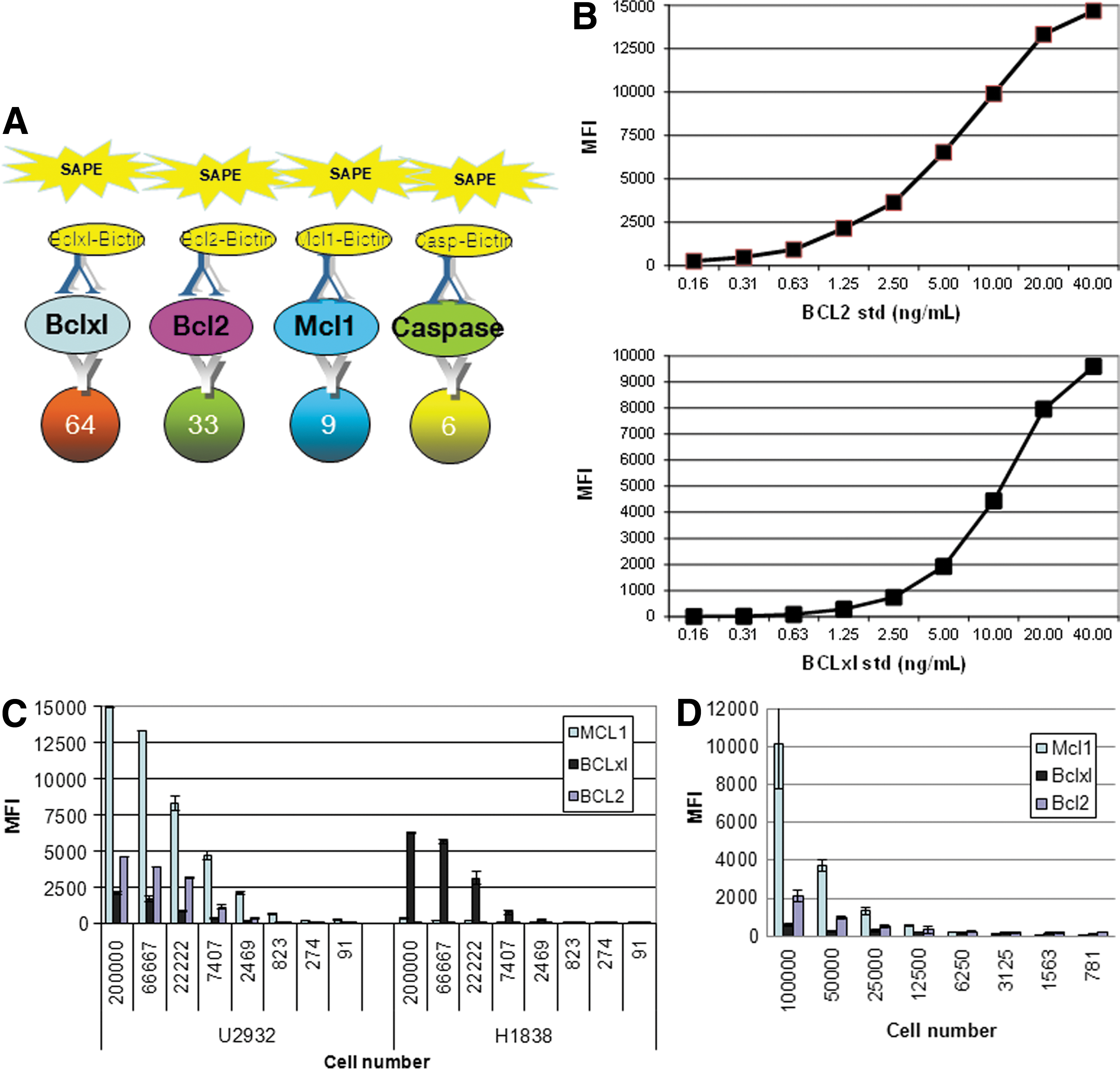
Specificity and dynamic range of the custom multiplex Luminex assay (quantitative 4-plex BCL2, BCLxL, MCL1, and caspase 3).
Recently, commercially available ELISA kits were developed for BCL2 and BCLxL (R&D Systems). Although these assays detect these proteins individually with high specificity, the goal was to develop a multiplex assay to detect these proteins simultaneously with wide dynamic range. We first conjugated the capture antibodies with microbeads. Figure 1B and C shows that these assays could detect BCL2 and BCLxL in a linear fashion. Dynamic range for BCL2 and BCLxL is 0.5–40 ng/mL and 1.25–40 ng/mL, respectively, which is similar to the ELISA format (data not shown).
Next, cell line lysates were titrated to determine the specificity and dynamic range of our custom BCL2, BCLxL, and MCL1 Luminex multiplex assay. From our genomic database, lymphoma line U2932 is found to express high levels of MCL1 and BCL2, while NSCLC line H1838 has high expression of BCLxl but not BCL2 (data not shown). Figure 1C and D shows that U2932 indeed expresses high levels of MCL1 and BCL2 but not BCLxL. We found that diluting cell lysates (Fig. 1C) vs. diluting cells in the plate before lysis (Fig. 1D) yield similar trends. In contrast, H1838 expresses high levels of BCLxL but not BCL2 and MCL1 (Fig. 1C). This data confirms our genomic data and the specificity of this assay. In addition, we show that we can detect the differences of these proteins in a wide cell number range. Intra-plate variability %CV performed using fixed number of U2932 cells with or without spike in recombinant proteins ranges from 20 to 37 (data not shown). The %CV is a little higher than expected due to low expression of some of these proteins in the cells and the assay was developed using the filter plate. We expect the %CV could improve substantially with magnetic beads. Taken together, our data suggest that this assay is highly specific and has high dynamic range of detection and therefore allowed us to proceed to use the assay for multiple applications. In this report, we will present three applications demonstrating how our custom multiplex assay was useful in drug discovery research.
Application 1: Basal Expression of BCL2 Determines Response to BCL2/BCLxL Inhibitor ABT-263 (Navitoclax)
One major application of this assay is to determine the basal expression of BCL2, BCLxL, and MCL1 in a single sample and correlate with response to certain compounds. An example of this is shown in Figure 2. Eighteen multiple myeloma lines were treated with BCL2/BCLxL inhibitor ABT-263 and EC50 was determined based on viability assay. Basal expression of BCL2, BCLxL, and MCL1 were measured with the Luminex assay. Although there was a good anti-correlation between response to ABT-263 (log EC50) and basal expression of BCL2 (R=−0.65), there was minimal correlation seen between the BCLxL or MCL1 and ABT-263 sensitivity (log EC50). Recent papers suggested that BCL2/MCL1 ratio predicts sensitivity to ABT-737, a similar compound to ABT-263, more accurately. 14,15 Toward this end, the BCL2/MCL1 ratio correlates with sensitivity to ABT-263 (log EC50) with coefficient of −0.70. This correlation was slightly stronger than BCL2 expression alone, suggesting both BCL2 expression and BCL2/MCL1 ratio are the best predictors of ABT-263 response. This data suggests that our assay is very useful in measuring protein expression of BCL2, BCLxL, and MCL1 in a single sample and correlate with response to compounds such as ABT-263. This assay would be highly applicable for identifying and developing stratification biomarkers to BCL2 inhibitors.
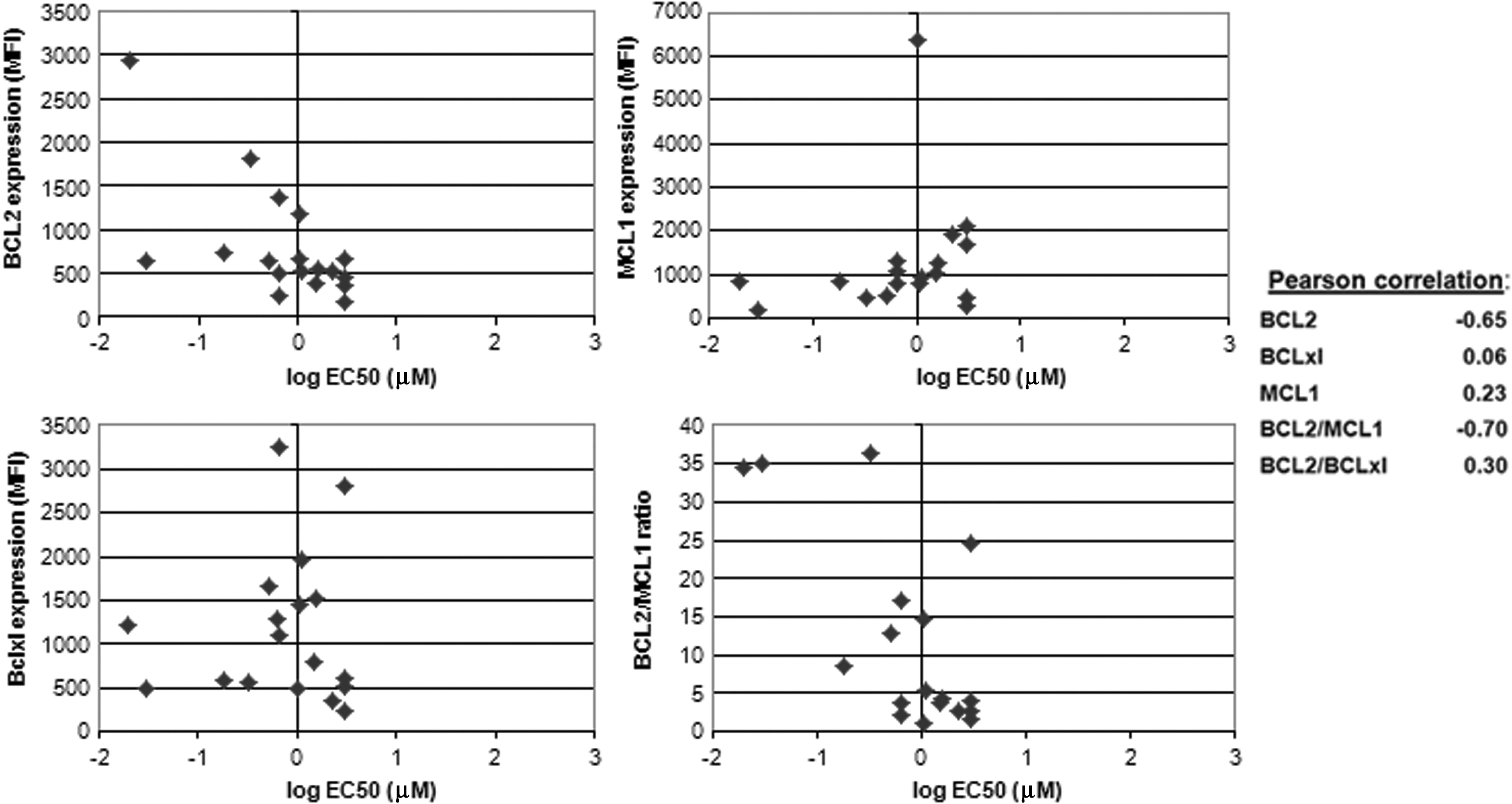
Application 1: Correlation of protein expression of BCL2 family members with response to ABT-263 in multiple myeloma cell lines. Basal protein expression levels of BCL2, BCLxL, and MCL were measured from 5 μg total protein from 18 multiple myeloma cell lines lysates using the custom multiplex Luminex assay. EC50 for each cell line was determined by measuring cell viability via Cell Titer Glo (Promega) after treatment with ABT-263 for 48 h. Cell viability response to drug (log EC50) and protein expression levels of BCL2, BCLxL, and MCL1 were plotted with Pearson correlation value determined by Microsoft Excel software (Redmond, WA).
Application 2: Increase Apoptosis via Induction of Active Caspase 3 Correlated with MCL1 Decrease in H23 Cells
Since the modulation of members of the BCL2 family could induce apoptosis, it would be advantageous to develop an assay that can measure apoptosis quantitatively. Caspase 3 is one of the “executioner” proteases involved in the apoptosis process and its activation is indicated by the cleavage of this protease. Recently, we showed that a few NSCLC cell lines depend on MCL1 for survival. 16 One of these cell lines, H23, undergoes apoptosis when MCL1 is silenced by siRNA. We reasoned that agents that decrease MCL1 should induce apoptosis in this line too. Cyclin-dependent kinase (CDK) inhibitors such as flavopiridol are known to inhibit multiple CDK family members including CDK9, an enzyme that plays a role in transcription elongation. 17,18 In addition, flavopiridol decreases MCL1 within 30 min of treatment due to the short half-life nature of MCL1 mRNA and protein. 17 We were interested in determining if levels of active caspase 3 increased as MCL1 decreased in H23 cells. In order to test this, we incorporated an existing active caspase 3 microbead assay into our 3-plex anti-apoptosis protein assay. Figure 3 shows that active caspase 3 expression increased as MCL1 protein expression decreased. No modulation of BCL2 and BCLxL expression was observed, possibly due to H23 cells expressing low levels of these proteins. 16 In addition, BCL2 and BCLxL have a longer half-life and no modulation would be expected in a 4-h period. Nevertheless, these data confirm that we have developed a multiplex assay that can detect BCL2, BCLxL, MCL1, and active caspase 3 simultaneously. This assay is particular useful to monitor changes in expression of BCL2, BCLxL, or MCL1 and induction of apoptosis.
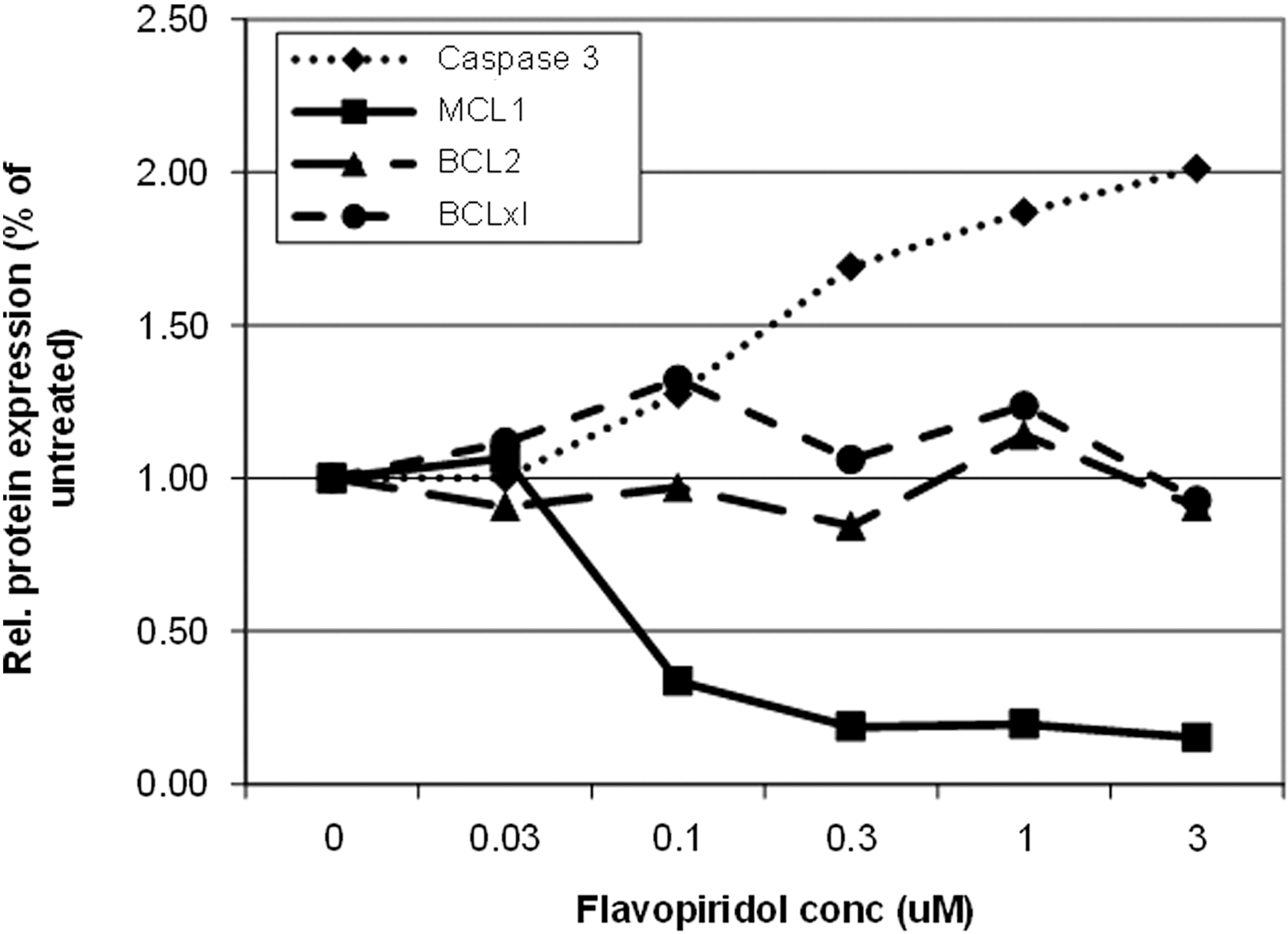
Application 2: Correlation between MCL1 decrease and induction of active caspase 3 in MCL1-dependent cell line H23 after treatment with cyclin-dependent kinase (CDK) inhibitor. H23 cells were treated with increasing concentration of flavopiridol for 4 h. Microbeads conjugated to BCL2, BCLxL, MCL1, and active caspase 3 antibodies were used to determine the levels of these proteins.
Application 3: Screening for MCL1 Modulating Compounds for Combination Therapy With ABT-263
ABT-263 is a first-in-class BCL2 family inhibitor that restores the ability of cancer cells to undergo apoptosis. However, many cancer cells are resistant to ABT-263 due to high expression of a BCL2 family member MCL1. 3,4 We reasoned that compounds that decrease MCL1 would synergize with ABT-263 in killing cancer cells. This cellular system would also facilitate the validation of our small molecule–based high-throughput screening approaches. In this regard, we performed a small molecule screen using a library of 120 known cancer inhibitors in four NSCLC cell lines in which MCL1 expression confers resistance to ABT-263. Four cell lines, chosen with known genomic abnormalities in order to identify potential pathways that regulate MCL1, were exposed to four concentrations of each inhibitor, and then screened for MCL1 protein levels using our custom Luminex multiplex assay. The results are presented in a heat map format prioritized based on the number of cell lines affected and the degree of MCL1 decrease (Fig. 4A). We found that around 30 compounds could decrease MCL1 protein expression, although to different degrees. Consistent with our previous study and others, 15,17 we found that CDK inhibitors flavopiridol and SNS-032 strongly reduced MCL1 protein levels in these cells (Fig. 4B). In addition, we tested if these compounds sensitize these resistant lines to ABT-263 by comparing cell viability of cells treated with CDKi alone to CDKi+/− ABT-263 in vitro. As shown in Figure 4D, ABT-263 has minimal effect on the four cell lines. Similarly, flavopiridol and SNS-032 have modest effect on these cell lines (except H1993), but these compounds strongly sensitized all four cell lines to ABT-263 with greater than 75% reduction in cell viability, suggesting that compounds that reduce MCL1 would combine with ABT-263.

Application 3: Small molecule screen identified CDK inhibitors and PF-562271 potently decrease MCL1 protein through CDK9 inhibition and potentiate ABT-263 killing.
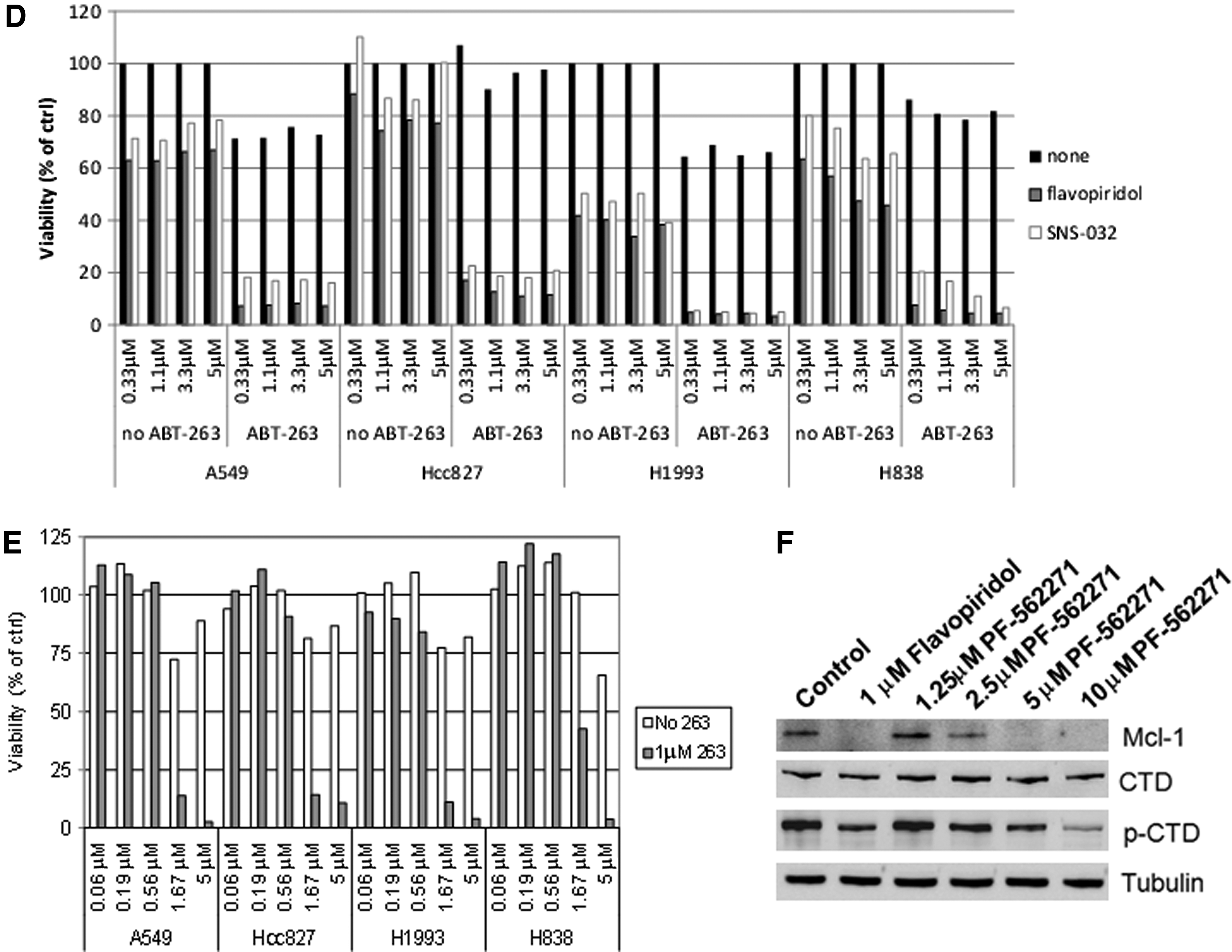
Surprisingly, PF-562271 was identified from the small molecule screen to strongly reduce MCL1 levels in all four cell lines (Fig. 4C). PF-562271 is a small molecule that inhibits focal adhesion kinase (FAK), a cytoplasmic tyrosine kinase that has been implicated in the formation and progression of solid and hematological tumors. 19 PF-562271 also synergized with ABT-263 in killing these cell lines, thus further supporting that reduction of MCL1 yields increased efficacy with ABT-263 (Fig. 4E).
Although PF-562271 displays greater than 100-fold selectivity against a long list of nontarget kinases, it was shown to inhibit multiple CDK-cyclin complexes in recombinant enzyme assays. 20 In light of its activity against CDKs, we hypothesize that PF-562271 inhibits the activity of CDK9. CDK9 is a transcriptional protein that activates RNA polymerase II to promote elongation by phosphorylation of its CTD. To investigate this potential mechanism of FAK inhibitor PF-562271, we performed Western blot analysis to determine the phosphorylation status of CTD of RNA polymerase II in A549 cells treated with flavopiridol or FAK inhibitor for 4 h. As shown in Figure 4F, p-CTD was strongly reduced in cells treated with flavopiridol or FAK inhibitor. In addition, MCL1 also decreases in a similar manner as the reduction of p-CTD in these cells. Our data unveiled a potential transcriptional effect of FAK inhibitor on cancer cells. In addition, our screening strategy may yield other potential candidates for combination with ABT-263.
In conclusion, we have developed a quick and convenient method to simultaneously measure protein expression of the anti-apoptotic proteins (BCL2, BCLxL, MCL1) and monitor induction of apoptosis (caspase 3) upon drug treatment. We have also shown that this assay could be used in high-throughput screening for compounds that modulate the expression of these anti-apoptotic proteins. Therefore, we believe our custom multiplex Luminex assay, with the advantages of the multiplex, high-throughput platform is a beneficial toolbox assay for drug discovery. Future improvement to the assay includes adding more plexes to detect additional apoptosis-related proteins, transforming the assay into a magnetic-based platform to improve washing, and automation.
Footnotes
Acknowledgments
We thank past and present members of the BCL2 project team at AbbVie for helpful discussions. We would like to thank Phil Hajduk (AbbVie) for compiling the bioactive library and Linda Traphagen's (AbbVie) high-throughput group for dispensing compounds into a 96-well format. We also thank Sai Sruthi Guttikonda (Abbstt) for excellent technical help and Tamar Uziel (AbbVie) for critical review of the manuscript.
Author Disclosure Statement
All authors are employees of AbbVie. The design, study conduct, and financial support for this research was provided by AbbVie. AbbVie participated in the interpretation of data, review, and approval of the manuscript.