
Abstract
The antifungal effects of 2-phenylethanol are clearly visible through its intervention in Candida morphogenesis. Chronic and recurrent vulvovaginitis, however, does not respond to this standard experimental therapy; therefore, the study presented in this article investigated the effect of common antifungal drugs (amphotericin B [AMB], fluconazole [FLU], and itraconazole [ITC]), in combination with 2-phenylethanol, on the Candida species isolated from cases of chronic and recurrent vulvovaginitis, thereby allowing the recommendation of a more appropriate treatment option. Forty isolates from patients with chronic and recurrent vaginal candidiasis were investigated in this experimental study. The specimens were examined by direct microscopy, culturing, and PCR to identify the species. The antifungal effects of 2-phenylethanol and conventional drugs, both alone and in combination, were determined in duplicate. Finally, the findings were analyzed. In this study, 40 strains of Candida species were identified, whose agents were Candida albicans (95%) and Candida africana (5%). After 48 h, the minimum inhibitory concentration (MIC) range of the 2-phenylethanol was 800–3,200 μg/mL. Also, in the final study on the MIC levels of common antifungal drugs, AMB (0.42 μg/mL) had the lowest MIC, FLU (40.51 μg/mL) had the highest MIC, and the combination of ITC and 2-phenylethanol had the lowest fractional inhibitory concentration index (FICI) of any of the combinations (FICI range, 0.26–1.03). Combining FLU and ITC with 2-phenylethanol can effectively increase their antifungal effect.
Introduction
The antifungal agent 2-phenylethanol, also called phenyl ethyl alcohol (PEA), is a colorless liquid with a rose-like odor, which is used as an aromatic compound with antimicrobial effects in perfumes and cosmetics, as well as in the food industry. PEA can be found in a variety of flowers, plant extracts, and many yeast species (such as Candida albicans) as their metabolites. 1,2 The antimicrobial effects of PEA were proven against Escherichia coli, Staphylococcus aureus, Enterococcus faecium, C. albicans, Candida dubliniensis, Saccharomyces cerevisiae, Kluyveromyces marxianus, Aspergillus niger, Penicillium notatum, Penicillium digitatum, Penicillium italicum, Botrytis cinerea, Rhizopus nigricans, and Neurospora crassa. 2 –5 Both the yeast and filamentous forms of C. albicans can produce PEA under both aerobic and anaerobic growth conditions, and it can also be produced by C. dubliniensis, Candida parapsilosis, and Candida tropicalis. Once its extracellular concentration reaches a certain level, this compound inhibits filamentous growth and germ tube formation in C. albicans. 3,4,6,7 The main role of PEA is to inhibit the morphological transition in C. albicans and C. dubliniensis. 4,8,9 The molecular mechanism by which PEA intervenes in morphogenesis remains incompletely characterized; it may damage membrane fluidity, causing ion leakage and reducing active transports, or it may destabilize the proteins involved in signal transduction and interfere with the morphogenesis signaling. PEA can play a role in inhibiting the expression of hyphae-specific genes, such as SIR2, the hyphal repressor, and TUP1. 1,6
The azoles (clotrimazole [CLO], fluconazole [FLU], itraconazole [ITC], ketoconazole) and polyenes (amphotericin B [AMB]) are the main conventional drugs used to treat candidiasis in infections such as recurrent vulvovaginitis. However, due to high toxicity effects, the severe side effects of polyenes, and the increasing resistance to azoles caused by their fungistatic rather than fungicidal nature, it has become expedient to develop new antifungal agents. Indeed, the number of antifungal drugs is very limited in comparison with antibacterial drugs. 10 –13
Recurrent vulvovaginal candidiasis (RVVC) is defined as four or more relapses of discrete episodes of vulvovaginal candidiasis per year. Patients suffering from chronic vulvovaginal candidiasis (CVVC) often appear to have a progressed stage of RVVC, perhaps indicating that RVVC and CVVC exist along a spectrum. 14 Most patients complain of vaginal itching, redness, soreness, burning, dyspareunia, and dysuria, and the discharge associated with candidal vulvovaginitis does not have an unpleasant odor in comparison with that of bacterial vaginitis. 15 Treatment of the infection depends on the patient's condition (immune status). For the prevention of recurrent cases, local and oral maintenance treatments are recommended. Local CLO 500 mg, oral ketoconazole 100 mg, or oral FLU 150 mg is administered for the treatment of RVVC. However, recurrence occurs in approximately half of all patients shortly after ending treatment. 15,16
Prolongation and recurrence of CVVC or RVVC can be bothersome for patients and can waste their time and money. However, few antifungal drugs exist, and the continuous consumption of these drugs causes fungal resistance. To improve the treatment options, we studied the effect of PEA, in combination with common antifungal drugs, on Candida species isolated from CVVC and RVVC.
Materials and Methods
This study was conducted experimentally from February 20, 2016, until December 20, 2016, on Candida species isolated from women with CVVC and RVVC who had been referred to Lolagar Hospital of Tehran.
Sampling
Sample size calculation
A sampling method based on easy accessibility was used for 6 months (from February 20 until August 20, 2016). Given the prevalence of RVVC in Iran, with the formula
Specimen collection
With patient consent, in accordance with the Declaration of Helsinki, and under gynecologist supervision, vaginal discharge samples were taken with sterile cotton swabs from women with symptoms who had been referred to the gynecology clinic at Lolagar Hospital. The swabs were placed into sterile Falcon tubes containing preservative (2 mL phosphate-buffered saline) (09-8912-100; Medicago, Canada), and an information form for each patient was completed and attached to the corresponding vaginal sample. The samples then were transferred to the mycology laboratory of the Iran University of Medical Sciences.
Specimen Processing
Direct microscopy
One/two drops of the vaginal sample from the Falcon tube, along with one drop of KOH 10%–20%, were applied to a slide and checked using an optical microscope. Oval-shaped or round yeast cells with false or true hyphae represent Candida species.
Culture
Samples were cultured on CHROMagar medium (CA220; CHROMagar™ Candida, France) and incubated for 2 days at 37°C. After the incubation period, the colors of the CHROMagar plates were recorded and samples carried out on the Sabouraud's dextrose agar slants (1054380500; Merck, Germany) (for purification and maintenance).
Polymerase chain reaction
The presence of the Candida species was diagnosed definitively with the PCR tests by using universal primers ITS1 (5′TCCGTAGGTGAACCTGCGG3′) and ITS4 (5′TCCTCCGCTTATTGATATGC3′) (TAG Copenhagen, Denmark) (to amplify the ITS1-5.8S-ITS2 area) and sequencing. Sequencing was done by Bioneer Company for all of the specimens (Republic of Korea). Briefly, isolated Candida species were cultured on a solid yeast extract peptone glucose agar (YEPD) medium (yeast extract, Y1625 [Sigma]; glucose, G8270 [Sigma]; peptone, 146447 [Merck]; agar, 101614 [Merck]; chloramphenicol, C0378 [Sigma, Germany]) and were incubated at 37°C for 24–48 h. According to the rapid DNA extraction protocol, DNA (template DNA) was extracted from a Candida colony. 19 The primers were purchased and, according to the manufacturer's instructions, mixed with sterilized distilled water and then utilized as follows (for each sample): 12/5λ Master Mix (180301; Ampliqon, Denmark) +1λ ITS1 + 1λ ITS4 + 9/5λ distilled water +1λ template DNA. In a negative control tube, instead of the template DNA, 1λ sterilized distilled water was added. PCR thermal cycles were performed using a thermo cycler (732-2773; Peqlab, Germany). Denaturation was performed for 35 cycles at 98°C (5 min and 30 s), annealing was performed for 1 cycle at 65°C (30 s), and extension was performed for 1 cycle at 72°C (30 s) and another cycle at 72°C (5 min); then, the PCR products were electrophoresed on agarose gel (A9539; Sigma, Germany), and images were recorded from the electrophoresis gels with PCR products.
Antifungal Susceptibility Test Alone
The antifungal drugs tested were AMB (A4888; Sigma, Germany), FLU (F8929; Sigma, Germany), ITC (I6657; Sigma, Germany) (common antifungals), and 2-phenylethanol (P13622; Aldrich, Germany). These susceptibility tests were performed using the microbroth dilution method (in duplicate), according to the CLSI M27-A3 protocol and the third informational supplement (M27-S3), 20 on 40 Candida species isolated from RVVC and CVVC patients, as follows.
Stock preparation (common antifungals)
The primary stocks of AMB, FLU, and ITC with a concentration of 128 μg/mL were prepared according to the CLSI M27-A3 protocol 20 for common drug powders, then weighed using an analytical susceptible balance (HR-200; Mettler Toledo Company, Columbus, OH) and solved using the appropriate amount of DMSO (102952; Merck, Germany). These stocks were filtered using a sterile syringe filter with a pore size of 0.22 μm (SFCA013022N; Membrane Solutions, China). Then, at the first weekday of each week of the study period, lower concentrations (from 128 to 0.03125 μg/mL) were prepared using serial dilution according to the CLSI M27-S3 protocol.
PEA stock preparation
To prepare a stock of liquid PEA (P13622; Aldrich) with a concentration of 6,400 μg/mL, according to the PEA minimum inhibitory concentration (MIC) for C. albicans in previous research, 21,22 calculations were performed according to a pharmacy technician's guidebook. 23 The calculated amount of 2-phenylethanol's primary stock was dissolved in DMSO and then filtered with a millipore filter (0.22 μm pore size). Thus, concentrations of 6,400 to 100 μg/mL were prepared in DMSO using serial dilutions.
Yeast suspension preparation
A yeast suspension was prepared in 0.85% sterile normal saline from a young Candida colony (24–48-h incubation at 35°C) using a spectrophotometer (Novaspec II Visible Spectrophotometer; Pharmacia LKB Biotechnology, Cambridge, England) with a wavelength of 530 nm; by changing the cell density, 85%–90% transmittance was achieved. Then, this 1 × 10⁶ cells/mL suspension was diluted in RPMI 1640 (R6504; Sigma, Germany) (with glutamine, without bicarbonate, and with phenol red as a pH indicator) in a 1:50 dilution followed by a 1:20 dilution; the final solution had 1 × 10³–5 × 10³ cells/mL.
MIC calculation for PEA and common drugs
Ninety-six sterile, well-capped microplates (351177; Becton Dickinson) were used for the MIC calculation. One hundred microliters of each common drug (of each concentration) was poured vertically on the plate. Then, 100 μL of yeast suspension was applied to all wells. The final drug concentrations were 128–0.03125 μg/mL for common drugs and 6,400–100 μg/mL for PEA. One hundred microliters of RPMI 1640 medium and 100 μL of yeast suspension were added to the positive control column, 200 μL of RPMI 1640 medium was discarded in the negative control column, and one well was considered a DMSO solvent control, containing 100 μL of RPMI 1640 medium plus DMSO and 100 μL of yeast suspension. The microplates were placed on the shaker (Sepand round shaker; Sepand, Iran) for 3–5 min before being placed in a 35°C incubator (IN30plus; Memmert, Germany); after 48 h, the microplates were read optically using a mirror. For each sample, the concentration in which no growth was observed was considered to be the MIC alone.
Minimum fungicidal concentration calculation
The contents of the MIC well and previous wells were cultured in Sabouraud's glucose agar with a sterile loop and checked after 48 h. The first concentration that had no growth was considered to be the minimum fungicidal concentration (MFC). Antifungal susceptibility was interpreted based on the MICs obtained from the isolates and according to the CLSI M27-S3 protocol.
Antifungal Susceptibility Testing in Combination
The strains resistant to conventional drugs were also investigated using antifungal sensitivity tests, in combination with PEA. These combination tests were carried out in duplicate as follows: for 4 isolates resistant to and 2 isolates sensitive to AMB, and 6 isolates resistant to FLU and ITC.
Antifungal susceptibility testing in combination was performed according to the antifungal combination protocol, 24 and the antifungal stocks and yeast suspension were prepared as in the antifungal susceptibility test alone.
MIC calculation for PEA in combination with common antifungals
For the MIC calculation, 96-well microplates were used, on which 50 μL of each common drug was poured vertically from columns 1 to 11. Then, 50 μL of PEA was poured horizontally from rows A to G. Fifty microliters of RPMI 1640 medium was added to row H and column 11, so row H contained common drugs alone, while column 11 contained PEA alone. The final concentrations of columns 1–11 of each common drug were 16–0.03125 μg/mL, except for FLU, the concentrations of which were 64–0.03125 μg/mL or 256–0.03125 μg/mL. The final concentrations of rows A to G were 6,400–100 μg/mL. Then, 100 μL of yeast suspension was added to each well. Two wells with 100 μL of RPMI 1640 medium and 100 μL of yeast suspension were considered positive controls, two wells with 200 μL of RPMI 1640 medium were considered negative controls, and one well containing 100 μL of RPMI 1640 medium plus DMSO and 100 μL of yeast suspension was considered a DMSO solvent control. In this way, the highest drug concentration was in the 1st vertical and horizontal well, and the lowest drug concentration was in the 10th vertical and 7th horizontal well. Then, the microplates were placed on a shaker for 3–5 min before being placed in a 35°C incubator; after 48–72 h, the microplates were read optically using a mirror. In column 11 and row H, in which drugs alone were poured, the first well in which no growth was seen was considered to be the MIC alone. In addition, according to the growth pattern in the antifungal combination protocol
24
(Fig. 1), MICs in combination, as well as any synergism, antagonism, or additive (no interaction) effects, were determined using the FICI (fractional inhibitory concentration index) formula.
X = drug A
Y = drug B

Checkerboard technique (Vitale, Afeltra, and Dannaoui). Interpretation of results based on fungal growth patterns in microplates (black wells): wells marked with A or B are the MICs of drug A or B alone, and wells marked with A + B are the MICs of the combination of two drugs. MIC, minimum inhibitory concentration.
The interpretation of the FICI results was as follows: ≤0.5, synergistic effect; >0.5 but <4, no interaction; and ≥4, antagonistic effect.
Statistical Analysis
Findings were analyzed using SPSS ver.16 software, as well as Mann–Whitney and paired sample t-tests. The confidence coefficient of these tests and means was 95%, and the differences for P values smaller than 0.05 were considered statistically significant.
Results
Patient Information
The ages of patients with RVVC or CVVC ranged from 19 to 57 years, with a mean age of 36.17 ± 10.25 years. Most patients (25%) were 31–35 years old. Most had a high school diploma and associate degree (42.5%), as well as an average economic status (52.5%). Most of the patients were housewives (87.5%) and nonsmokers (95%), and all patients were living in a city. In this study, the predisposing factors for CVVC and RVVC among all age groups were not being under medical care for the treatment of CVVC or RVVC (82.5%), as well as not using any antifungal medicines until being referred to a doctor, or discontinuing antifungal treatment after a short period of time (80%). Also, the majority of patients (97.5%) had no history of genetic disease or abortion (75%), had 1 or 2 children (67.5%), and had a history of 2 child deliveries (32.5%).
Findings of Specimen Processing
Forty Candida species were isolated from CVVC and RVVC patients. After performing identification (culture and PCR) tests, the isolates were identified as being 95% C. albicans (38 cases) and 5% Candida africana (2 cases) (Fig. 2).
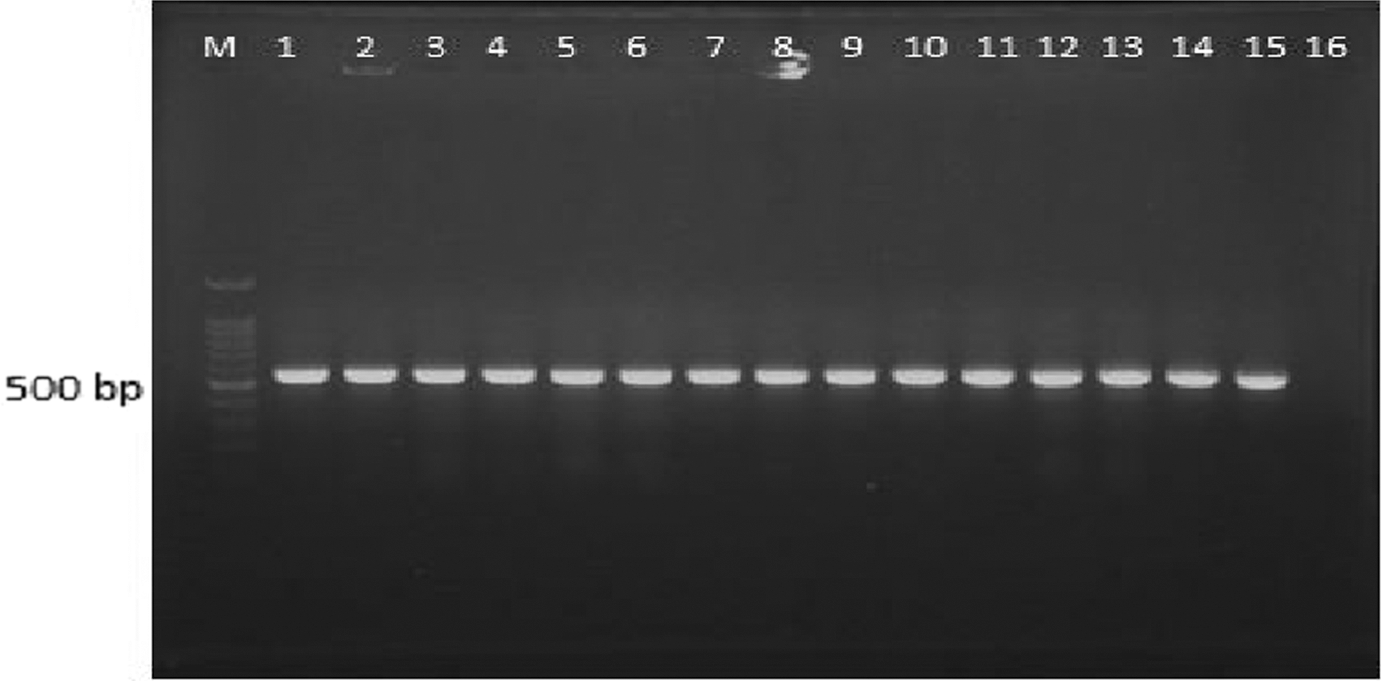
Candida albicans and Candida africana isolated from the patients, identified by the PCR test with universal primers ITS1 and ITS4. Column M; marker 100 bp, Column 1; positive control of standard C. albicans (PTCC 5027), Columns 2 to 11 and Columns 14 to 15; C. albicans, Columns 12 and 13; C. africana and Column 16; negative control.
Findings of Antifungal Susceptibility Tests
A significant difference existed between the MIC mean of PEA and common drugs (AMB, FLU, and ITC) (P = 0.0001). The susceptibility testing revealed that the MIC values for PEA ranged from 800 to 3,200 μg/mL, and the MFC values ranged from 1,600 to 6,400 μg/mL, with MIC50 and MIC90 of 3,200 μg/mL. The MIC and MFC values of AMB ranged from 0.0625 to 2 μg/mL, with MIC50 of 0.25 μg/mL and MIC90 of 0.5 μg/mL. The MIC and MFC values for FLU ranged from 0.25 to 256 μg/mL, with MIC50 of 16 μg/mL and MIC90 of 64 μg/mL. Finally, the MIC and MFC values for ITC ranged from 0.0625 to 64 μg/mL, with MIC50 of 16 μg/mL and MIC90 of 32 μg/mL (Tables 1 and 2). Susceptibility was interpreted according to the CLSI M27-S3 protocol and a fundamental medical mycology textbook. 20,25 The most effective antifungal agent used in this study was AMB, to which 36 isolates (90%) were sensitive (MIC ≤1 μg/mL), followed by FLU (18 sensitive isolates, 45%) (MIC ≤8 μg/mL) and ITC (2 sensitive isolates, 5%) (MIC ≥1 μg/mL) (Table 3).
Minimum Inhibitory Concentration Ranges of 2-Phenylethanol, Amphotericin B, Fluconazole, and Itraconazole Against Strains Included in the Study
MIC values are expressed in μg/mL.
Number of isolates.
GM expressed in μg/mL. In parenthesis, MIC90 for all the strains and C. albicans.
GM, geometric means; MIC, minimum inhibitory concentration.
Minimum Fungicidal Concentration Ranges of 2-Phenylethanol, Amphotericin B, Fluconazole, and Itraconazole Against Strains Included in the Study
MFC values are expressed in μg/mL.
Number of isolates.
GM expressed in μg/mL
MFC, minimum fungicidal concentration.
Antifungal Susceptibility of the Strains Included in the Study
Susceptibility values are expressed in μg/mL.
Number of isolates.
R, resistant; S, susceptible; S-DD, susceptible-dose dependent.
Finally, for the combination of PEA and common antifungals, the mean FICI of the PEA and AMB, PEA and FLU, and PEA and ITC combinations was obtained as 1.16 ± 0.37, 0.78 ± 0.36, and 0.82 ± 0.31, respectively (Table 4). Also, there were significant differences between the mean MIC of FLU and ITC alone and in combination with PEA (P = 0.016, P = 0.004), but no significant difference existed between the mean MIC of AMB and PEA in combination (P = 0.137). Furthermore, it was found that the highest FICI results were related to the PEA and FLU combination (Table 5).
Interactions of Antifungal Drugs with 2-phenylethanol Against Some Resistant Candida Strains Included in the Study
Number of interactions.
FICI, fractional inhibitory concentration index; GM, geometric mean.
MICs of Antifungal Drugs and 2-phenylethanol Against Some Resistant Candida Strains Included in the Study
MIC values are expressed in μg/mL.
GM expressed in μg/mL
Discussion
The current experimental study was performed to identify a new antifungal agent to improve the treatment of CVVC and RVVC. According to the findings of this study, the MIC range of PEA on C. albicans and C. africana species isolated from CVVC and RVVC patients was 800–3,200 μg/mL after 48 h (Table 1). The MIC of PEA was reported in a study by Paulus as 2,500 μg/mL against the C. albicans yeast form, and the mean of the MICs obtained in our study was 2,580 μg/mL, which correlated with what Paulus reported. 21 The MIC range of PEA was reported as 650–7,670 μg/mL against allergic molds affecting humans by Kosalec et al. in 2007. 22 In 2013, Han et al. claimed that the concentration of 3–4 g/L (3,000–4,000–3,000 μg/mL) of PEA completely prevented the growth of the S. cerevisiae yeast form. 3 The MICs from studies conducted by Kosalec et al. on molds 22 and Han et al. on S. cerevisiae 3 were similar to the MICs obtained in the current study on C. albicans and C. africana. Contrary to the current study, in a 2011 study by Chauhan et al., PEA had no significant effect on the growth and stability of C. albicans cells in the yeast phase up to 4% (40,000 μg/mL), and a significant reduction in persistence (40%–50%) was detected in 6%–8% (60,000–80,000 μg/mL) of PEA. 6 These observed differences may be due to differences in the strains that were used, as well as differences in the source of PEA (biosynthetic or synthetic origin) used in these studies.
In our previous study, PEA demonstrated the highest synergism effect in combination with CLO, with a mean FICI of 0.28 ± 0.084 against resistant isolates of CLO in CVVC and RVVC patients. 26 In the present study, PEA was able to improve the effect of FLU and ITC against isolates resistant to FLU and ITC in CVVC and RVVC patients (P = 0.008, P = 0.002), but it could not change the effect of AMB (P = 0.068). It is likely that the intervention of PEA in Candida morphogenesis, especially C. albicans, or in the biosynthesis of macromolecules (e.g., DNA, RNA, and proteins) could improve the effect of azoles in the inhibition of ergosterol biosynthesis, particularly in resistant organisms (CLO, FLU, and ITC). 5,27
In the present study, FLU and ITC were not so effective in preventing the development of Candida species isolated from CVVC or RVVC patients; they were only effective in high concentrations. Among Candida species isolated from these patients, 45% and 5% were sensitive to FLU and ITC, respectively, and in our previous research, 17.5% of the mentioned isolates were sensitive to CLO. 26 In another study on Candida species isolated from CVVC patients, 42% of isolates were sensitive to FLU, which correlated with our study. 28 The results of other similar studies contrast with those of the present study in that all or most of the isolates were susceptible, dose dependent, and sensitive to FLU, 29 –34 and in some other studies, most of the isolates were resistant to FLU. 35 –38 The reasons for these differences may be related to variations in the population studied, changes in sensitivity patterns due to the continuous use of the drug, and differences in host immunity. In a study by Lakshmi et al., most of the isolates were resistant to ITC, and in another study by Mohamadi et al., 62% of isolates were resistant to ITC, which correlated with the present study. 35,38 Unlike in our study, the results of a study by Zafar et al. showed that 47.6% of isolates were sensitive to ITC; in other similar studies, most of the isolates were sensitive or susceptible and dose dependent to ITC. 30,32,36 The reason for these differences is probably related to variations in the population studied or changes in the sensitivity pattern of the antifungals.
Conclusion
According to the results obtained in this study, combining FLU and ITC with 2-phenylethanol can effectively increase their antifungal effect. Administering common antifungal agents (FLU or ITC) with 2-phenylethanol could potentially be useful in the treatment of chronic and recurrent candidal vulvovaginitis in animal models. As the reported oral and cutaneous LD50 of PEA for rats and guinea pigs was 1,790 mg/kg and 10–5 mL/kg, respectively, 21 the MIC range of 800–3,200 μg/mL does not seem problematic. However, toxicological tests and research in animal models should be conducted in this field.
Footnotes
Acknowledgments
We thank the Vice Chancellor of Research and Technology of the Iran University of Medical Sciences and the staff members for their cooperation and financial support of this project. This study was part of a research project approved by the Iran University of Medical Sciences, by code 27077-30-05-94.
Ethical Aspects
The patients were informed about the study, and after voluntarily agreeing to participate, they signed a free and informed consent. The study was approved by the Research Ethics Committee of the Iran University of Medical Sciences, under registration number IR.IUMS.REC1394.9313385003.
Disclosure Statement
No competing financial interests exist.