
Abstract
Introduction:
The United States Environmental Protection Agency (US EPA) has issued a directive to reduce mammalian studies to 30% by 2025. The application of new approach methodologies is being implemented to provide information on chemical hazard and risk assessment. In vitro methods, such as the MucilAir™ assay, are used to detect airway damage and acute irritation potential in agrochemicals.
Materials and Methods:
Four agrochemicals, including Adepidyn™ and two developmental chemicals (DC 1 and 2), were evaluated using the MucilAir assay to determine the need for future in vivo inhalation studies. Chlorothalonil (CTN), which exhibits irritant properties in mucous membranes in vivo was assessed in the MucilAir assay to inform chronic inhalation risk. Evaluation of irritation potential and tissue damage were determined through transepithelial electrical resistance (TEER), lactate dehydrogenase (LDH) release, resazurin reduction, or histology. Each agrochemical was applied to the MucilAir for 24 hours at six concentrations (Adepidyn: 0.005–0.5 mg/L; chemicals no. 1–2: 31–317 mg/L) and 10 concentrations (CTN, 2–200 mg/L). As controls, vehicle, positive (sodium dodecyl sulfate, 4 mM) and untreated air–liquid interface groups were tested in parallel.
Results:
The control treatments confirmed correct assay functioning for each endpoint. Exposure to Adepidyn or DC 1 and 2 did not cause a change in TEER or LDH release. There was no change in resazurin reduction. Altogether, the irritation potential panel and histological evaluation indicated no evidence of toxicity. A benchmark dose (BMD) level of 0.00730 mg/cm2 was derived for CTN based on TEER, LDH release, and resazurin endpoints.
Discussion:
A good concordance of response across endpoints and human tissue donors was observed. A wide range of concentrations were tested to derive points of departure for risk assessment through BMD modeling.
Conclusion:
The MucilAir assay evaluating chemically induced airway cytotoxicity is fit for purpose for respiratory tract hazard screening and refining inhalation risk assessment of agrochemicals.
Introduction
Evaluation of inhalation toxicity is a frequent requirement in the regulation of agrochemicals. Assessing the toxicity of substances, including agrochemicals has relied on both in vivo and in vitro testing for a number of decades. This testing provides information about potential risk and enables informed decisions regarding safety. However, the demand to find and implement new approaches that encompass the Three Rs principles of laboratory animal welfare (replacement, reduction, and refinement) continues to increase to inform safety determinations from chemical exposure.1,2 Health effects due to inhalation of substances are of considerable interest and form the basis for the development and use of new approach methodologies (NAMs).
NAMs are defined as in silico and in vitro methods that can provide information on chemical hazard and risk assessment.3,4 NAMs, including alternatives to animal testing approaches, may be able to provide a large amount of data, decrease animal testing, which is time consuming and expensive and fill critical information gaps. 5
Regulatory agencies support the development of NAMs in efforts to obtain the data on the safety of chemicals.6,7 In the United States, the Frank R. Lautenberg Chemical Safety Act for the 2lst Century outlines the implementation of alternative testing methods to promote the development and timely incorporation of new scientifically valid test methods and strategies that are not based on vertebrate animals. 8 These methods include computational toxicology and bioinformatics, high-throughput screening methods, testing of substances in chemical categories, in vitro studies, system biology and new/revised methods identified by the Interagency Coordinating Committee on the Validation of Alternative Methods (ICCVAM), and the Organization of Economic Cooperation and Development (OECD).8,9
For a NAM to be available for regulatory use, the method must receive approval by international validation bodies or a regulatory agency. The method may be appropriate alone or used within an integrated approach to testing and assessment. The development of overarching guidelines and frameworks are critical steps in the acceptance of NAMs for regulatory use. The framework launched by the Health and Environmental Sciences Institute (HESI) to guide in the evaluation of NAMs for human safety risk assessment is based on the foundation of the NAM's context of use. 10 The evaluation framework provides criteria (core principles) for guidance in evaluating a NAM for a regulatory purpose. The level of evaluation is dependent upon use for prioritization, hazard screening, and risk assessment. Overall, this presents a problem formulation strategy for the appropriate evaluation of NAMs and helps to establish confidence in NAMs. 11
In vitro point of departure (POD) assessments have been shown to exhibit a good correlation with existing in vivo toxicity knowledge for phenol embryotoxicity, 12 developmental toxicity induced by retinoic acid, 13 and aristolochic acid-mediated nephrotoxicity. 14 Determination of in vitro POD for site-specific effects can be adequately represented by a benchmark dose (BMD) model of in vitro data for use in safety assessment.15–17
Conventional methodologies for the characterization of the toxicity of repeated inhalation exposure typically use laboratory rodents.18,19 Extrapolation of dosimetry and toxic responses from animal models to humans for safety assessment is complex and multifaceted due to differences in respiratory anatomy and physiology, the distribution of epithelial tissue types within the respiratory tract, breathing parameters, and biochemistry, all of which make the extrapolation of risk assessment endpoints for safety assessment challenging. 20
At the cellular level, the differences between species are less pronounced allowing for the use of in vitro methods for direct comparison of interspecies responses. For example, in all mammalian species, the ciliated pseudostratified epithelium that lines the different parts of the respiratory tract along with basal, goblet, neuroendocrine, serous, clara, and intermediate cells are the same. 21 In humans, the pseudostratified respiratory epithelium extends well into the lung, into the tertiary bronchioles, unlike the rodent where it only extends into the trachea and large bronchi. This distribution makes the rodent upper respiratory tract and the in vitro human respiratory cell assay well suited to represent the adverse effects that could occur in the human respiratory tract. 21
In vitro models of the respiratory tract range in complexity from simple two-dimensional monocultures, to three-dimensional structures, including lung-on-a-chip that contain multiple cell types representative of the in vivo architecture.22,23 MucilAir™ is a three-dimensional morphological and functionally differentiated model of the human respiratory epithelium, composed of basal stem cells, mucous-producing goblet cells, and ciliated cells grown on a semiporous membrane. 24 This model accurately reflects the structure of the ciliated pseudostratified respiratory epithelium found in the respiratory tract of humans, rats, and other mammals. MucilAir has been shown to replicate the main physiological and barrier functions of the airway epithelial cells, including mucociliary clearance, making it an appropriate model for in vitro assessment of human respiratory irritation represented by direct cytotoxicity.25–28
The MucilAir assay provides the opportunity to measure a variety of membrane and cell damage endpoints. The ones selected for the present work include transepithelial electrical resistance (TEER), which measures the integrity of tight junctions between cells in the membrane, lactate dehydrogenase (LDH) release serves as an indicator of membrane damage as a marker of cytotoxicity, and resazurin reduction as a measure of metabolic activity of the MucilAir epithelial structure.
The present study was designed to evaluate the utilization of the MucilAir assay, for hazard screening and to derive an inhalation endpoint-based POD to be used for risk assessment. The assay is currently under evaluation by the United States Environmental Protection Agency (US EPA) to confirm that it is an acceptable method for refining inhalation risk assessment. 29 Adepidyn™ (classified by US EPA in toxicity category IV) and two early stage/developmental chemicals (DC 1 and 2) were evaluated for irritancy potential using the MucilAir assay as a form of hazard screening. This assay was useful in determining if additional in vivo testing was needed. The MucilAir test system was used to evaluate chlorothalonil (CTN), a broad-spectrum fungicide, which is a respiratory irritant and is classified by the US EPA in toxicity Category II following acute 4-hour inhalation exposure. 30 Furthermore, the evaluation was utilized to establish that a NAM was suitable to inform an inhalation risk assessment in lieu of a whole animal inhalation study.
Materials and Methods
A formulation, Bravo Suspension Concentrate 720 containing fungicide, CTN (CTN content 54.7% w/w, design code A12531C), Adepidyn, and two developmental chemicals (DC 1 and 2) were supplied by Syngenta Ltd. Sodium dodecyl sulfate (SDS) was used as a positive control compound (Sigma-Aldrich Ltd.) Sodium chloride (0.9%, w/v; saline) solution was obtained from Baxter International (Deerfield, IL). Bravo Suspension Concentrate 720 without CTN was used as a vehicle control for the CTN study. Two experimental designs were conducted: Experiment 1: use of MucilAir for Hazard screening (Adepidyn, DC1 and DC2) and Experiment 2: use of MucilAir to derive an endpoint for the inhalation risk assessment (CTN). The two designs are summarized in Table 1.
Experimental Design Summary of Use of MucilAir
LDH, lactate dehydrogenase; SDS, sodium dodecyl sulfate; TEER, transepithelial electrical resistance.
Previous range-finding studies were conducted to determine the CTN MucilAir assay concentration range (data not shown). CTN, a known respiratory irritant, was then used as a benchmark to set concentration ranges for assessing any flags of chemical-related toxicity for Adepidyn, DC 1, and 2. Serial dilutions were used accurately to create solutions, which resulted in concentration curves. The dilution factor at each step was kept constant resulting in a geometric progression of the concentration in a logarithmic fashion.
MucilAir assay
Experiment 1: use of MucilAir for Hazard screening
The MucilAir test system (product code EP02) and medium (EP05MM) were obtained from Epithelix Sàrl (Geneva, Switzerland). MucilAir is derived from human airway cells collected from healthy donors. The MucilAir tissues used in this study were a pooled donor variant prepared from 14 healthy donors (Table 1). Tissues were cultured at the air–liquid interface (ALI) on Costar Transwell® polyester membrane units, at 37°C and 5% CO2, in a humidified incubator using optimized chemically defined media (700 μL/well). In this study, Adepidyn and two developmental chemicals (DC 1 and 2) were evaluated using the MucilAir assay in independent experiments. Evaluation of irritation potential and tissue damage was determined through the following endpoints: TEER, LDH release (an indicator of cytotoxicity), resazurin reduction, and histology review. There were no interferences with the assessment of TEER, LDH, or resazurin endpoints when evaluating Adepidyn and two developmental chemicals (DC 1 and 2).
The final media change was ∼24 hours before dosing to ensure that the predose media collected at 0 hour, and the 24 hours postexposure media samples for the LDH release assay corresponded to equivalent release time periods. Before dosing, tissues were transferred to fresh prewarmed culture media (700 μL/well). Dilutions of Adepidyn and each developmental chemical were prepared in sodium chloride (0.9%, w/v; saline), saline was also used as the buffer for TEER measurements and preparation of resazurin solutions.
Each agrochemical was applied to the four MucilAir replicates (tissue units) at six concentrations (Adepidyn: 0.0005–0.5 mg/L; DC 1 and 2: 31–317 mg/L) for 24 hours ±15 minutes. Vehicle control (saline), positive control (SDS, 4 mM), and untreated ALI control groups were tested in parallel. The ALI group went through all the same procedures and measurements as the treated groups but did not have any dose or vehicle applied. Baseline measurements for TEER were measured before dosing (day-3) and LDH release was measured at 0 hour; TEER, LDH release, and resazurin reduction were measured at 24 hours as described in Figure 1.
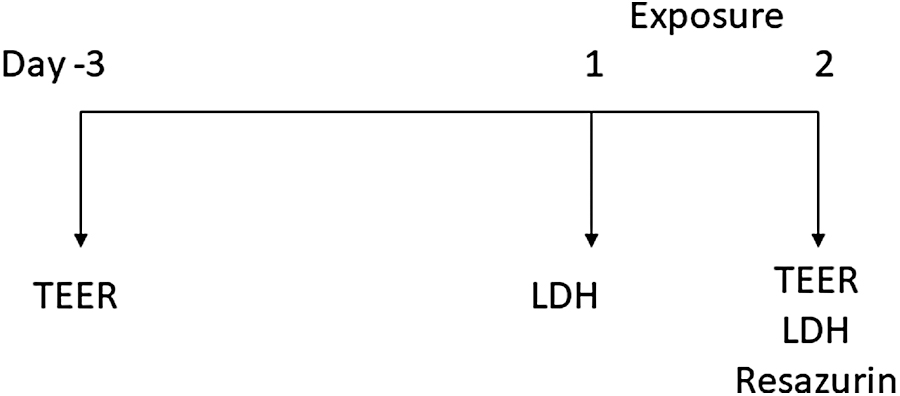
Experimental design for studies examining effects of Adepidyn™ and developmental chemicals 1 and 2. Cells were assessed for TEER, LDH, and resazurin reduction. A baseline measurement for TEER was obtained day 3 and LDH on day 1. Exposure to Adepidyn or developmental chemicals 1 and 2 began on day 1. TEER, LDH, and resazurin reduction measurements were assessed immediately after 24 hours of exposure. LDH, lactate dehydrogenase; TEER, transepithelial electrical resistance.
Dosing solutions (30 μL) were applied to MucilAir tissues by pipetting directly into the center of the apical surface. The internal diameter of the tissue culture inserts is 6.5 mm (33.18 mm2). The MucilAir units were exposed to dosing solutions for 24 hours in a humidified incubator set to maintain a temperature of 37°C in a 5% CO2 atmosphere. At the end of the 24-hour exposure period, any media that had permeated into the apical chamber was pooled with the basal media. The apical surface of tissues was rinsed three times with saline (0.5 mL per rinse). Tissues were transferred to saline for measurement of TEER and spent culture media were collected for measurement of LDH release. TEER and LDH release assays were conducted as for predose.
Experiment 2: use of MucilAir to derive an endpoint for the inhalation risk assessment
The MucilAir test system (product code EP01) and medium (catalog no. EP04MM, batch no. MM00205) was also obtained from Epithelix. Tissues were cultured at the ALI on Costar Transwell polyester membrane units, in a humidified incubator at 37°C and 5% CO2 using optimized chemically defined media (700 μL/well). The samples evaluated in this study were obtained from both male (n = 2) and female (n = 3) healthy donors (n = 5 total, ages 46–71) with no pathology reported. The condition of MucilAir tissues was then determined by viewing representative tissues (n = 4 per plate) by microscopy to verify epithelial structure and correct cilia function.
Tissues were maintained in the same manner as noted in the previous section. Vehicle control (saline), positive control (SDS, 4 mM), and untreated ALI control groups were tested in parallel. Evaluation of irritation potential and tissue damage was determined through the following endpoints: TEER, LDH release, and resazurin reduction. There were no interferences with the assessment of TEER, LDH, or resazurin endpoints when evaluating CTN in the Bravo 720 SC formulation.
Dilutions of the Bravo 720 SC formulation were prepared in sodium chloride (0.9%, w/v; saline), saline was also used as the buffer for TEER measurements and preparation of resazurin solutions. CTN is present as solid particles suspended in the Bravo 720 SC formulation, with these particles exhibiting a median diameter of ∼2.4 μm. Care was taken to thoroughly resuspend the Bravo 720 SC before use. Bravo 720 SC was vortex mixed (ca 5 minutes) before preparation of the stock solution, and solutions were mixed by shaking and vortex during the preparation of the serial dilutions (Table 2).
Preparation of Dosing Solutions for MucilAir Assay
CTN, chlorothalonil.
Dosing solutions (30 μL) were applied to MucilAir tissues by pipetting directly onto the center of the apical surface, taking care not to scratch the surface with the pipette tip. Gentle tapping of the unit was used to ensure test item was dispersed across the apical surface. The internal diameter of the tissue culture inserts is 6.5 mm (33.18 mm2). As such, applied doses of 2, 5, 8, 13, 20, 32, 50, 79, 126, 200 mg/L solution was then converted to surface concentration (in mg/cm2) for calculation of dose metrics.
MucilAir units were exposed to dosing solutions for 24 hours in a humidified incubator set to maintain a temperature of 37°C in a 5% CO2 atmosphere. Ten dilutions of the formulation were prepared and MucilAir tissues from five different donors were exposed to each dilution for 24 hours. Six MucilAir replicates (tissue units) were exposed to each dose level or control for each donor. At the end of the exposure period, the apical surface of tissues was rinsed three times with saline (ca 1.5 mL in total) to remove residual dosing solution. Concentrations of Bravo 720 used in this study were normalized to the CTN concentration of the formulation (54.7% w/w) and reported concentrations in this study reflect the concentration of CTN rather than the concentration of Bravo 720. TEER and LDH release was measured at 0 hour; TEER, LDH release, and resazurin reduction were measured at 24 hours.
Measurement of LDH release
The release of LDH from cells was measured using the CytoTox ONE™ Homogeneous Membrane Integrity Assay (Catalog No. G7891) following the manufacturer's protocol (Promega, Southampton, UK). MucilAir tissue culture supernatants were collected and allowed to cool to ambient temperature (>20 minutes) before adding Substrate Mix (100 μL) to each well. The reaction was allowed to proceed at ambient temperature for 10 minutes with plates protected from light with foil, and the reaction was stopped by adding 50 μL Stop Solution to each well.
Fluorescence was measured at an emission wavelength of 590 nm with excitation at 544 nm within 2 hours of stopping the reaction. An additional set of control wells for background fluorescence, containing only culture media, were also measured. Measured fluorescence values were corrected for the fluorescence of cell culture media by subtracting the mean emission of background (i.e., MucilAir assay medium only) controls included in each assay.
Release of LDH from treated tissues was expressed as a percentage of the benchmark LDH content of healthy cells measured in untreated cells disrupted with Lysis Solution.
Resazurin reduction cytotoxicity assay
The metabolic activity of cells was assessed by measuring the ability of MucilAir tissues to reduce resazurin to resorufin and dihydroresorufin using the manufacturer's supplied protocol (Epithelix Sàrl, Geneva, Switzerland). Following TEER measurement at the 24-hour time point, MucilAir tissues were transferred to 24-well plates containing resazurin solution (6 μM in saline; 500 μL/well) and a further aliquot of resazurin solution was applied to the apical surface of the tissues (220 μL/well) (Fig. 2). Plates were then incubated for 1 hour ±5 minutes in a humidified incubator set to maintain a temperature of 37°C in a 5% CO2 atmosphere. After incubation, duplicate samples (100 μL) were collected from the apical chamber for analysis. Reduction of resazurin to resorufin by viable cells was measured by fluorescence (544ex/590em). Readings were corrected by subtracting the mean value of background controls (unreacted resazurin solution only). The resazurin reduction of treated MucilAir tissues was expressed relative to saline vehicle control using the calculation as given below:
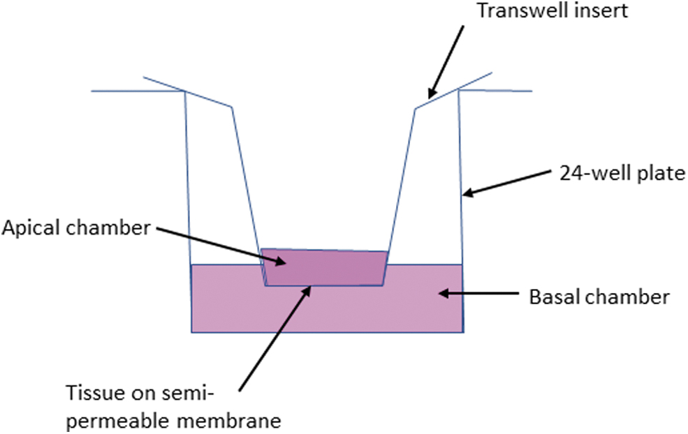
Schematic representation of the experimental procedure for resazurin reduction assay. Following TEER measurement at the 24-hour time point, MucilAir™ tissues were transferred to 24-well plates containing resazurin solution (6 μM in saline; 500 μL/well) and a further aliquot of resazurin solution was applied to the apical surface of the tissues (220 μL/well).
Histology
Following the resazurin reduction assays conducted for hazard screening purposes, tissues were fixed with 10% neutral buffered formalin for 24–48 hours, embedded in paraffin wax, then sectioned (two strips of membrane per block), stained with Hematoxylin and Eosin and mounted onto microscope slides. Slides/images were evaluated using a semiqualitative analysis by a board-certified veterinary pathologist and assessed on the extent and severity of cilia damage, degeneration, necrosis, and atrophy.
BMD modeling methods
A BMD modeling method was used to determine the POD for risk assessment endpoints from MucilAir data with the data generated in Experiment 2. To determine the appropriate statistical model to describe the data in this study, Hill and polynomial models were evaluated. Our assessments of model fit statistics strongly favored the Hill model based on the Akaike's information criterion (AIC) values. These values were determined from dose–response data simulated by randomly generating from a normal distribution with mean and standard deviation (SD) matching the historical dose–response data produced by the conducting laboratory. The Hill model produced the best fit for the data with lower AIC values (137 and 190) with four to five simulated replicates than the alternative cubic (polynomial of three degree) model with AIC values of 168 and 220 using the same number of replicates, respectively.
The Hill model was fitted of the following form to the dose–response data of each donor for each endpoint:
where the parameter γγ represents the baseline level of response for the control group and parameters v and k and n determine the change rate of the response with dose level, and the variance of the random error term ɛɛ (dose) is assumed to be a constant. In the BMD software, the power parameter n has a limit of 18 and by default <1.
The SD BMR is defined as the dose representing a change of one SD of the control group in the response and was used to derive BMD and the BMD lower limit of the 95% confidence interval (BMDL) values reported in this study. This approach is considered health protective in cases where a precise understanding of the quantitative biological linkage between two endpoints is not available and were generally lower than endpoints set on the basis of a no observed adverse effect dose level, indicating that the use of BMDL values as POD for risk assessment is a health-protective approach for deriving reference doses.17,31 The EPA BMD software (US EPA, version 2.6.0.1) was used for model fitting and calculating BMDs.
Results
Treatment with the vehicle control resulted in consistently high TEER, very low levels of LDH released, and tissue morphology characteristic of healthy tissues. In contrast, treatment with the positive control SDS resulted in a reduction in TEER to close to background/membrane-only levels, very high levels of LDH released into culture media, a notable reduction in resazurin reduction compared with the respective vehicle groups, and total loss of the membrane upon pathological evaluation. These responses confirm for both experiments of the MucilAir assay, the vehicle was not adversely impacting tissue condition, and the model consistently responded as expected to the respiratory irritant, SDS. Collectively, this confirms that the test system and these assays were performing satisfactorily.
Experiment 1: The use of MucilAir for hazard screening
Before dosing with Adepidyn (day-3), the mean TEER reading was similar across all concentrations tested and ranged from 519 to 582 Ω × cm2 (Fig. 3). Following dosing with Adepidyn at 24 hours, the mean TEER reading varied from 348 to 412 Ω × cm2 (Fig. 3). Although, modest reductions were observed in Adepidyn-treated groups (∼27%–38%), these reductions were similar to the reduction observed in vehicle control group (27%) and were not statistically significant due to variability across replicates/units (Fig. 3A). The mean percentage of LDH release, indicative of cellular cytotoxic membrane damage at 0 hour varied from 2.05 to 6.17. Adepidyn treatment at 24 hours, the mean percentage of LDH release was extremely low at each concentration (<4%) and decreased in a concentration-dependent manner (Fig. 3B). No histologic alterations were present in treated wells (data not shown).
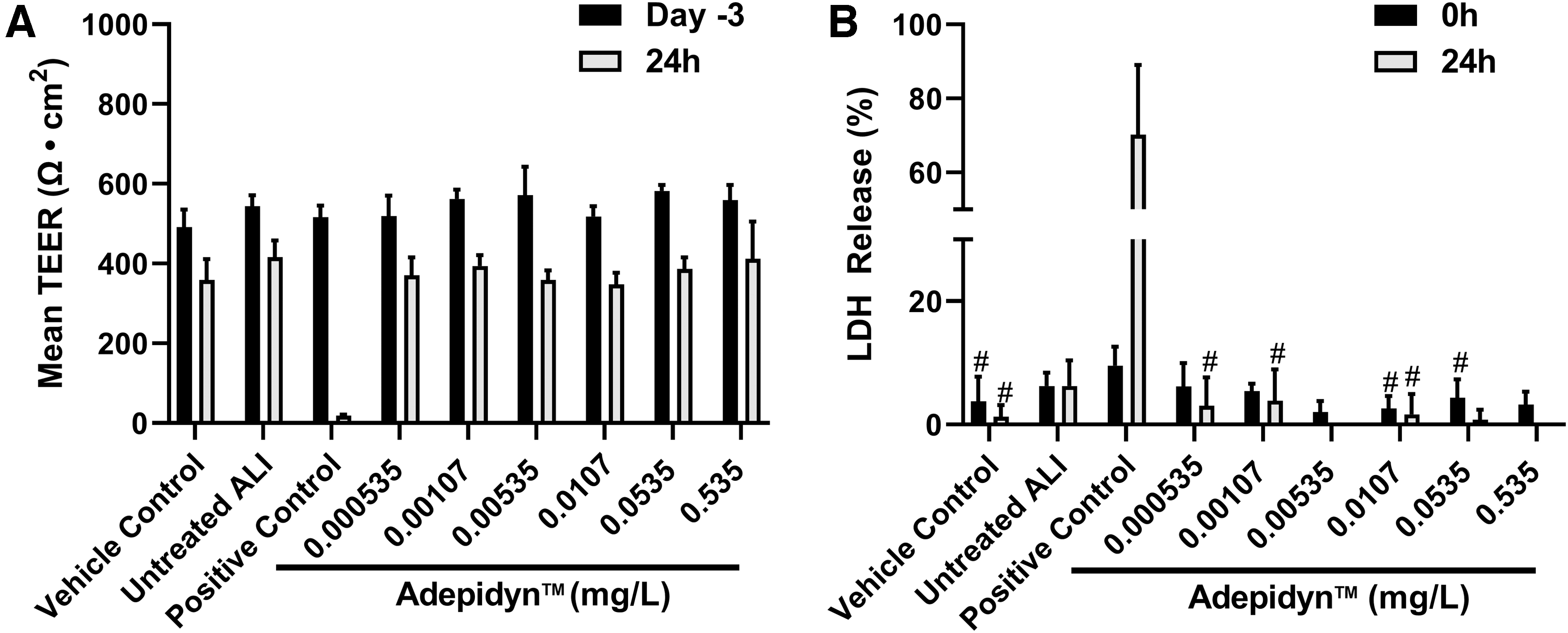
Effect of Adepidyn and controls on
The mean TEER readings observed were 386–482 Ω × cm2 before dosing with DC1. After 24 hours of dosing with DC1, TEER readings were not changed at any of the concentrations tested when compared with baseline measurements (Fig. 4A). The mean percentage LDH release (%) at 0 hour for DC1 concentrations were 0–0.26. At 24 hours, LDH release increased to values of 0.67%–1.81%. These values were a small percentage (5%) of the positive control at the same timepoint (Fig. 4B). The resazurin reduction response to 24 hours' treatment with DC1 responded in a similar manner to the vehicle control. These mean values ranged from 97% ± 26% to 124% ± 23% (Fig. 4C).
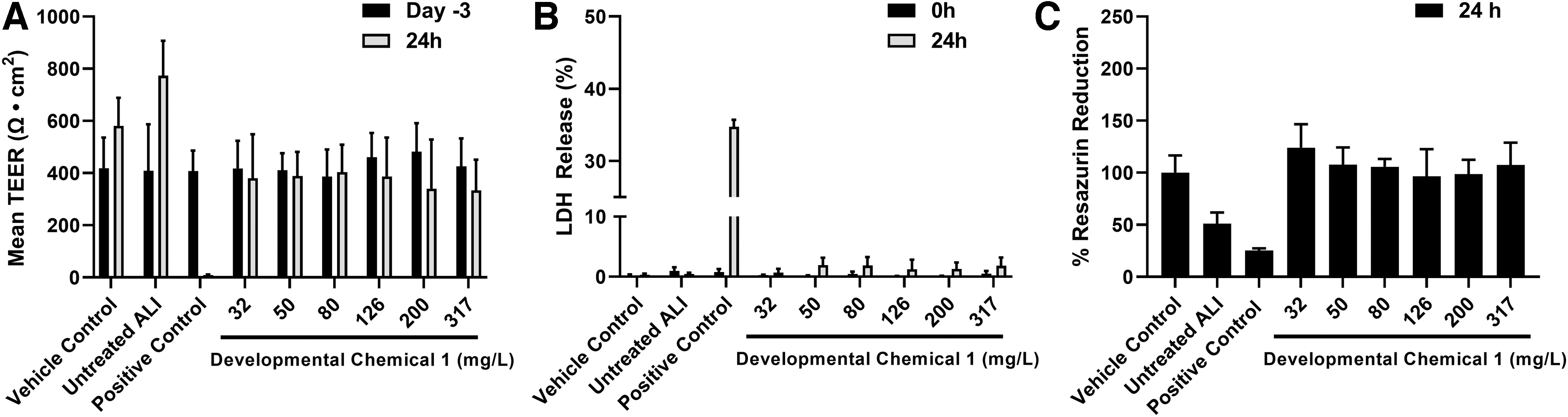
Effect of developmental chemical 1 and controls on
Before treatment, the mean baseline readings for TEER were 259–325 Ω × cm2 with similar variation observed during the Adepidyn and DC2 MucilAir assays. After dosing for 24 hours, minimal changes were observed in any of the DC2 treatment groups (Fig. 5A). At 0 hour, LDH release (%) ranged from 0.71 to 1.08 for the DC2 treatment groups. There was little to no change in LDH release at the end of the treatment period (24 hours) (Fig. 5B). The resazurin reduction response to DC2 treatment after 24 hours yielded no significant changes when compared with the controls. The mean resazurin reduction (%) was 79 ± 18–129 ± 14 (Fig. 5C). No histologic alterations were present in the treated wells. Based on these results, DC 1 and 2 induced no chemical-mediated damage at any of the tested concentrations.
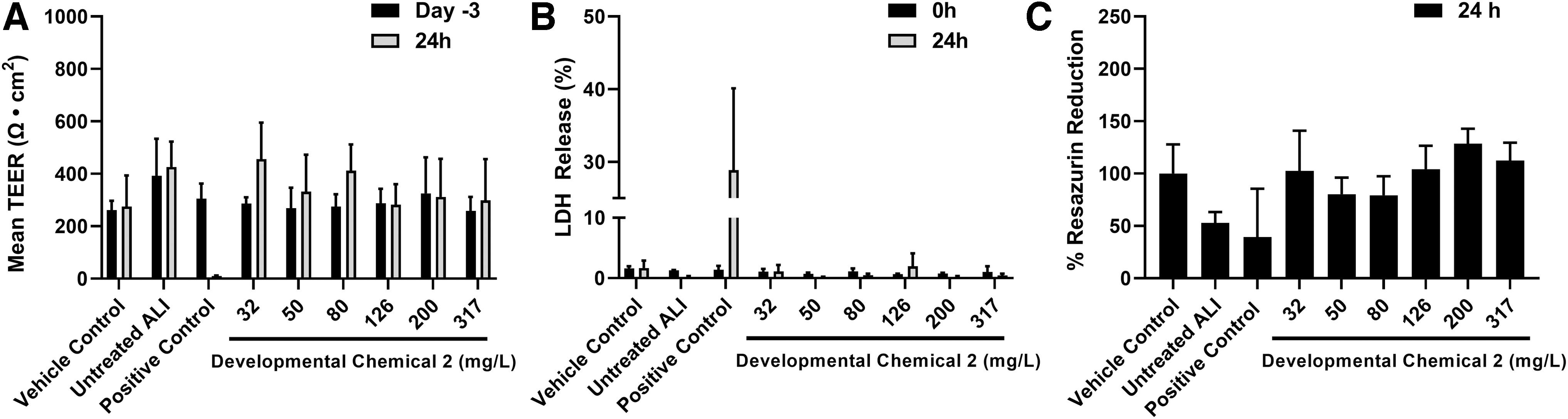
Effect of developmental chemical 2 and controls on
Experiment 2: The use of MucilAir to derive an endpoint for the inhalation risk assessment
Before dosing with Bravo 720 with or without CTN, the mean TEER reading for all donors was 2021 Ω. Tissues from all donors responded in a similar manner to negative control treatments. In all negative control tissues, a reduction in TEER (∼38%) was observed following 24 hours of incubation. In all positive control tissues, a considerable (ca 95%) reduction in TEER was observed following 24 hours of incubation. Each concentration decreased the mean TEER reading but did not reach statistical significance (Table 3).
Transepithelial Electrical Resistance, Lactate Dehydrogenase Release, and Resazurin Metabolism Across MucilAir in All Donors (n = 5 Total) Before and After Bravo 720 SC (24 Hours) Treatment
Mean and SD calculated from data where negative values have been corrected to zero.
SD, standard deviation.
The mean percentage LDH release for the donors varied from 0.71% to 2.40% of the appropriate positive controls. LDH release was low (<4%) at 0 hour for all donors. All tissues responded in a similar manner to negative control treatments and were within the normal range. In the positive controls, LDH release increased considerably (ca 200%) for all donors after 24 hours of treatment. All donors showed a dose-related increase in LDH release following challenge with Bravo 720 SC, with marked responses observed at CTN concentrations of 125.89 and 199.53 mg/L. The values increased from 0.84% and 1.09% to 16% and 169% (Table 3).
The resazurin reduction response to 24 hours treatment with Bravo 720 SC with CTN as a percentage of the negative control treatment followed a similar pattern for all donors. The percentage of resazurin reduction decreased in a concentration-specific manner. The most pronounced change in percentage resazurin reduction was observed in the two highest concentrations (125.89 and 199.53 mg/L). After positive control treatment, resazurin reduction was decreased to 0%–14% of the negative controls (Table 3).
The calculated BMDL values ranged from 40 to 125 mg/L with an overall geometric mean of 81 mg/L (Table 4). For comparison to in vivo deposited doses, the geometric mean was 0.00730 mg/cm2.
Chlorothalonil Benchmark Dose Lower Limit Values Calculated from MucilAir Data
BMDL, benchmark dose lower limit of the 95% confidence interval.
Discussion
The US EPA's Office of Pesticide Programs (EPA/OPP) has evaluated NAMs to supplement or replace traditional toxicity testing to improve hazard and exposure evaluations along with risk assessment practices following potential exposure to pesticides. The US EPA OPP's regulatory goal is to have sufficient information to support the safe use of crop protection products while avoiding the unnecessary use of resources associated with whole animal studies. 32 A part of this effort has been the incorporation of NAMs to determine if adequate knowledge is available to support a weight of evidence evaluation and allow waiving a traditional test guideline study.32,33 The use of a relevant human cell-based in vitro technology to address safety questions which may previously have required a live whole animal approach offers a scientifically robust alternative, consistent with commitments to reduce and refine animal testing. 34
The present study was conducted using MucilAir, a relevant human cell-based in vitro assay/NAM, for hazard screening to evaluate agrochemicals, including Adepidyn and two early stage/developmental chemicals (DC 1 and 2) for potential markers of irritation, including TEER, LDH release, resazurin reduction, and conducting histological evaluation. Furthermore, the MucilAir assay was also used to evaluate the potential for CTN to act as a cytotoxic surface irritant to the respiratory epithelium and to select a POD for human health risk assessment in lieu of using a whole animal study (OECD test guideline study 413) consistent with the US EPA's guidance on fulfilling data requirements. 33
Our results indicate that the MucilAir assay can be used for hazard screening for agrochemicals with the markers of irritation and histological evaluation confirming no evidence of toxicity at any concentration tested for Adepidyn and DC 1 and 2. This assay was also used for known respiratory irritant, CTN, through the quantitative evaluation of three endpoints/markers of effect (TEER, LDH release, and resazurin reduction) and identifying the BMD Level of 0.00730 mg/cm2 derived for CTN for use in calculating a human equivalent concentration for use in risk assessments. 29
Previous evaluations have demonstrated that 3D in vitro models of respiratory epithelium are able to predict in vivo respiratory irritancy.35,36 MucilAir is a functionally differentiated, 3D model of the respiratory epithelium derived from human nasal epithelial samples that secretes mucus and has electrically tight junctions. Nasal, tracheal, and bronchial lumens are all lined by ciliated pseudostratified respiratory epithelium with goblet cells. Nasal epithelium can be used as a surrogate for bronchial epithelium and is easier to obtain from nasal brushings and is the same cell type. Comparative studies between MucilAir nasal and bronchial epithelial sources have shown that respective responses to xenobiotics are equivalent. 37 This is also the case for functional assays such as transepithelial permeability of xenobiotics. 27 As such, the MucilAir model is considered to be representative of respiratory irritancy observed in vivo.
Inhalation of a sufficient concentration of a respiratory cytotoxic irritant can result in a necrotic and inflammatory injury to the epithelium. Repeated exposure to concentrations that cause a cytotoxic response can result in an altered epithelial differentiation to a more resistant tissue type, presenting histologically as squamous metaplasia. 38 This response is typically reversible and is considered an adaptive response to the repeated acute injury. 38 This similar acute cytotoxic response has been reported with other respiratory irritant chemicals, such as ammonia, formaldehyde, hydrogen sulfide, and glutaraldehyde.39–46 As necrotic injury to the airway epithelium is the key event in the adverse outcome pathway that will result in outcomes due to repeated exposure such as metaplasia, 38 prevention of this initial injury following acute exposure is protective of downstream effects from repeated exposure. Therefore, the ability to assess potential necrotic injury to the epithelium through the MucilAir model to agrochemicals is appropriate for hazard screening (Fig. 6).
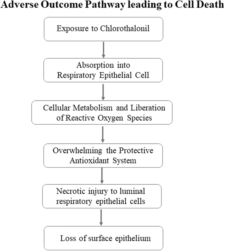
AOP illustrating exposure to CTN leading to cell death. AOP, adverse outcome pathway; CTN, chlorothalonil.
When assessing cytotoxic and irritancy potential, Adepidyn did not affect TEER and LDH release after 24-hour treatment. Histological evaluation further confirmed there was no evidence of toxicity at any concentrations tested. Screening for cytotoxic and irritancy potential of the two developmental agrochemicals, DC 1 and 2 included the standard battery of parameters (TEER, LDH, and histological evaluation) and evaluation of resazurin reduction. Similar to responses observed in Adepidyn, there was no chemical-mediated changes at any of the tested concentrations of DC 1 and 2. Testing Adepidyn and the two developmental chemicals demonstrated the MucilAir assay can be used as a hazard screening tool for assessing the potential of respiratory toxicity for developmental agrochemicals. The data derived from conducting the MucilAir assay for hazard screening helped to determine if any further in vivo testing would be needed and refine experimental design to identify respiratory hazards in the safety assessment of future agrochemicals.
The evaluation framework for NAMs for human health safety assessment presents three steps as determined by problem formulation. 10 The initial step, to be determined before following the framework, is establishing a problem formulation foundation. Building the foundations includes creating the problem statement, “a NAM can be developed and would be suitable to inform inhalation toxicity in lieu of a whole animal inhalation study.” Within the present study, the goals were to examine the MucilAir assay based on the evaluation framework in efforts to address the proposed problem statement and confirm the fitness of the method. Based on the application of the framework presented in Parish et al., the assay meets the fit-for-purpose criteria to address both hazard screening for developmental agrochemicals (Adepidyn and DC 1 and 2) and risk assessment (CTN). 29
TEER, LDH release, and resazurin reduction were evaluated in MucilAir tissues from five individual human donors to determine irritancy potential following treatment with Bravo 720 formulation containing CTN. The three endpoints responded similarly to CTN challenge, consistent with in vivo data (unpublished) demonstrating that CTN exhibits an irritant effect due to direct cytotoxicity in the respiratory epithelium. Small, but statistically insignificant, variations were observed for measured endpoints across donors, suggesting low interdonor variability in sensitivity to the effect. This is consistent with previous investigations of respiratory effects in humans suggesting differences in sensitivity are largely attributable to physiological, rather than histological, differences between individuals. 47 These endpoints were affected by CTN at similar concentrations consistent with previous work examining the same endpoints when challenged with respiratory irritants. 35 As MucilAir tissues were exposed to CTN for 24 hours, the MucilAir POD values derived in this study are considered to represent a worst-case measurement of respiratory toxicity and are therefore, health protective.
BMD modeling to determine the POD values for acute cytotoxicity in the respiratory tract has been previously used to evaluate inhalation toxicity data to determine reference exposure levels for ammonia and formaldehyde,48,49 methyl isocyanate, 50 as well as a number of other inhaled toxicants. 51 As described previously, an analysis of BMD methodologies has indicated the use of a BMD of one SD from control generally produces lower endpoints than those set on the basis of a no-observed adverse effect dose level, indicating that the use of BMDL values as POD for risk assessment is a highly health-protective approach for deriving reference doses. 17
Conclusion
In conclusion, the cytotoxic response to the respiratory epithelium can be assessed using the MucilAir assay, a three-dimensional model of the human airway epithelium formed from differentiated primary human cells. No evidence of irritancy was detected at any tested concentration for Adepidyn and developmental agrochemicals, DC1 and 2. Upon evaluation of CTN in the Bravo 720 formulation, a cytotoxic effect of the respiratory epithelium was observed based on both LDH and resazurin assays, consistent with in vivo data 29 in a prior report. Using BMD modeling of the responses provided a benchmark response that can be used as the POD to inform a human health risk assessment from inhalation exposure. The MucilAir assay evaluating chemically induced airway cytotoxicity is fit for purpose for both respiratory tract hazard screening and refining inhalation risk assessment of agrochemicals.
Footnotes
Acknowledgments
The authors thank Drs. Paul Mosquin and Donald Brambilla (RTI International, Research Triangle Park, NC), Dr. Clive Roper (Charles River Laboratories, Edinburgh, United Kingdom) in the conduct of this work, and Drs. Sheung Ng and Amber Goetz for article review.
Author Disclosure Statement
M.M.H., B.P.-D., D.C.W., and A.C. are employees of Syngenta Crop Protection, the developer and producer of CTN (including the Bravo 720 SC/A12531C formulation of CTN), developmental Compounds 1 and 2, and Adepidyn. Samuel Constant is an employee of Epithelix Sàrl, the developer and producer of the MucilAir system.
Funding Information
This study was funded by Syngenta Crop Protection, LLC.