
Abstract
Significance:
The endogenous hydrogen sulfide (H2S) pathway produces an array of biological effects that vary depending on the bodily region. In addition, the H2S pathway's relevance often changes depending on a healthy or disease state. There is abundant evidence pointing to a key role for this pathway in male and female genito-urinary diseases, suggesting it as a possible target for new therapeutic approaches.
Recent Advances:
The tissue-specific localization of the H2S enzymes in the genito-urinary tract has allowed for a better understanding of its role in the body's pathophysiology. Indeed, in humans, cystathionine-γ-lyase (CSE) plays a major role in corpus cavernosum whereas cystathionine-β-synthase (CBS) plays a role in bladder functioning. The prostate epithelium expresses CBS and CSE, but stromal CSE only. In the uterus, up- or downregulation of CBS and CSE varies strongly depending on the female's hormonal cycle or pregnancy.
Critical Issues:
There is still the need to better define the male and female's sexual hormonal roles in regulating the H2S pathway, particularly in human pathological conditions. The lack of a correlation between human and animal data should be carefully considered when planning preclinical studies. The unmet need for selective enzymatic inhibitors and the different methodologies for H2S measurements still represent a critical issue in this research field.
Future Directions:
It is feasible that the L-cysteine/H2S pathway can represent an alternative therapeutic target in genito-urinary tract disorders. The research should focus on erectile dysfunction and preeclampsia, characterized by vascular defect, as well as on bladder disorders where the urothelium is compromised. Antioxid. Redox Signal. 27, 654–668.
Penile Erection
P
ED is now defined as the consistent or recurrent inability of a man to attain and/or maintain a penile erection that is sufficient for sexual activity. It is now clear that endothelial dysfunction modifies the balance between vasodilating and contracting mediators that allows the passage from the flaccid to the erected status. To date, PDE5 inhibitors are the mainstay in the treatment of ED and a link between ED and cardiovascular diseases has been established as well. Indeed, several clinical studies have shown that ED is an early clinical sign of cardiovascular disease (34). Therefore, it is now widely accepted that if a patient has early signs of ED, and is a naive patient of cardiovascular pathology, he must be referred to a cardiologist (34). This theory relies on the hypothesis that, as penile artery caliber is smaller, compared with those of coronary arteries, ED often precedes the other relevant cardiovascular signs.
Hydrogen Sulfide and Penile Function and Dysfunction
Hydrogen sulfide and its role in penile pathophysiology
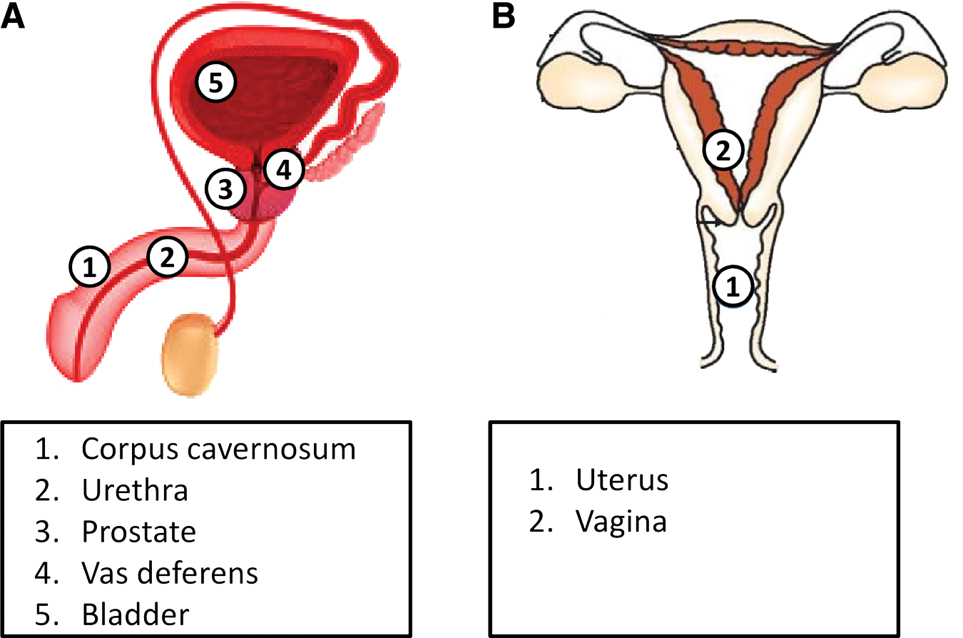
It is well established that hydrogen sulfide (H2S) plays a role in the regulation of vascular homeostasis (72). The presence of the H2S pathway in the male genito-urinary tract is summarized in Figure 1A. To date, the role of H2S in the corpus cavernosum (CC) function has been investigated in animal as well as human tissues (15, 22, 74). The first evidence, published by Srilatha et al., showed that the intra-cavernous injection of sodium hydrosulfide (NaHS) increases penile length and cavernous pressure in primates, which was measured by using a laser Doppler flow meter and cutaneous probe (62). All three enzymes involved in the H2S biosynthesis process, i.e. cystathionine-β-synthase (CBS), cystathionine-γ-lyase (CSE), and 3-mercaptopyruvate sulfurtransferase (MPST), are expressed and able to convert L-cysteine into H2S in mice (5, 73). The CC mice strips are responsive to either NaHS or L-cysteine (5, 73). Indeed, both L-cysteine or an H2S donor relax the CC mice strips in a concentration-dependent manner. The removal of endothelium as well as the blockade of CSE, even more than CBS, inhibits the L-cysteine-induced relaxation (5). Therefore, at the present stage, CSE-derived H2S appears to be the major player in mouse CC.
The same evidence applies to rats where CSE, but non-CBS, is expressed and H2S production is reduced by the CSE inhibitor (35). In addition, it has also been shown that CSE inhibition causes a reduction in perfusion pressure after cavernous nerve stimulation. This finding has led to the hypothesis that in rats the endogenous H2S plays a facilitating role in non-adrenergic-/non-cholinergic (NANC)-mediated penile tumescence (62). Regarding the effect of H2S donors, it has been proved that all cause the relaxation of CC tissues that are harvested from humans, rabbits, mice, and rats in an endothelium-independent manner (5, 21, 35, 61, 62). A possible link with the NO pathway has also been investigated. It has been shown that incubation with a nitric oxide synthase (NOS)- or a guanylyl cyclase (GC) inhibitor before the H2S donors' challenge, in mice CC strips, leads to an enhancement of the relaxant effect, implying that the NO/cyclic guanosine monophosphate (cGMP) pathway negatively modulates the effect driven by the exogenous H2S (5). In line with this finding, NO-mediated NANC nerve relaxation has been shown to be potentiated by CSE inhibition (35). There are at least two feasible explanations for this: (i) H2S can modulate NOS activity; (ii) a chemical reaction between NO and H2S can occur. Both hypotheses have been investigated (66) but at present, the molecular basis of this crosstalk has not yet been defined. Further and more detailed molecular and functional studies are required.
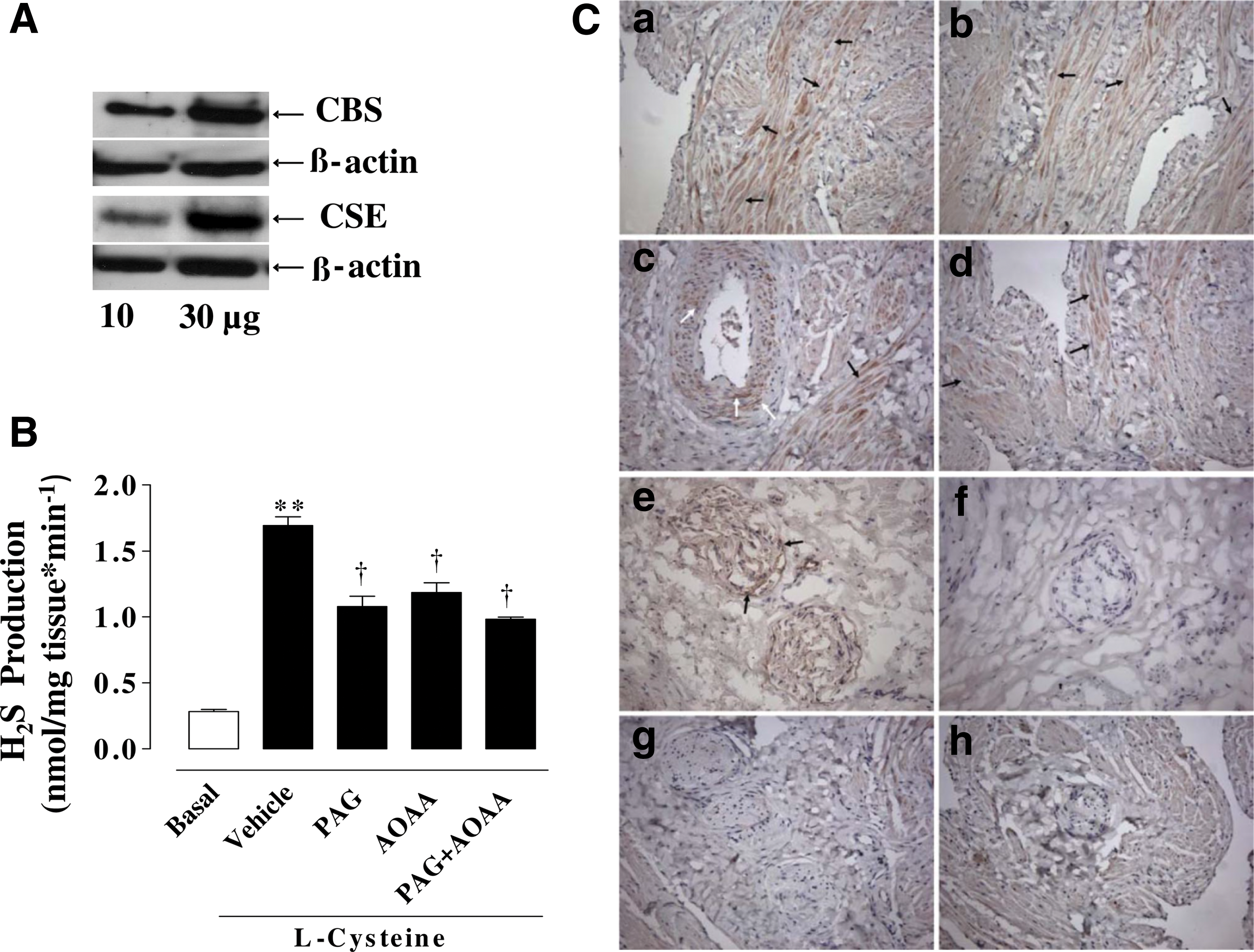
The first evidence of H2S involvement in human CC was published in 2009 (21). In this article, a functional role for the H2S pathway in human CC was demonstrated. The human CC expresses both CBS and CSE, the main enzymes involved in H2S biosynthesis. The two enzymes are localized within the muscular trabeculae and the smooth-muscle component of the penile artery. However, a more robust signal is reported for CSE as compared with CBS in these two compartments. Interestingly, immunohistochemistry studies have demonstrated that only CSE is expressed on peripheral nerves. Thus, CSE seems to be the enzyme that is mainly involved in the modulation of the H2S pathway in human CC function. The human CC tissue generates detectable basal amounts of H2S, and human homogenates can convert L-cysteine into H2S (Fig. 2). In vitro studies performed by using human CC strips clearly demonstrate that the H2S plays a functional role since both H2S donor (exogenous H2S source) and L-cysteine (the substrate) cause a concentration-dependent relaxation. In addition, experiments performed by using peripheral nerve electrical field stimulation (EFS) of human penile tissue show that the endogenous H2S contributes to penile homeostasis (21). Indeed, EFS of human CC strips under resting conditions causes an increase in tension that is significantly potentiated by inhibiting CSE and/or CBS. A role for H2S pathway in erectile function has also been confirmed in vivo, where an intra-cavernous injection of either NaHS or L-cysteine increases the intra-cavernous pressure in rats (21). The effects of the H2S pathway in different species and tissues are summarized in Table 1.
CC, corpus cavernosum.
H2S and potassium channels
ATP-dependent potassium channels (KATP) have been defined as one of the major H2S molecular targets (80). A role for KATP channels has also been defined in the H2S-relaxant effect in human CC (21). Generally, potassium (K) channels mediate relaxation in human, rabbit, and rat CC strips, thereby contributing to vascular penile homeostasis (51, 60). An impairment of the functioning of these channels has been shown in vitro by simulating the diabetic condition in human CC strips (68). The adenylyl cyclase (AC)/cyclic adenosine monophosphate (cAMP)/protein kinase A (PKA) signaling is the major molecular target of these channels (53), and studies performed by using rabbit CCs have demonstrated that the NaHS-relaxant effect can be inhibited by blocking AC (61). The same applies to mouse CC, confirming that there is an important role for cAMP as a downstream signal (5). In this same study, indirect proof of the involvement of KATP channels obtained by using gliblencamide, a KATP inhibitor, has been reported (5). Besides, it has also been defined that voltage-dependent K channel (KV) and inward rectifier K channel (KIR) but not big conductance calcium (Ca+2)-dependent K channel (BKCa) and small conductance Ca+2-dependent K channel (SKCa) are involved (5).
H2S and testosterone
Another important player in CC function is the male hormone testosterone. It is known that there is a link between KATP channels and testosterone (15). Indeed, the testosterone-relaxant effect on human CC strips involves KATP channels, but not BKCa, KV, or KIR, which are responsible for the endothelium-independent effect; whereas endothelium-dependent relaxation is ascribed to NO (4). By using rat aorta, it has been shown that the testosterone-relaxant effect is non-genomic, androgen receptor mediated and involves H2S as a final mediator. Of note, more recently, it has been reported that testosterone-induced relaxation involves the formation of a ternary complex between the androgen receptor, CSE, and heat shock protein 90 (8). Thus, H2S acts as a downstream signal after testosterone binding to the androgen receptor and it is likely that the same mechanism occurs in the highly vascularized CC. In other words, since aging in man is associated with lower testosterone levels, it is feasible to hypothesize that there is a consequent reduction in H2S levels that contributes to the impairment of erectile function. This hypothesis is further supported by Srilatha et al., who have shown that H2S deficiency is a biomarker for age-related ED, independent from NO (64). In this context, it is important to stress that H2S can also act as an endogenous inhibitor of PDE (10). This effect implies that H2S, by itself, can increase cGMP signaling, just as a PDE5 inhibitor does. Overall, these data suggest that there is a close link between aging, testosterone, H2S, and ED. Indeed, reduced testosterone levels are associated with a decline in H2S production, with a subsequent impairment in cGMP levels, thus leading to ED. In addition, the finding that PDE5 inhibition, produced by sildenafil, leads to an increase in H2S production in mouse CC strongly supports this hypothesis of the mechanism (23). Thus, in CC, H2S has at least two mechanisms: (i) inhibition of PDE and (ii) activation of KATP channels that can be modulated by testosterone after its interaction with the androgen receptor. Further studies are needed to better define the physiopathological significance of this interaction and how this positive feedback is switched on or off. However, a consideration that can be made by looking at the available data is that in CC exists an interplay between H2S and NO/cGMP and that the on/off of signaling may be reciprocally regulated and influenced by testosterone. In the literature, evidence exists proving that an interplay between the NO and the H2S pathway does exist. For example, H2S in rat CC can enhance endothelial NOS (eNOS) expression, increasing the NO formation (50). In line with this finding, it has been reported that H2S activates PI3K/Akt and increases eNOS phosphorylation in vasculature (66). This mechanism may also occur in penile vasculature but so far, there is only one study that has partially addressed this issue, showing a compensatory role for the H2S pathway (73). Indeed, by using pharmacological approaches and mice genetically ablated for eNOS (eNOS−/−), it has been demonstrated that the lack of eNOS-derived NO causes an increase in L-cysteine-induced relaxation in mouse CC as well as an upregulation of MPST and CSE expression, but not CBS (73).
All these findings suggest a clinical relevance for H2S signaling and the possibility that this pathway represents a therapeutic target in ED patients with diseases that are associated with endothelium dysfunction such as diabetes, hypertension, and metabolic syndrome. Notably, in rat CC smooth muscle, a reduction in H2S production has been demonstrated that is ascribed to a downregulation of cysteine aminotrasferase/MPST and d-amino acid oxidase/MPST, as well as to low activities of CBS and CSE in diabetes (77).
An altered expression of CSE and changes in H2S levels have been shown in acute inflammation, atherosclerosis, diabetes, hypertension, hyper-homocysteinemia, and obesity, which are other pathological conditions that are often associated with ED (16, 17, 25, 70, 76, 78).
H2S, Prostate, and Vas Deferens
The presence and role of H2S in vas deferens and prostatic tissue have been reported (Fig. 1A). The vas deferens transports sperm from the epididymis to the ejaculatory ducts and, therefore, has an important function in the reproduction process. Both CBS and CSE enzymes have been found in man as well as in rat and mouse vas deferens (46). Furthermore, it has been shown that H2S (NaHS) causes in vitro vas deferens smooth muscle relaxation that is reversed by iberiotoxin or tetraethylammonium. This result indicates the involvement of BKCa, also suggesting a redox-mediated mechanism (47). The same authors have excluded the involvement of a transient receptor potential (TRP), KATP channels, and NOS in NaHS-induced relaxant effect (47).
H2S signaling has been found in human prostatic tissues as well as in cell cultures (39). The presence of the H2S pathway has been confirmed by using biopsies from cancer-free human prostates (33). Guo et al. have demonstrated that both CBS/CSE protein levels and enzymatic activities are higher in the androgen-dependent prostate cancer cell LNCaP than in the other cell lines (39). In addition, CBS/CSE expression in human prostate stromal and epithelial compartments has been assessed. In particular, prostate epithelium expresses both CBS and CSE, as opposed to stromal where only CSE is expressed (39). Notably, the researchers also found that dihydrotestosterone downregulates CBS and CSE expression in the androgen-dependent prostate cancer cell LNCaP. These results may support the finding that testosterone binding to the androgen receptor involves the H2S pathway. MPST has been found in human prostatic tissue (33, 39), whereas Zhao et al. showed that MPST is not expressed in both human prostate adenocarcinoma and normal prostate tissue (79). This difference may be due to the detection methods employed. In addition, it was also demonstrated that the expression of CSE, but not CBS, is reduced in prostate cancer tissue when compared with the control (79). In the same study conducted in an in vitro model of anti-androgen-resistant prostate cancer cells, the CSE expression is lower when compared with the parental cell line, with a parallel reduction in H2S production. The key role for CSE was supported by the finding that the aging CSE knock-out mice show a higher cell proliferation rate in prostate tissue, with a concomitant reduction of 80% in H2S production (79). Thus, the CSE/H2S system may represent either a diagnostic tool or a therapeutic target for the treatment of prostate cancer.
H2S and Female Sexual Function
Few data are reported on the possible involvement of the H2S pathway in female sexual physiology. The presence of the H2S pathway in the female genito-urinary tract is schematized in Figure 1B. In 2009, a pilot study was published suggesting that the H2S pathway plays a physiological role in the female sexual apparatus. The authors showed the effect of exogenous H2S in vaginal and clitoral cavernosal smooth muscle strips of rabbits. By using H2S donors and specific inhibitors, it was shown that the H2S vasodilatory effect involves cAMP, NO, cGMP, and KATP channels. Notably, the inhibition of H2S-induced relaxation is observed only when a combination of both AC and GC inhibitors is used, suggesting that both nucleotides may concur to H2S effect (63). Thus, the H2S pathway seems to be involved in female sexual function as well and it appears that in women, the link between H2S and the NO pathway is even closer, suggesting a clear gender difference. There is a lack of data on the role of female hormones in H2S pathways in sexual function. However, an attempt to demonstrate the link between estrogen (ERT) and the H2S pathway in vasculature has been recently published (45). In fact, it has been demonstrated that ERT stimulates the biosynthesis of H2S in the uterus and mesenteric artery, specifically by upregulating CBS expression. Unraveling the molecular basis of the crosstalk between NO and H2S and the role of female hormones may help to define the still unknown and complex aspects of female sexual dysfunction.
H2S and Bladder Function and Dysfunction
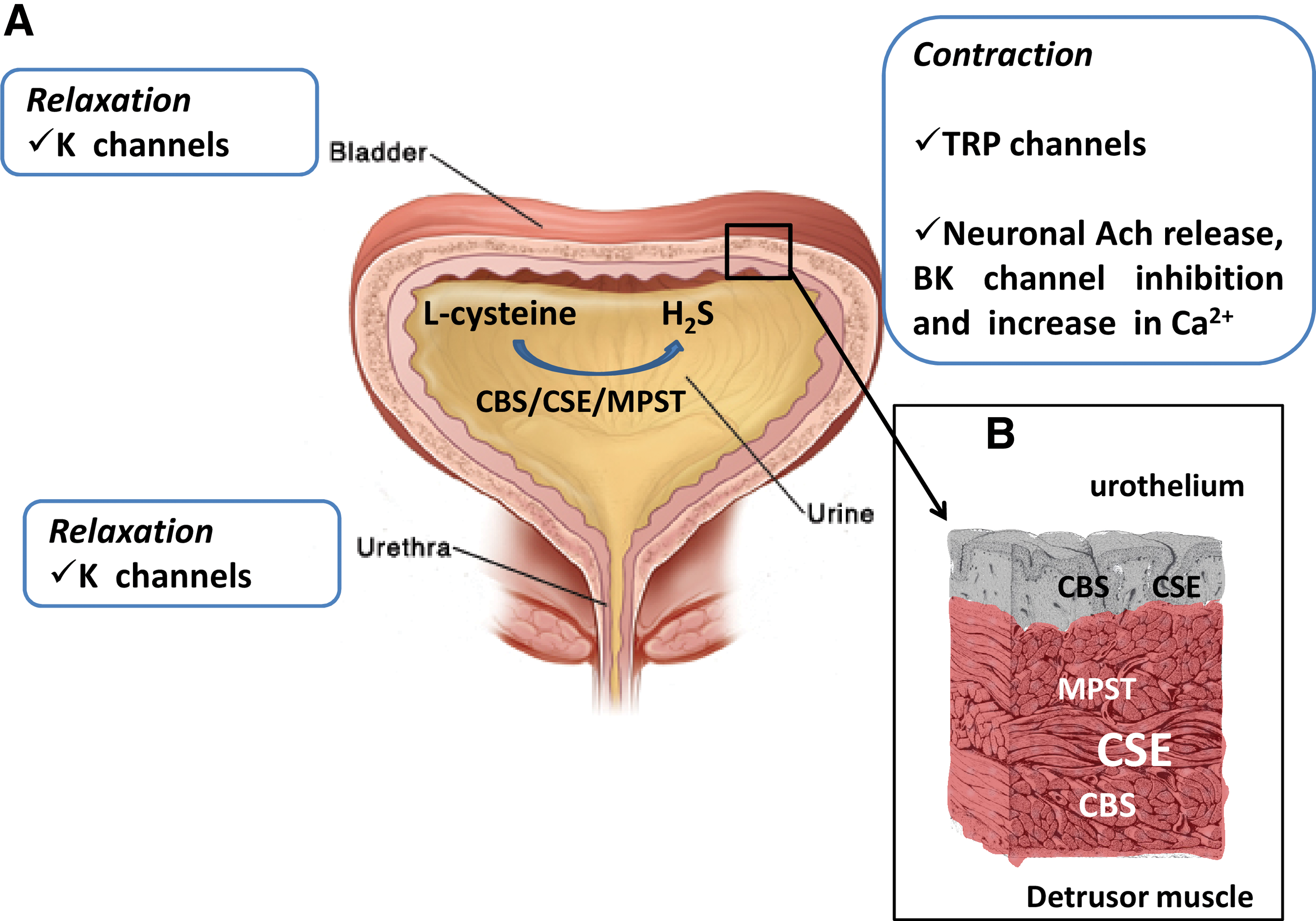
The urinary bladder has two functions: (i) the storage of urine that requires the bladder to relax and fill with an increase in intravescical pressure and (ii) voiding, which needs contraction of the detrusor muscle with concomitant relaxation of the bladder outlet and urethra (1). The bladder's tone is finely controlled by an interaction between the somatic, parasympathetic, and sympathetic nervous systems. Several signaling molecules contribute to control bladder homeostasis, acting in either an autocrine or a paracrine manner. Many neurotransmitters, including acetylcholine, norepinephrine, dopamine, serotonin, excitatory and inhibitory amino acids, adenosine thriphosphate, NO, and neuropeptides, are involved in the regulation of micturition (14). H2S is endogenously produced in the urinary bladder of trout, mice, pigs, rats, and humans (24, 27, 31, 33, 49). The three enzymes CBS, CSE, and MPST involved in H2S biosynthesis are expressed in the rat bladder. CBS and CSE are expressed in the human bladder, but a contribution of MPST in humans cannot be excluded (31, 33). In the mouse bladder, up to now, only the CSE presence has been shown (49). Overall, it is clear that H2S is endogenously produced in the urinary bladder and that the enzymes responsible for its biosynthesis are constitutively present. Indeed, homogenates of bladder can generate detectable amounts of H2S in basal or stimulated conditions in different species, including humans (24, 27, 31, 33, 46). The H2S response in the urinary bladder seems to vary depending on the species and the experimental conditions used. NaHS (an exogenous source of H2S) or L-cysteine (the substrate) relaxes rat and human bladder strips in a concentration-dependent manner (31, 33). The effect and the enzyme localization (urothelium/detrusor) are summarized in Figure 3.
H2S and ion channels
In trout bladder, NaHS or Na2S inhibits spontaneous contractions and relaxes pre-contracted strips (24). In pig ureter and bladder neck, H2S induces relaxation via KATP-channel activation, favoring the release of sensory neuropeptides (27, 28). Of note, the relaxing effect reported in the pig's bladder neck suggests a contribution of H2S in the urine outflow as well. The involvement of KATP in the H2S-relaxing effect has been reported in rats and the human bladder (33) but not in trout (24). In this context, it is important to note that KATP and, more generally, K channels have been recognized as therapeutic targets for overactive bladder, lower urinary tract symptoms (LUTS), and other urological diseases (37).
A contracting effect of H2S has recently been demonstrated on the detrusor muscle in guinea pig, and a novel mechanism of H2S-induced bladder contraction has been proposed (30). Indeed, the H2S donor GYY4137 increases the in vitro spontaneous phasic and nerve-evoked contraction of the detrusor smooth muscle and these effects are reduced, respectively, by blocking neuronal voltage-gated Na+ channels or by antagonizing the muscarinic (M) receptor (30). In the same study, a contribution of L-type Ca-dependent channels and BK channels has also been shown (30). Thus, it has been proposed that H2S promotes neuronal acetylcholine release from the bladder nerve in a Ca+2-dependent manner and, in corollary, H2S inhibits BKCa channels, causing membrane depolarization, leading to detrusor muscle contraction.
In line with this evidence, Patacchini et al. show a contractile effect induced by H2S in the rat urinary bladder (54). The authors report the involvement of the sensory neurons in a ruthenium red-sensitive but not capsazepine-dependent manner in the H2S effect (54). Although the H2S molecular target on rat sensory neurons remains to be further clarified, the involvement of: (i) the transient receptor potential vanilloid receptor 1 (TRPV1) at a domain independent from those bound by vanilloids, (ii) a ruthenium red-sensitive TRP cation channel co-expressed with TRPV1 on primary afferent neuron terminals, and (iii) the transient receptor potential ankyrin 1 (TRPA1) have all been suggested (54, 65). Therefore, the TRP channels family may play a key role in the mechano-sensory transduction of the urinary bladder.
The TRP family consists of 28 channels that can be subdivided into six different classes: TRPV (vanilloid), TRPC (canonical), TRPM (melastin), TRPP (polycystin), TRPML (mucolipin), and TRPA (ankyrin) (26). TRPV1 seems to regulate the frequency of bladder reflex contractions, either through direct excitation of sensory fibers or through urothelial-sensory fiber crosstalk involving the release of neuromediators from the urothelial cells (3, 7). The expression of TRPA1 on bladder C-fiber afferents and urothelial cells together with the finding that intravesical TRPA1 activators elicit contraction strongly suggest a key role for TRPA1 in sensory transduction in the bladder (14). Of particular interest is the finding that organ sulfur compounds, that is, allyl isothiocyanate or allicin that are H2S donors, are also TRAP1 activators. These organic H2S donors can contract a rat's urinary bladder, and their mechanism of action involves stimulation of sensory neurons with a subsequent release of neuropeptides and prostanoids (2). However, it is also important to underline that polysulfides are able to activate TRPA1 channels more effectively than H2S in rat astrocytes (44). The authors assume polysulfides as possible H2S-derived bioactive molecules that stimulate TRP channels. This is a relevant consideration for a future perspective study that could provide new insights into the biology of H2S and therapeutic development in bladder diseases involving these substances. Moreover, further studies are necessary to better define how cells differentially utilize H2S and polysulfides.
A role for the H2S pathway has also been shown in the ureter. An involvement of the TRPA1 receptor has been demonstrated by using a pig intra-vesical ureter (29). Indeed, the antagonist of either TRPV1 or TRPA1 reduces both H2S- and EFS-induced ureter relaxation, also implying that endogenous H2S contributes to the homeostasis of the ureter tone (29). This hypothesis is supported by the finding that the EFS-induced relaxation in ureter strips is significantly reduced by CSE (29). Interestingly, CBS inhibition did not affect EFS-induced ureter relaxation (29). This latter result is in line with the literature findings describing a greater expression of CSE in the ureter, suggesting that CSE-derived H2S plays a major role in the regulation of ureter tone. Taking into account all this evidence, it is feasible to hypothesize that H2S can act as a TRPA1 and/or TRPV1 activator and could be involved in the physiopathology of urinary disorders. However, to date, no clear evidence exists showing the involvement of TRPA1 and/or TRPV1 in the human urinary bladder, as opposed to experimental animal models that are reviewed herein. The only study conducted in the human urinary tract showing the involvement of TRPA1 has been performed on the urethra. The urethra, together with the smooth muscles within the bladder, controls the storage and the voiding of urine. This functional study demonstrates that H2S does not affect the basal tone but, instead, relaxes the pre-constricted human urethral strips and the effect involving TRPA1 (38). The presence of all the three enzymes synthesizing H2S CBS, CSE, and MPST has been reported in both human and rat urethra (33).
H2S and cGMP
Recently, a link among the H2S pathway PDE, cGMP, and cAMP in the human bladder has been found (31). Sildenafil directly relaxes human bladder strips, and this effect is accompanied by an increase in H2S production. The involvement of the H2S pathway in the sildenafil effect is further suggested by the finding that H2S production and the relaxant effect in vitro are reduced by blocking CBS and CSE. Interestingly, in this same article, it was shown that this effect is mimicked by the incubation of human samples with the stable analogue of either cGMP or cAMP, that is, 8-bromo-cGMP (8-Br-cGMP) and dibutyril-cAMP (d-cAMP), respectively (31). The fact that cyclic nucleotides cause an increase in H2S formation has been deeply investigated by using human urothelium and a urothelial cell line (18).
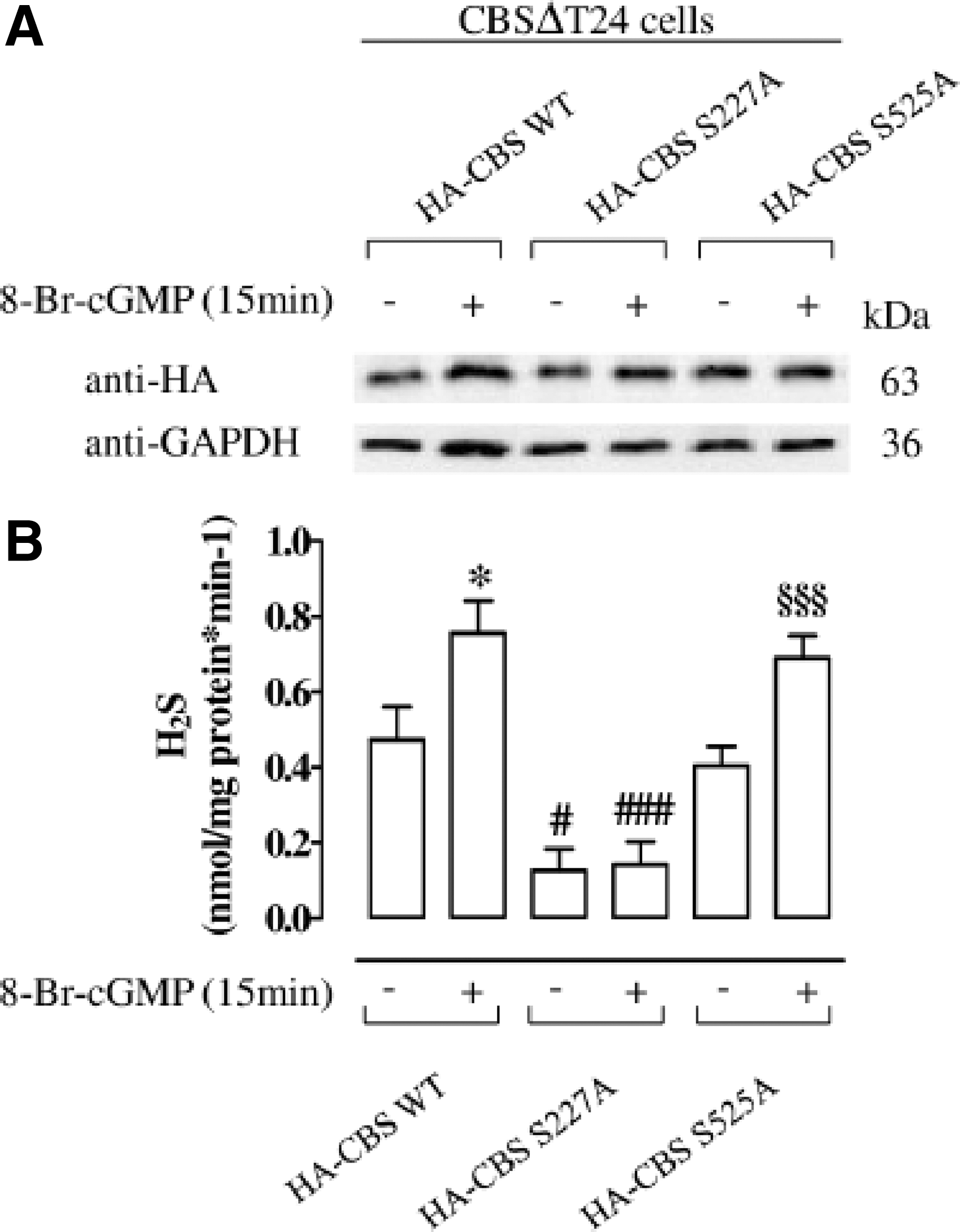
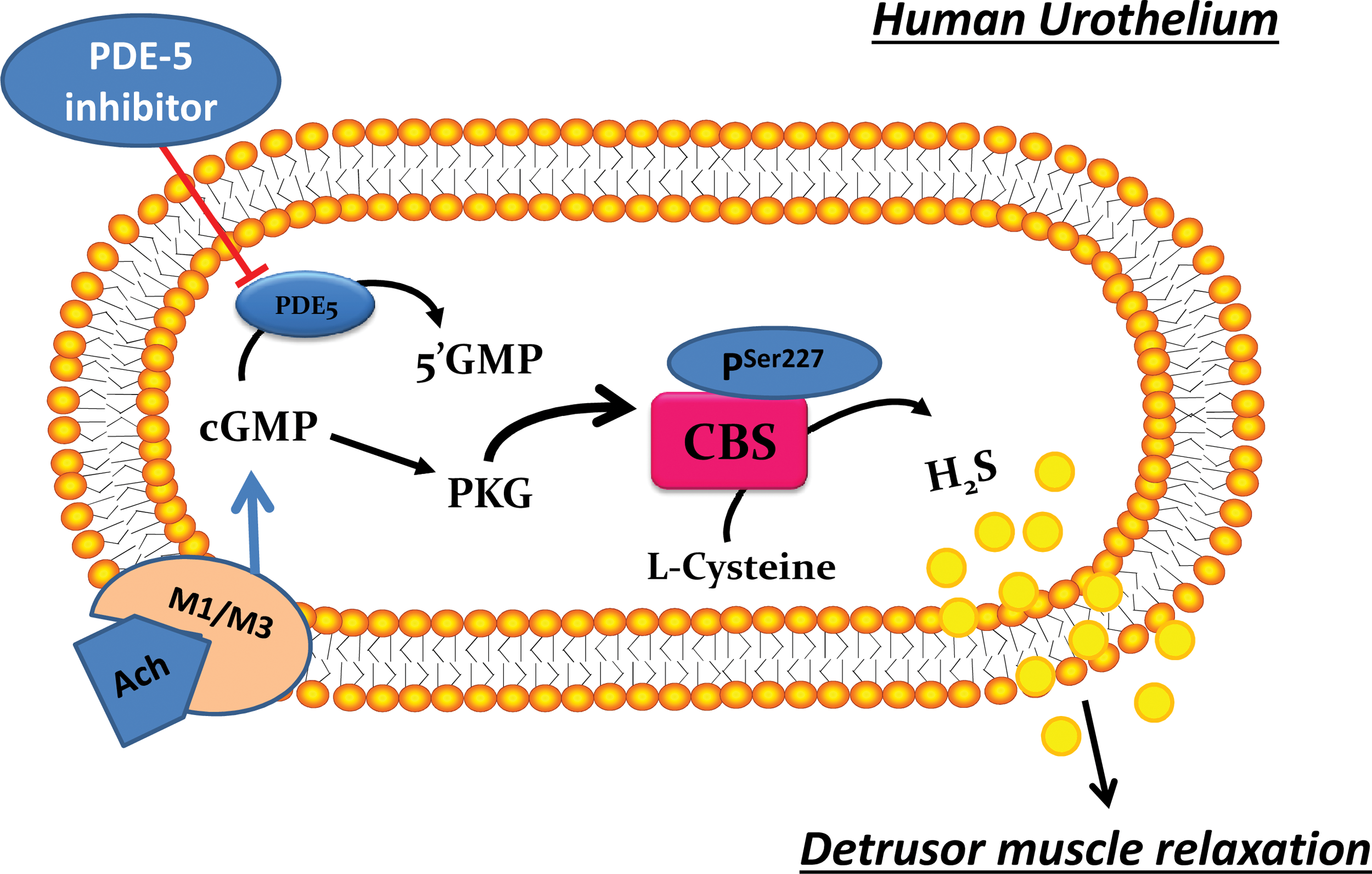
The internal face of the detrusor smooth muscle wall of the urinary bladder is covered by the urothelium, separating muscle cells from the urine. It is now well established that the urothelium is more than a passive barrier with very low permeability since it offers a sensory function that controls the degree of bladder filling and composition of the urine (6). Thus, the urothelium dynamically participates in controlling urinary bladder tone. On this basis, some years ago, the presence of an un-identified urothelium-derived relaxing factor (UDRF) was postulated (41). The human urothelium expresses both CBS and CSE, both of which are functional. Indeed, homogenates of human urothelium are able to convert L-cysteine into H2S. Therefore, H2S released by the urothelium exerts its action on the underlying detrusor muscle, contributing to bladder homeostasis. This key role for urothelium-derived H2S is supported by the finding that carbachol-induced contraction in bladder strips with urothelium is significantly less pronounced as compared with that obtained in its absence. So far, the nature of UDRF has not yet been identified. On this basis, it is feasible that H2S acts as a UDRF. Indeed, the carbachol-induced contraction in the presence of urothelium in the human bladder strip is enhanced by the H2S pathway blockade (19). Cellular studies performed by using the human urothelial cell line T24 further support this role for H2S. This cell line expresses both CBS and CSE to the same extent as does the human urothelium (18). Their stimulation in vitro with the stable analogous of cGMP, for example, 8-Br-cGMP, but not with the stable analogous of cAMP, for example, d-cAMP, causes a time-dependent increase in H2S production. The lack of effect of d-cAMP is due to the almost total absence of the PKA expression in the human urothelium as well as in T24 cells. The increase in H2S production induced by the stable analogous of cGMP is abrogated by protein kinase G (PKG) inhibition. This increase has been shown to be driven through phosphorylation at a specific site of CBS, that is, at serine227 (pCBSser227) in a PKG-dependent manner (18). This phosphorylation has also been shown by using human bladder biopsies (18). The phosphorylation of CBS shifts the enzyme in a more active status, causing an increase in H2S production (Fig. 4). The key players in bladder function are the M receptors. A link between M and CBS phosphorylation has also been demonstrated (19). The presence of M receptors in the human urinary bladder has been well described in different areas of the bladder, including the urothelium (9). Although the M receptors' downstream signal in the detrusor muscle is well defined, its role in the urothelium is still unclear. It has been recently shown that the activation of the M receptor, in particular M1 and M3, in the human urothelium, involves H2S as a downstream signal (19). In particular, it has been demonstrated that cGMP is formed on M stimulation and through the PKG phosphorylates CBSser227, leading to an increased formation of H2S. This evidence may also have clinical relevance since changes in the expression of the M receptors driven by pro-inflammatory stimuli have been described in urothelial cells (36). The link between cyclic nucleotides and the H2S pathway in the human urothelium is shown in Figure 5. Overall, the results of these studies may help to give further insights into the mechanism of action of two drugs, for example, tadalafil and mirabegron, which have been recently approved by the FDA for use in LUTS. Tadalafil is a PDE5 inhibitor that is being currently used in ED, whereas mirabegron is a selective β3 agonist. Both drugs can elevate cyclic nucleotide levels (cAMP or/and cGMP), and, therefore, it is feasible that the H2S pathway could be involved in their mechanism of action. Defining this issue may help to better understand the mechanism of action as well as additional molecular targets that have been described as being engaged by H2S. Gai et al. report that CBS, CSE, and MPST are present in the bladder specimens of patients who are affected by urothelial cell carcinoma. In addition, they found the highest expression of CBS, CSE, and MPST and, in parallel, the highest H2S production in the carcinoma urothelial cell line that fully correlated with the malignant degree (32). This finding may suggest H2S as a feasible diagnostic marker, but further studies need to better clarify its role in urothelial carcinoma in humans.
H2S and the Uterus
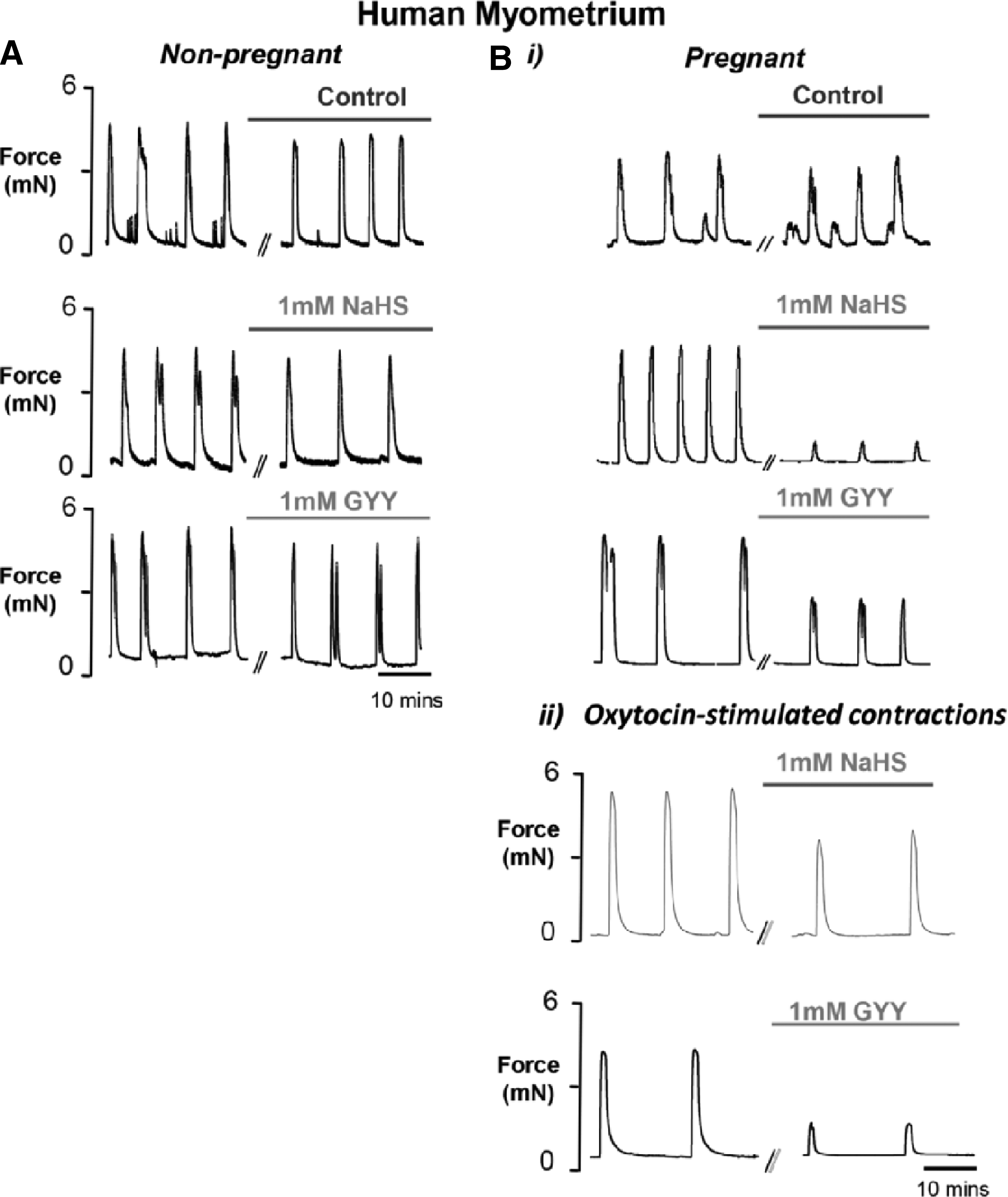
The myometrial contractility during estrus, pregnancy, and parturition is controlled by several complex events. The mediators and signaling molecules involved are different and often exert opposite activities depending on the status of the uterus. The involvement of the H2S pathway was first investigated by Sidhu et al. The authors demonstrate that L-cysteine and NaHS produce significant concentration-dependent decreases in a rat uterine's spontaneous contractility, suggesting H2S as a novel tocolytic agent. Indeed, the effect is defined as specific since other related amino acids such as D-cysteine, L-serine, DL-methionine, and DL-homocysteine are ineffective (58). The authors did not investigate the mechanism of action that would be addressed by studies published several years later. At present, it has been demonstrated that H2S can be endogenously produced and the enzyme expression of CBS and CSE has been reported in both rat and human uterine tissues. By means of Western blotting, the expression of CBS and CSE has been detected in all rat intrauterine tissues, as well as in human placenta, myometrium, amnion, and chorion. The same authors also demonstrate that the enzymes efficiently convert L-cysteine into H2S (55). A contribution to the understanding of the functional role of this gaseous transmitter in the uterus' contractility in relation to the status (non-pregnant/pregnant) has been given by You et al. In this study using immunohistochemistry, it was demonstrated that CSE and CBS are mainly localized in smooth muscle cells of human pregnant myometrium biopsies obtained from pregnant women undergoing cesarean section at term. Moreover, the mRNA and protein expression of CBS, as well as the H2S production rate, is downregulated in laboring tissues when compared with non-laboring tissues. Interestingly, the authors demonstrate the ability of L-cysteine to decrease in the amplitude of spontaneous contractions in both non-laboring and laboring myometrium strips. This effect is more pronounced in non-laboring strips. On the other hand, L-cysteine at a high concentration (10−3 M) increases the frequency of spontaneous contractions and induces tonic contractions. This evidence has led the authors to hypothesize that, since H2S synthetic enzymes are downregulated during labor, H2S could be one of the factors involved in the transition of pregnant uteri from quiescence to contractile state after the onset of parturition (75). The effect of GYY4137, a synthetic H2S donor, on spontaneous and oxytocin-stimulated contractility is evaluated in human and rat myometrium, throughout gestation, and in non-pregnant condition compared with NaHS (57). In human pregnant tissue, GYY4137 and NaHS significantly reduce oxytocin-stimulated and high-K depolarized contractions as well as spontaneous activity (Fig. 6). The authors, to reach an effective and lasting concentration of H2S, used a high concentration of NaHS (1 mM) since it is fast volatilized and oxidized. Ion channels have been widely reported as H2S targets on smooth muscle and which type of channel is involved is a matter of a specific district and organ considered. In particular, direct effects on Ca2+ entry via L-type Ca+2-dependent channels and K channels contribute to the relaxant effects of H2S. Indeed, the same authors provide evidence that the effect observed on the myometrium is mainly due to an interaction with Ca+2- instead of K channels. In addition, the H2S inhibitory effects increase as gestation advances, but they are abruptly reversed in labor. These data confirm that H2S contributes to uterine quiescence from mid-gestation until labor, mainly through L-type Ca+2-dependent channels by reducing Ca2+ entry and thereby myometrial contractions (57). Thus, this pathway represents an attractive target for a therapeutic approach for disorders of myometrial contractility.
The development of drugs that are able to increase or decrease the H2S contribution, depending on the status of the uterus and/or on the trimester of the pregnancy, is of clear interest in this field of research. In this regard, it has been demonstrated that sildenafil relaxes rat and human myometrium during preterm labor but the underlying mechanism is still unclear (11). The involvement of H2S in sildenafil's effect has been recently demonstrated by using mouse uteri. Indeed, it has been shown that sildenafil significantly increases H2S production in mouse uteri, and this effect is abrogated by CBS or CSE inhibition. In parallel, L-cysteine, NaHS, or sildenafil, but not D-cysteine, reduces spontaneous uterus contractility in a functional study. Inhibition of either CBS or CSE reduces this latter effect, and a major involvement of CSE rather than CBS has been recognized. This latter evidence is confirmed by experiments performed by using tissues harvested from CSE−/− mice, where the increase in H2S production mediated by L-cysteine or by sildenafil is blunted. Evidence for a crosstalk between H2S and NO has also been found in this specific tissue/organ since sildenafil's effect on the increase in the cGMP level is significantly reduced by CSE inhibition (52).
Sildenafil and other PDE inhibitors have been widely used in therapy in humans for many years, and their safety has been extensively demonstrated. Therefore, the therapeutic advantage of using this class of medication also in woman who serves as a tocolytic agent could possibly be exploited. The effect of the H2S pathway on the uterus' contractility depending on the status and the species is summarized in Table 1.
Another important issue to highlight is that a major cause of preterm labor in pregnant women is the intra-amniotic infections mediated by inflammatory processes. The role of H2S in inflammatory responses has been widely studied. The available data highlight a physio-pathological role in either promoting and/or resolving inflammation, depending on the status (physiological vs. pathological) and on the district/organ involved (16, 20, 76). The role played by H2S in infectious preterm birth has been studied in a mouse model of lipopolysaccharides (LPS)-induced preterm birth. LPS significantly increases leukocyte infiltration in the uterus, stimulating the expression of interleukin 1β (IL-1β), IL-6, tumor necrosis factor α (TNF-α), chemokine (C-C motif) ligand 2 (CCL2), and chemokine (C-X-C motif) ligand (CXCL15), and in the myometrium. Interestingly, administration of an H2S donor as NaHS delays the onset of the labor induced by LPS in a dose-dependent manner. NaHS prevents leukocyte infiltration into intrauterine tissues, inhibits the production of pro-inflammatory cytokines in the myometrium, and decreases the levels of these cytokines in maternal circulation. These effects have been ascribed to the signal regulation of extracellular signal-regulated kinases (ERK) and the nuclear factor kappa-light-chain-enhancer at the activated B cells (NF-κB) level. Thus, H2S can be considered a potential novel therapeutic target in infection-related preterm labor (48).
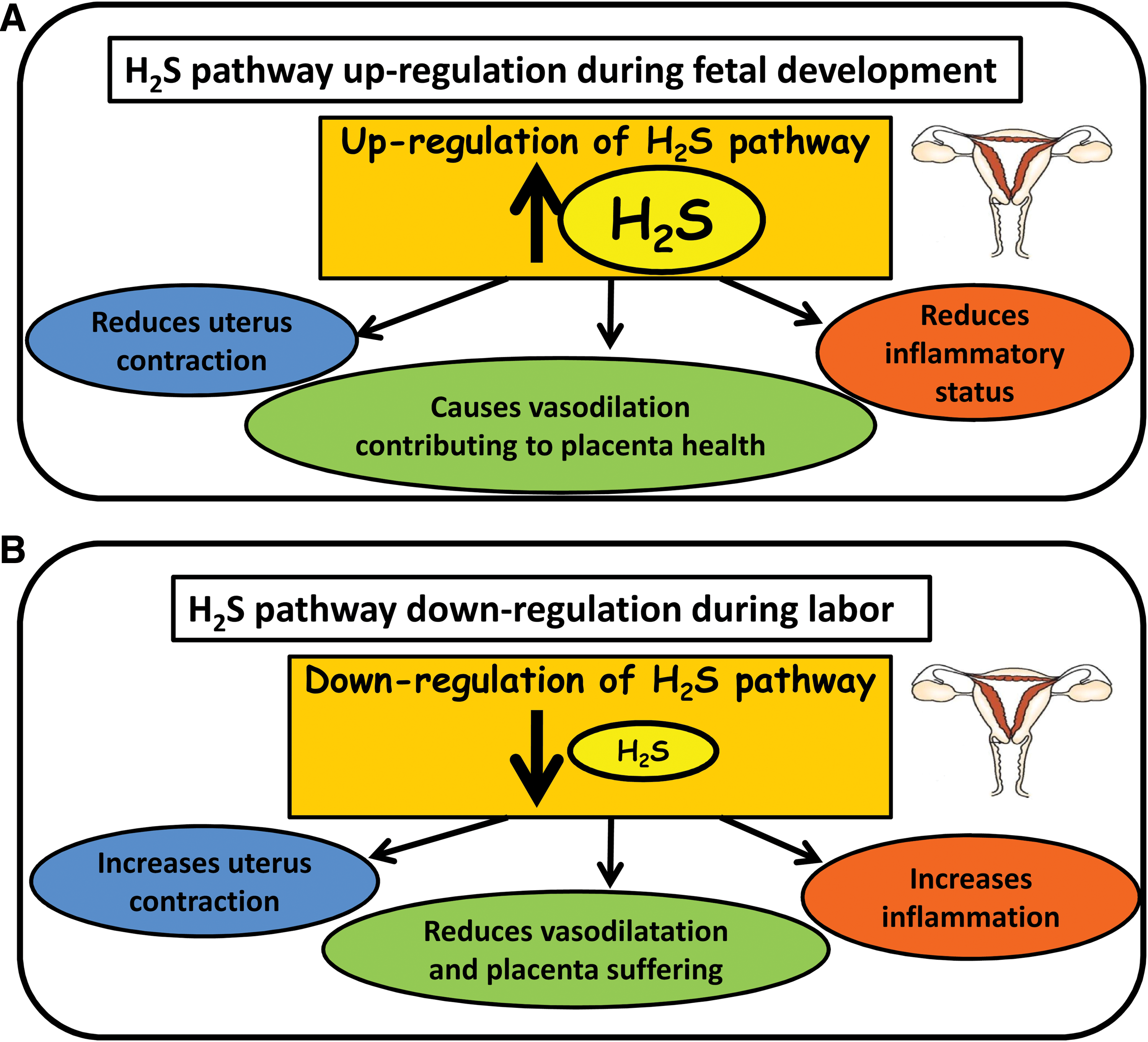
In conclusion, all these data strongly support the concept that H2S could be considered a natural tocolytic agent for two related properties, such as vasorelaxation (reduction of contraction amplitude and frequency) and anti-inflammatory effect (Fig. 7).
Another interesting matter is that the concentration of oxygen can modulate the synthesis of H2S. This is an important issue in pregnancy since oxygen tension is a key factor in the physiological development of the fetus. Interestingly, under low oxygen conditions, the production of H2S is significantly elevated in human placenta, rat liver, uterus, and fetal membranes. This increase in human intrauterine tissues could play a role in pregnancy pathophysiology. In fact, low oxygen conditions can reproduce a “pre-eclampsia” (PE)-like condition whereby poor placental function is present, resulting in intrauterine growth restriction (IUGR) and other placental dysfunctions. CBS and CSE activity has been shown to be modulated by oxygen. CBS contains a heme group in its structure, which could bind oxygen, affecting enzyme function. Conversely, CSE does not contain heme, so an alternative or a novel mechanism could be involved. Thus, it has been proposed that H2S under hypoxic conditions could have a role in PE (55). Hypoxia can also bring about other pre-eclamptic features, such as the release of pro-inflammatory cytokines and oxidative stress (59).
Hyperhomocysteinemia during pregnancy has been implicated in adverse outcomes, such as neural tube defects, PE, spontaneous abortion, and premature delivery. The adaptive changes in methionine metabolism during pregnancy in humans have been determined in pregnant and non-pregnant healthy women (13). Uncomplicated pregnancy in humans is associated with a higher rate of trans-sulfuration in early gestation and a higher rate of trans-methylation of methionine in late gestation. Studies in humans have also shown the absence of trans-sulfuration activity in the fetal liver and therefore the inability of the fetus to synthesize cysteine, a well-known key component of glutathione and one of the most important substrates for the enzymes synthesizing H2S (43). Hence, trans-sulfuration in the maternal compartment becomes an important source, other than protein breakdown and diet, of cysteine for the fetus. Plasma cysteine concentrations in pregnant women are significantly lower than those in non-pregnant women. Moreover, during pregnancy, L-cysteine is significantly lower in the third trimester than in the first trimester. Homocysteine concentrations in pregnant women in their third trimester are significantly different from those in non-pregnant women. In general, it seems that during a non-pathological pregnancy, the plasma concentration of both cysteine and homocysteine declines until the labor starts. These results are in line with the finding that there exists a high rate of trans-sulfuration during the first trimester that elevates plasma homocysteine concentrations, followed by a decrease during the third trimester, leading to a lowering in plasma homocysteine concentrations (13). The biological significance of the high rate of trans-sulfuration during the first trimester compared with the third remains unclear and speculative. The activity of CBS is low, and that of CSE is absent in the fetal liver in humans. Therefore, the high rate of trans-sulfuration in the first trimester could be aimed at providing cysteine and glutathione to the fetus. The need for glutathione by the developing embryo has been widely discussed and represents a key point to counteract the development of fetal malformations (40). A high rate of trans-sulfuration would also result in an obligatory requirement for methionine and is consistent with the high rate of neural tube defect and fetal growth retardation in women with a lower dietary intake of methionine.
To date, no evaluation of plasma H2S levels during non-pathological pregnancy has been addressed, and this lack of data does not allow for the right definition of the physiological role of H2S during the three pregnancy trimesters. In our opinion, this is a key issue since it could help to clarify the role played by changes in L-cysteine levels in relation to the synthesis of glutathione and H2S, and thus in H2S activity as well, either during normal pregnancy or in pathological disorders that are associated with pregnancy. A paper that analyzes a possible role of this pathway at the onset/development of PE was published in 2012. The authors demonstrate that CBS and CSE are mainly localized in the endothelium within the fetal vessels of the chorionic- and stem villi and that CBS/CSE protein expression is significantly downregulated in all placenta after spontaneous delivery compared with cesarean delivery. Regarding the role of the H2S pathway in PE, the major finding is that CBS mRNA expression is significantly downregulated in placental villous tissue obtained from pregnancies complicated by an early onset of PE, but not in the late onset of PE when compared with matched controls. On the other hand, there are no differences in CBS/CSE protein expression between PE and delivery matched controls. Unfortunately, H2S levels are not evaluated in this study; therefore, a clear correlation cannot be defined (42). However, a study evaluating plasma levels of H2S and the role of the H2S pathway in PE was published in 2013 (69). The study was focused only on one stage of pregnancy, that is, labor. The authors showed that the plasma level of H2S was significantly decreased in women with PE and this was associated with reduced placental mRNA CSE expression determined by immunohistochemical staining, but no Western blot analysis was reported. These results were confirmed by using a mouse preclinical animal model and the CSE inhibitor D,L-propargylglycine (PAG). The mice treated with D,L-PAG displayed a reduced placental growth factor production from the first trimester (8–12 weeks gestation). In particular, administration of D,L-PAG to pregnant mice induced hypertension and liver damage, promoted abnormal labyrinth vascularization in the placenta, and decreased fetal growth. Moreover, D,L-PAG in vitro treatment, using human placental explants, caused an inhibition of placental growth factor. The researchers found that GYY4137 reverts soluble endoglin levels and soluble fms-like tyrosine kinase-1 (sFlt-1) and restores fetal growth, demonstrating that the effect of the CSE inhibitor is attributable to the inhibition of H2S production. sFlt-1 is the soluble form of the vascular endothelial growth factor (VEGF) receptor that binds and reduces free circulating levels of the proangiogenic factors VEGF and placental growth factor, blunting the beneficial effects of these proangiogenic factors on maternal endothelium. Thus, sFlt-1, in particular, is considered a key factor of maternal hypertension, proteinuria, and multisystem organ injury during PE. Knockdown of CSE by small-interfering RNA in human umbilical vein endothelial cells increases the release of soluble endoglin and sFlt-1. On the other hand, adenoviral-mediated CSE over-expression in human umbilical vein endothelial cells inhibits their release. Administration of D,L-PAG to pregnant mice induces hypertension and liver damage, promotes abnormal labyrinth vascularization in the placenta, and decreases fetal growth. Treatment with GYY4137 reverts sFlt-1 and soluble endoglin levels and restores fetal growth, demonstrating that the effect of the CSE inhibitor is to be ascribed to the inhibition of H2S production. Thus, endogenous H2S is required for healthy placental vasculature, and a decrease in CSE/H2S activity could contribute to the pathogenesis of PE.
Overall, at the present stage, there is evidence that the H2S pathway has a relevant role, not only in the fine control of uterus contractility but also in the vascular function at the placental level. To date, a proper experimental model that accurately reproduces the human pathology of PE is not available, and this represents a major hurdle in further defining the role of the H2S pathway. However, the involvement of the H2S pathway in pregnancy-induced hypertension has been addressed by using a preclinical animal model that developed hypertension involving the NO pathway. The hypertension was induced by daily treatment with N(G)-nitro-L-arginine methyl ester (L-NAME), an NOS inhibitor. NaHS (50 μmol/kg/twice daily for 7 days) significantly reduces the L-NAME increase in blood pressure at the gestational days 18 and 20. In this model, the increased levels of circulating sFlt-1 were reduced by NaHS treatment. In this way, researchers demonstrate that on gestation day 21, NaHS blunted the increases in circulating plasma level of both sFlt-1 and VEGF induced by L-NAME. Thus, it could be hypothesized that the reduction in circulating plasma sFlt-1 and VEGF levels observed with NaHS treatment may prevent the concomitant damages to mother and fetus that would have been otherwise caused by VEGF signaling. The authors attempt to demonstrate that sFlt-1 increase could originate from circulating mononuclear cells that are released simultaneously with proteases and reactive oxygen species, leading to vascular endothelial injury. NaHS does not affect changes in myeloperoxidase activity and antioxidant capacity caused by L-NAME, leading the authors to discard this hypothesis. To explain the NaHS beneficial effect, the NO level was measured. Treatment with NaHS increases NO levels in hypertensive pregnancies, but not in normotensive pregnant rats, thereby suggesting that the NaHS effect is secondary to an increase in NO. Several studies in recent years have addressed the possible crosstalk between H2S and NO in specific district/organs. Therefore, these results support the presence of this crosstalk in the vascular dysfunction (hypertension) associated with PE (56).
The beneficial effects of ERT in the cardiovascular system are widely accepted (71). The possible relation between ERT and the H2S pathway has been examined in the uterine artery. In particular, this study has examined the vascular reactivity of uterine artery in comparison to carotid and mesenteric arteries harvested from ovariectomized rats undergoing ERT replacement therapy. ERT significantly stimulates mRNA and CBS protein expression without altering mRNA and CSE expression in both uterine and mesenteric arteries but not in carotid arteries. The physiological significance of ERT in regulating uterine blood flow during the follicular phase of the ovarian cycle, during pregnancy, and in postmenopausal women's health, and the link between ERT and H2S-derived CBS strengthen the role of this gas in uterus function (45).
Conclusion and Future Perspectives
A growing body of evidence clearly shows that the H2S pathway plays an important role in the male and female genito-urinary tract. As presented in this review, it appears that the enzymes responsible for H2S biosynthesis are specifically and differently distributed within the male and female genito-urinary tract. A clear gender difference seems to be present in the role and regulation of this pathway. In simpler terms, one enzyme predominantly contributes in a gender-specific manner to H2S formation compared with the others. For example, CSE-derived H2S has a major role in CC function in both man and animal models. Moreover, the evidence that the H2S donors relax corpus cavernous in an endothelium-independent manner suggests a backup role for this pathway when the endothelium is damaged and NO production is impaired. Therefore, drugs that are able to release H2S may represent a valid alternative approach to PDE5 inhibitors for those patients who do not respond to this class of drugs. Undoubtedly, it will be very important to better define the role of testosterone in modulating H2S levels and in determining how this impacts ED, particularly in aging men. At the same time, a better knowledge of how ERTs modulate the enzyme expression and H2S levels in health versus disease may help to define new therapeutic strategies, not only in the pathologies discussed in this review but, in our opinion, also in uterine and breast cancer that so far have been poorly studied.
It is our belief that an important emerging role for this pathway has been established in bladder pathophysiology. The recent finding that H2S is released by the urothelium on M receptor stimulation opens a new scenario for understanding the possible causes underlying bladder disorders. In particular, the finding that H2S formation can be modulated by cyclic nucleotides may help to give some insights into the mechanisms of action of drugs already in therapy. Indeed, many drugs currently used in therapy act by modulating cGMP/cAMP; therefore, a defect in H2S biosynthesis can be modulated by elevating nucleotides signaling and vice versa. With regards to woman H2S donors or drugs that can indirectly enhance H2S levels, these could be developed as tocolytic agents or perhaps used to replace the H2S contribution in pathological conditions such as in PE and preterm labor.
In conclusion, the clinical and therapeutic importance of modulating the H2S pathway/levels in different disorders that affect the genito-urinary tract is becoming increasingly clear. In this regard, a positive benefit of H2S donors compared with NO donors and their possible use in therapy is that they do not affect blood pressure and hemodynamic parameters, which has been demonstrated in a clinical study by injecting Na2S intravenously into human volunteers till a maximum dose of 0.2 mg/kg (67).
Footnotes
Acknowledgment
The authors thank Juliet Ippolito, BA, Vassar College, MPhil, University of Dundee, for English language editing.