
Abstract
Significance:
In the host–microbe microenvironment, bioelectrical factors influence microbes and hosts as well as host–microbe interactions. This article discusses relevant mechanistic underpinnings of this novel paradigm. It also addresses how such knowledge may be leveraged to develop novel electroceutical solutions to manage biofilm infection.
Recent Advances:
Systematic review and meta-analysis of several hundred wound studies reported a 78.2% prevalence of biofilms in chronic wounds. Biofilm infection is a major cause of delayed wound healing. In the host–microbe microenvironment, bioelectrical factors influence interactions between microbes and hosts.
Critical Issues:
Rapid biological responses are driven by electrical signals generated by ion currents moving across cell membranes. Bacterial life, growth, and function rely on a bioelectrical milieu, which when perturbed impairs their ability to form a biofilm, a major threat to health care. Electrokinetic stability of several viral particles depend on electrostatic forces. Weak electrical field strength, otherwise safe for humans, can be anti-microbial in this context. In the host, the electric field enhanced keratinocyte migration, bolstered immune defenses, improved mitochondrial function, and demonstrated multiple other effects consistent with supporting wound healing. A deeper mechanistic understanding of bioelectrical principles will inform the design of next-generation electroceuticals.
Future Directions:
This is an opportune moment in time as there is a surge of interest in electroceuticals in medicine. Projected to reach $35.5 billion by 2025, electroceuticals are becoming a cynosure in the global market. The World Health Organization reports that more than 50% of surgical site infections can be antibiotic resistant. Electroceuticals offer a serious alternative.
Introduction
A recent article in Time magazine discusses why it is time to take electrified medicine seriously (66). Bioelectrical cues guide subcellular, prokaryotic as well as eukaryotic cellular behavior (1, 48). The influence of electric principles in eukaryotic biology traverses a wide range of physical and physiological behaviors in plants and animals. Electrical properties of microbial life have been leveraged to benefit humans in many ways. The use of microbial cells to produce electricity was first achieved in the early 20th century (70). Microbial fuel cells (MFCs) rely on microbes as catalysts to generate electric power from organic matter (10).
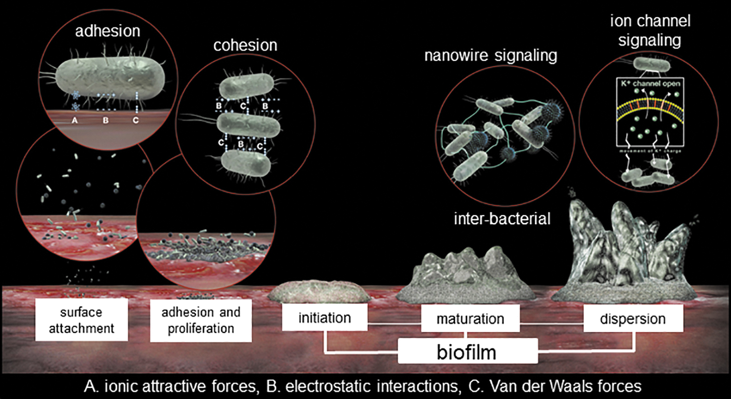
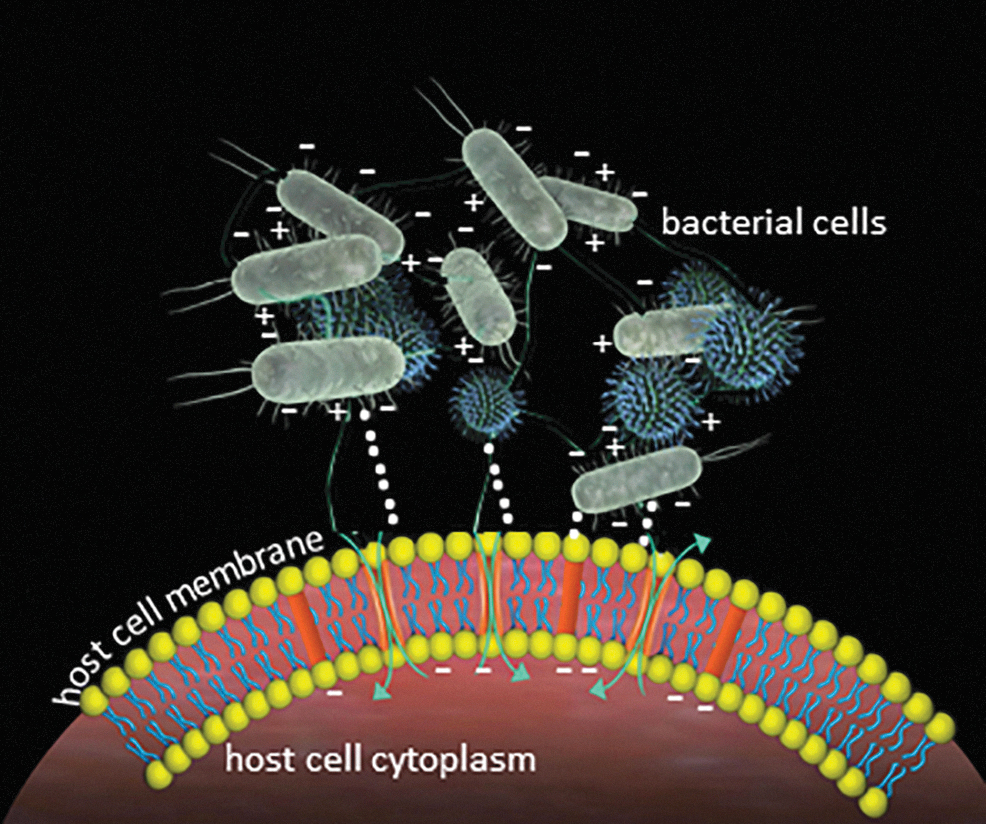
In bacterial biology, bioelectrical mechanisms influence fundamental processes (Fig. 1), including (i) adhesion to surfaces (electrostatic interactions) (75), (ii) cohesive interactions to build communities (matrix–extracellular DNA [eDNA], eDNA-protein, and matrix–protein held together by weak physicochemical interactions such as electrostatic forces, van der Waals interactions, hydrogen bonds, and ionic forces) (46), (iii) intra- and interspecies communication (ion channels) (72), and (iv) physical interactions between cells (conductive nanowires) (54). Certain plants utilize electrical activity to induce long-distance defensive signaling akin to synaptic activity in animal neurons (60). Electrostatic forces modulate the structure and function of some viral strains (77). In the animal kingdom, electrical mechanisms drive fundamental activities such as (i) biosensing for navigation and detection (birds, monotremes, and aquatic animals), (ii) foraging for food (aquatic animals and bumblebees), (iii) self-defense (electric eels), (iv) neuromuscular, auditory, and cardiac functioning, and (iv) wound healing (eyes and skin) (8, 79).
Electric Factors in Biology
Interaction between electricity and physiology was established in the late 1700s. Luigi Galvani, an Italian scientist, was in an open market where he noted that lightning was able to induce twitching of frog legs on sale. Frog muscle research gave rise to the field of electrophysiology. This discipline has evolved, making room for emergent areas. One such area of interest, central to the scope of this article, addresses the role of electrical factors in wound healing (64), cell migration, and management of relevant wound infection (6). In the interest of simplicity of discussion, in this work, electrical factors are split into electric current, electric field, electrostatic force, and redox electrochemistry. During healthy living, physiological processes and mechanisms are known to be sensitive to each of these components (6, 7).
Bacterial Biology
The study of electrical factors in bacterial biology is deeply rooted in animal neuroelectrophysiology (57). In the 18th century, electrical stimulation experiments performed by Luigi Galvani demonstrated that living cells utilize the flow of electrochemical species to guide biological function. In this context, ion channels, in particular, have been the subject of many lines of investigation directed to understand how the neuronal network communicates and guides processes from development to everyday responses such as movement. The works of Hodgkin and Huxley provided the first quantitative description of the electrical events underlying generation of action potentials (34), thereby revolutionizing our understanding of neuronal activity. In that vein, works of Roderick MacKinnon established the fundamental importance of potassium ion channels in all life forms, from prokaryotes (bacteria and archaea) to eukaryotes (63). The foundation for such work was established by research performed using simpler microbial models such as Escherichia coli (bacteria) (55) and Saccharomyces cerevisiae (yeast) (30). Bioelectricity has largely been the concern of electrophysiology, with the central focus being neuromuscular excitation leading to movement/response to external stimuli (11). Putative roles for bioelectricity in development, regeneration, and wound healing have been proposed a long time ago (81). Although bacteria have been used as tools to dissect integral biochemical and physiological cellular bioelectric responses, particularly as they relate to gated ion channel pathways in neuromuscular excitatory responses, a deeper understanding of these and other electroactive pathways in the bacterial/microbial lifecycle has only recently started to unfold (72). Specific questions of interest are how and why bacteria utilize bioelectrical principles for their daily functions. The quest for answers to these questions has led to the rapidly growing interest in the field of bacterial electrophysiology (53, 72, 74). In the context of the biofilm mode of growth (immobilized clusters/microconsortia of synergistic bacteria encapsulated in the polymeric electroactive matrix) (16), these principles seem to be amplified and guide diverse functions, including, but not limited to, biofilm formation (72), intra- and interbiofilm communications (53, 72, 74), and survival in harsh environments.
Electrophysiological processes within bacterial biofilms are currently understood to function in three ways: (i) direct electrical contact/transfer via nanowires and/or membrane-bound cytochromes; (ii) passive diffusion of electroactive metabolites such as flavins and phenazines; and (iii) active long-range signaling via voltage-gated ion channels (VGICs).
Direct electrical contact/transfer: In nature, microbial biofilms generate energy for growth by cycling carbon and other elements. For example, bacterial species, such as Geobacter spp. and Shewanella spp., extract and transfer electrons to insoluble and soluble electron acceptors using electroactive membrane components such as conductive appendages (e.g., pili [or nanowires]) and heme-containing c-cytochromes (52). Nanowires serve as electrical conduits to extracellular electron acceptors such as insoluble metal oxides or electrodes. From a practical viewpoint, such electroactive bacterial biofilms act as electrochemical reactors in the treatment of wastes (agricultural, industrial, and human) and as materials and devices for bioenergy (MFCs) and for bioremediation (52). A 2013 study was the first to demonstrate the presence of nanowires in bisphosphonate-related osteonecrosis of the jaw, a clinically relevant biofilm-mediated disease. The significance of this exciting observation remains to be further elucidated. Before this observation, electroactive physical structures had been primarily studied in environmental biofilm isolates (54).
i) Passive diffusion of electroactive metabolites: Some bacteria utilize soluble redox-active metabolites or capacitive particles to enable electron transfer between cells at a distance. Some examples of these metabolites include (i) flavins (produced by S. oneidensis) (50), (ii) phenazines such as pyocyanin (PYO; produced by Pseudomonas sp.), and (iii) quinolones such as the pseudomonas quinolone signal. PYO is a well-known, biofilm quorum-sensing (QS) mediator of Pseudomonas sp. that could also enable electrical responses in biofilms. PYO enhances electric current production by mixed microbial biofilms in MFCs (73). From a clinical perspective, the redox-active PYO promotes virulence by impairing eukaryotic electron transport, host cellular respiration, energy metabolism, and other critical cellular functions (31).
ii) Active long-range signaling: In 2015, Prindle et al. described an ion channel-mediated, electrical signaling-based, cell-to-cell communication process (72) that serves as a resource sharing mechanism between neighboring biofilm communities to enable survival during reduced nutrient supply. Using the Bacillus subtilis model system, it was demonstrated that potassium (K+) ion channels conduct long-range electrical communications within biofilm communities that are dependent on a quorum/threshold of biofilm mass for measurable electrical oscillations. These waves form a positive feedback loop, creating a wave of depolarization that coordinates metabolic states throughout the biofilm community. Interestingly, interspecies communication was noted between Bacillus spp. and Pseudomonas spp., dependent on the release of K+ as well as the membrane potential of the motile cell (36). Since Bacillus sp. is not known to have an Na+ ion channel system, this ionic species did not have an effect on biofilm growth dynamics. However, it does not preclude the possibility of Na+, Ca2+, Cl−, and ammonium ions enabling electrical connectivity within and between bacterial species.
Bacterial electrical biomembrane—VGICs
Electrical signaling through cellular membranes enables rapid response. In this form of communication, inducible gene expression, biochemical synthesis, specific receptors, or complex signaling pathway activation is not required (72). In Bacillus species, the cellular machinery driving electrical communication is a VGIC specifically responsive to K+. VGICs are multisubunit protein complexes that undergo conformational changes in response to changes in membrane potential. Sodium (Nav), potassium (Kv), calcium (Cav), and chloride (Cl−)-specific VGICs are present in microbes. Na+, K+, and Ca2+ channels have fundamental similarities in structure and function.
The chemical basis of electrical signaling
Rapid biological responses are typically driven by ion-generated electrical current moving across cell membranes, initiated and propagated by VGICs. VGICs contain a tetramer of transmembrane subunits or domains (S1–S6) made up of a voltage sensor and a pore module. The S4 segment has a symmetrical arrangement of charged residues, including arginine or lysine, making this domain function as the voltage sensor of the channel (3). Upon membrane depolarization, a sliding helix mechanism drives outward movement of the voltage sensor, causing voltage-dependent activation and opening of the intracellular gate. The selectivity filter conducts hydrated ions rapidly and is selectively guided by a unique negatively charged site. The collapse of an asymmetric pore-caused voltage-dependent inactivation terminates ion conductance (12).
Measuring electrical activity in biofilms
Patch-clamping
The classical electrophysiological clamping setup employing glass microelectrodes is not applicable to microbes because of the size of these organisms. The patch-clamp recording method developed by Neher and colleagues (32) overcame this shortcoming. Giant spheroplasts (large cytoplasmic bags devoid of cell wall) of E. coli were used for patching the inner membrane where the ion channels are found. Initial studies using this methodology identified mechanosensitive channels (56).
Array-based measurements
Multielectrode array systems, previously used for studying the neuronal electrical network, have been applied to study whole bacteria in biofilm communities (Bacillus licheniformis and P. alcaliphila) and planktonic growth (E. coli HEC30) (58). Electrical activity in the form of action potentials corresponded to maximum biofilm growth. Planktonic bacteria showed electrical activity, but with significantly lower amplitude strength compared with the biofilm. As bacterial cells increase in the developing biofilm, interactions between the individual cells create a network similar to neuronal networks. It is possible that the cohesiveness within the biofilm promotes a stronger electrical activity, which could play an important role in the emergence of collective behaviors such as sensing and communication with other cells for survival in a harsh environmental milieu.
Use of electroresponsive dyes
Radioactively labeled tetraphenylphosphonium ion (TPP+) or fluorescent dyes such as thioflavin T can be used to measure membrane potential changes. Membrane potential-dependent protein localization also serves as a measurement for membrane depolarization (80).
Conducting polymer-based electrochemical biosensors
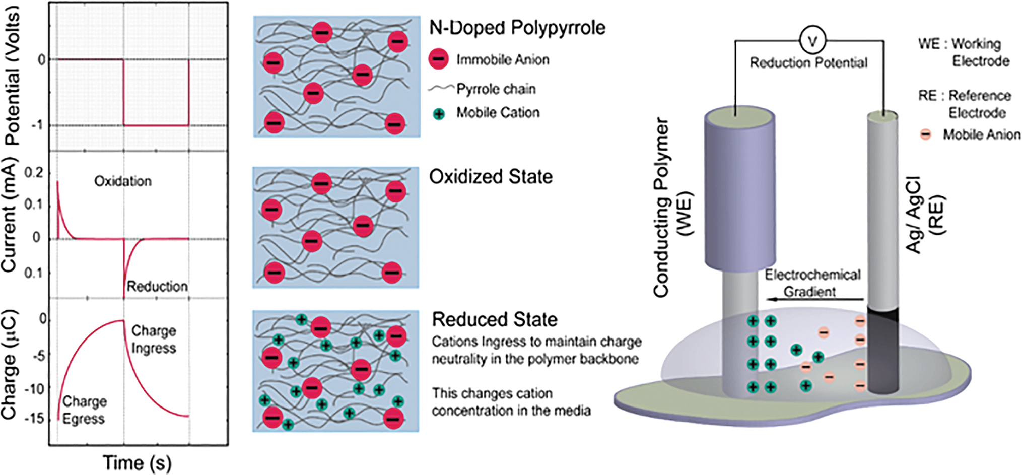
Conducting polymers (CPs) are a unique category of organic polymers that exhibit electrical conductivity and redox activity (51). Some of the most commonly applied CPs, poly (3,4-ethylene dioxythiophene), poly(aniline) (PANI), and poly(pyrrole) (PPy), have low toxicity and excellent long-term environmental stability in aqueous and in vivo environments. CPs can be doped with an appropriate antibody, oligonucleotide, enzyme, and bulky dopant molecules (such as dodecyl benzene sulfonate [DBS], dodecyl sulfonate, and bis (trifluoromethane) sulfonimide) or autodoped with small mobile ions to serve as recognition elements (24). The resulting electron transfer from the dopant to the polymer serves as the transduction pathway for detection via potentiometric or amperometric methods. Among CPs, PANI and PPy are widely used as analytical cations, gas sensors, and biosensors to varying degrees of success (Fig. 2). PPy doped with a bulky anionic dopant such as DBS [PPy(DBS)] enables the precise detection of the concentration of monovalent or divalent cations in solution and therefore is used as an electrophysiology sensor (84). The PPy(DBS) electrophysiology sensors can be directly applied to monitor biofilm ionic activity by culturing the cells directly between the electrodes in the sensor. This allows the biofilm to become a part of the control volume, and ionic activity can be directly measured using methods outlined for CP electrophysiology sensors (24).
Purpose of electroactive pathways
Biofilm establishment and growth
Using B. subtilis as the model organism, independent groups have identified a role for potassium in regulating biofilm formation. Altering expression levels of surfactin, kinase, and K+ transport regulator, all of which impact K+ intracellular levels, results in modifications in biofilm formation (49). Potassium uptake and efflux systems have also been implicated in P. aeruginosa biofilm formation, and production of QS regulated virulence factors such as PYO.
Bacterial adhesion and cohesion
Electrostatic forces enable adhesion of bacterial cells (Fig. 3). Studies with titanium implant surfaces in relation to oral bacteria have shown that modification of the titanium implant surface significantly alters the early adherence of bacteria on the surface and thus biofilm formation, which eventually affect health outcomes (4). Ionic strength and pH of the suspending solution together with the potentials of bacteria and the surface drive bacterial adhesion. The resultant electric interactions play an important role in bacterial adhesion (69). The extracellular matrix (ECM), comprising eDNA, polysaccharides, and proteins, is essential for biofilm formation (23). Electrostatic attractive and repulsive interactions, ionic attractive forces, hydrogen bonds, and van der Waal's interactions are among the weak physicochemical interactions that may maintain the multicellular structures that allow bacteria to cooperate metabolically and to be recalcitrant to antibiotics or immune cells (23).
Communication
Bacteria use a cell density-dependent collective behavior to release chemical signals that drive survival (9). In 2017 (47), it was demonstrated that artificial cells can sense and send QS molecules. Electrical signaling is recognized as an efficient cell-to-cell communication process. Ion channel-based electrical signaling attracts distant motile cells based on the membrane potential and the cell's modulation of tumbling frequency. Such long-range electrical signaling serves as an advanced communication mechanism, which is completely generic. Interestingly, cross-species communication is thereby enabled. A question that arises here is what are the long-term consequences and/or benefits of interspecies attraction and communication via electrical signaling? It also remains to be understood if the QS and electrical systems may impact each other and how that impact may be affected.
Resource sharing
Biological systems frequently deal with resource limitations. Time-sharing is a strategy where users take turns consuming resources. In such cases, different systems may compete with each other. Glutamate starvation in B. subtilis biofilm communities causes collective growth rate oscillations. A negative feedback loop guided by biomass increase leading to glutamate stress drives these oscillations. This stress, in turn, influences biofilm growth. Ion channel-mediated electrical signaling coordinates this phenomenon (72). The metabolic oscillations in biofilm communities are synchronized in their growth dynamics by electrical signals. This further increases competition by synchronizing demand for limited nutrients.
Flagellar motility
Transient changes in membrane potential cause motility changes. Comparable effect was demonstrated in a recent study demonstrating that K+ signaling from the biofilm and the membrane potential of planktonic/motile bacteria are both determinants of flagellar motility. This motility is more directional when the motile organism is further away from the biofilm (K+ signal).
Defense mechanisms
The biofilm extracellular polymeric substance (EPS) restricts penetration of antimicrobials, causing antimicrobial tolerance. EPS may also serve as a diffusion barrier to antibiotics. The eDNA component of EPS displays cation-chelating properties, thus inducing resistance to host-derived or therapeutic antimicrobials. Positively charged antibiotics such as tobramycin are sequestered in the biofilm periphery via ionic interactions with negatively charged matrix components. Tobramycin penetration into the biofilm was enhanced by addition of cations.
Virulence mechanisms
Redox-active PYO is toxic to eukaryotic hosts (45) and other microbes. PYO induces the production of reactive oxygen species, such as the superoxide anion radical, augmenting virulence (27). PYO induces oxidative stress in cellular systems, which manifests as premature cellular senescence. PYO may influence the intracellular redox state by decreasing carbon flux through central metabolic pathways (71).
Eukaryotic Biology
Bioelectric properties in development
Electrical fields have been detected both extracellularly and intracellularly (59). Endogenous electric fields exist within extracellular spaces and influence cell behavior in development and wound healing. Studies of amphibian (toad and axolotl) and avian (chicken) embryos demonstrate that endogenous electric fields (normal polarity and magnitude) are necessary for development of neural and other tissues. Scrambling of physiological electrical cues results in gross developmental abnormalities caused by interference in patterning and cell migration in the embryo (35).
Bioelectric properties of human organ systems
Neuromuscular system
Nerve fibers act as communication cables connecting and transmitting electrical impulses that guide the body's response to multiple stimuli. It is estimated that each neuron produces ∼70 mV of electric potential, while muscle cells produce about 95 mV. This potential, in the form of adenosine triphosphate (ATP), powers electrogenic pumps that are translated to active outputs.
Cardiac system
The sinoatrial node located in the right atrium controls the rhythm of our heartbeat and pumping of blood to the rest of the body. Utilizing electrical signals to set the pace, it is the body's natural pacemaker (13).
Skin
In 1849, Emil du Bois-Reymond first observed that the human skin was electrically active (22). This was further corroborated by Neumann and Blanton (61a) who demonstrated a connection between cutaneous electrical activity and sweat glands. Electrical impedance is lowest in the palms with abundant sweat ducts. A 1 Hz to 1 MHz range of electrical strength is estimated from the skin surface into the underlying dermis and subcutaneous tissue. The ability of the human skin to self-repair allows it to function as a protective barrier. The intact mammalian skin has positive transepithelial potentials between 10 and 60 mV. During epithelial wound healing, there is induction of electric current of magnitude ∼10–100 mA/cm2 caused by a transepidermal voltage gradient created by the epithelial sodium ion pumps. With gradual decrease in electric field strength, there is progressive coverage of wound area with epithelial cells. The presence of an endogenous electric field may have critical roles in cutaneous wound healing (37). Cellular outcomes such as cell migration, cell division, leukocyte infiltration, nerve sprouting, endothelial cell remodeling, and associated angiogenesis, within 500 μm–1 mm of the wound edge, are known to be influenced by the electrical voltage gradient (7, 64).
Electric principles in cell migration
Exposure of cells to physiological electric fields affects cell orientation, migration, protein synthesis and distribution, and activation of signaling pathways such as CDC42p, Rho/Rac,PI3K/PTEN, and phosphatidylinositol phosphate (86, 88), as well as epithelial sodium channels (85). A local, direct electric field (dc electric field) of low magnitude (10–400 mV/mm) guides the motility of living cells through a process called galvanotaxis (61). Several lines of evidence support that changes in electrical parameters may influence the function of host cells such as keratinocytes, fibroblasts, neutrophils, macrophages, lymphocytes, and endothelial cells, all of which are relevant to wound healing (7). Under the influence of an electric field, changes in cell membrane plasticity, cytoskeletal rearrangements, and alterations in interactions of the cell with its microenvironment enable the cell to move forward. Such movement is further facilitated by electric field-induced, intracellular signaling events involving several growth factors, for example, epidermal growth factor, vascular endothelial growth factor, (87), and hepatocyte growth factor, and protein kinases such as protein kinase C, cGMP-dependent protein kinase, and mitogen-activated protein kinase (65). These signaling events directly regulate cell polarization and migration (7, 88).
Immune cell function and inflammation
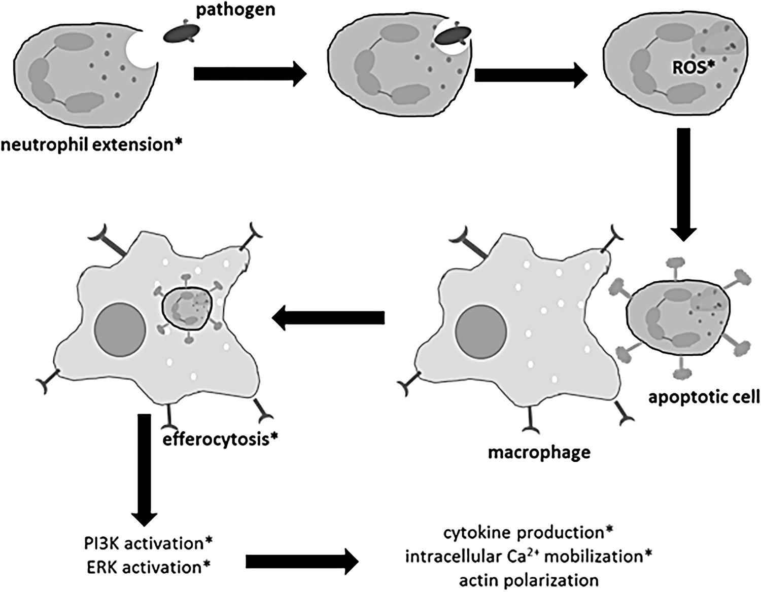
Immune cells play a major role in host defense and infection management. Electric fields stimulate immune cell function. For example, membrane-mediated Ca2+ signaling processes are responsive to electric fields. Neutrophils represent the first cells that arrive at the site of injury to defend the body against microbial pathogens. Application of an external electric field activates the respiratory burst of neutrophils, neutrophil extension, metabolic resonance, and DNA damage (42). Monocytes represent the next blood-borne immune cells that extravasate to form macrophages and migrate to the site of injury to bolster host defenses (25). The phagocytic activity of macrophages in dead cell clearance can be enhanced by external electric fields (33). Such intervention causes changes in cellular signaling, for example, PI3K and ERK activation. The pattern of cytokine release thus changes as do the intercellular Ca2+ response and actin polarization (Fig. 4). Bioelectric modulation of ATP-sensitive potassium channels influences macrophage polarization and is likely to modify macrophage plasticity.
Wound healing
Endogenous electric fields (∼100–200 mV/mm in the skin and cornea) provide directional cues to guide the tissue repair response. Electric fields guide cell migration in diverse cell types involved in the healing response, including keratinocytes, macrophages, neutrophils, and fibroblasts. Furthermore, supportive actions for the healing process, including generation of ATP, increased secretion of collagen by fibroblasts for ECM restoration, and increased blood flow and capillary density, are also responsive to electric fields. Membrane receptors such as EGFR, VEGFR, and integrins, integral to the wound healing process, are redistributed and activated in response to endogenous electric fields (87). Activation of any of these receptors by electric fields triggers downstream signaling cascades directly relevant to wound healing (7, 87, 88). Recent studies testing mechanisms underlying the action of an electroceutical wound care dressing demonstrated responsiveness of key signaling pathways accelerating keratinocyte migration (7), a key cellular component in wound reepithelialization. The electric field enhanced keratinocyte migration by three mechanisms: (i) hydrogen peroxide generation (a potent driver of redox signaling), (ii) phosphorylation of redox-sensitive insulin growth factor receptor (IGF1R), and (iii) reduction of protein thiols and increase in integrin αν expression. Electric fields also increased the keratinocyte mitochondrial membrane potential supporting an energy-demanding migration process. In this context, therefore, exogenously applied electric fields could mimic the effect of an endogenous electric field, possibly stimulating and guiding all the above cellular behaviors to enhance wound healing.
Electroceutical wound care therapies
In biofilm-infected cutaneous wounds, wound healing is compromised. Although the affected wound may close, barrier function of the repaired skin is deficient, as measured by elevated transepidermal water loss (8, 26, 76). Treatment of wounds with electric field-based antimicrobial dressings corrected such deficiency and restored functional wound healing. Specific biofilm-repressed molecular pathways, including the adherens junction protein, E-cadherin, essential for in vivo epidermal barrier function, were rescued by such dressing. Furthermore, electric field-based wound care dressing managed biofilm-induced persistent inflammation (8). A clinical trial testing this Food and Drug Administration (FDA)-cleared dressing in a setting of burn wounds is currently in progress (NCT04079998).
Several other forms of electroceutical interventions have been tested in wound care (Table 1) (2, 5 –7, 14, 15, 18, 20, 28, 39, 41, 43, 62, 68, 78, 82, 83). Unlike electric field-based dressings discussed above, the notion of electric stimulation devices in wound care relies on direct application of electric current to stimulate the wound tissue. Most of such devices that rely on application of electric current have underperformed in wound care. Such suboptimal performance can be attributed to the lack of consideration of the complex mechanistic implications of electrical factors, as addressed in this work. In wound care, tested electric stimulation devices employ a range of variables, including high voltage, current, mode, and length of time of application. These devices employ wired electrodes for direct application of much higher current to the wound tissue compared with the dressing discussed above. A low- or high-frequency pulsed electrical current that stimulates the peripheral nerves, called transcutaneous electrical nerve stimulation, has been tested for pain control (44). The frequency rhythmic electrical modulation system varies the pulse, frequency, duration, and voltage during application. The Fenzian system, an electronic biofeedback system utilizing degenerate waves, has been used in the treatment of acute wound healing and scar problems in the skin. Pulsed current is a common mode used in electrotherapeutic trials. Short voltage pulsed current devices such as Aptiva Ballet (Lorenz Therapy System) or Naturepulse (Globe Microsystems) report increase in circulating vascular endothelial growth factor and nitric oxide in response to stimulation. Limited studies claim improved wound closure in the treatment of chronic venous and diabetic ulcers. The silver iontophoresis stimulator electrotherapy device is an iontophoretic system utilizing low-intensity direct current to deliver silver ions to target sites within the body to fight infections and promote wound healing. This device claims applications for treatment of antimicrobial-resistant bacterial infections as well as fungal and yeast infections.
Electrical Treatment Modalities Available for Wound Management
BPC, biphasic pulsed current; DC, direct current; FDA, Food and Drug Administration; HVPC, high-voltage pulsed current; MAPK, mitogen-activated protein kinase; MPC, monophasic pulsed current; PEMF, pulsed electromagnetic field; TENS, transcutaneous electrical nerve stimulation; WED, wireless electroceutical dressing.
Wireless electroceutical dressing (WED) is an FDA-cleared wireless dressing with a matrix of embedded elemental silver and elemental zinc. When in direct contact with a conductive medium, redox chemical reactions drive the transfer of electrons from zinc to silver (6 –8), generating an electric field at the dressing surface, which promotes keratinocyte migration (7) and biofilm disruption (6, 8). When tested in a preclinical, porcine experimental model of long-term wound biofilm infection involving an intact host immune defense system, WED was effective in preventing biofilm formation and disrupting established biofilm infection and associated pathological complications (8). Furthermore, WED effectively managed biofilm-induced persistent inflammation and promoted restoration of skin barrier function following injury (8). WED may be viewed as a first-generation wound care dressing, representing a translationally viable option to disrupt wound biofilm infection in vivo.
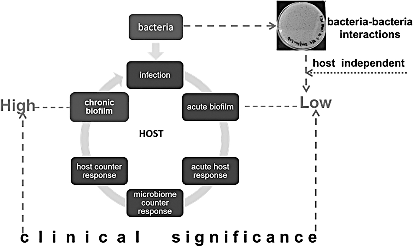
Therapies marketed as antibiofilm may not necessarily by useful in fighting wound infections, especially if they have been tested primarily in in vitro or short-term in vivo models (26). Such approaches are powerful in understanding microbiological processes, but limited in addressing biofilm mechanisms in the context of host infection. Although the Wound Healing Society recommends the porcine model as the most relevant preclinical model of cutaneous wound healing (29), short-term infection studies even in these models disallow prolonged interactions between polymicrobial biofilm-forming pathogens and the host. Short-term models therefore have limited power to understand long-term, clinically relevant host–biofilm interactions inclusive of host immune system responses that shape an acute-phase infection into a pathogenic chronic biofilm (Fig. 5). The translational relevance of antibiofilm therapies will be better tested in the context of live, long-term immune-competent models that capture the persistent nature of biofilm-infected chronic wounds (26).
Electroceuticals against antimicrobial resistance
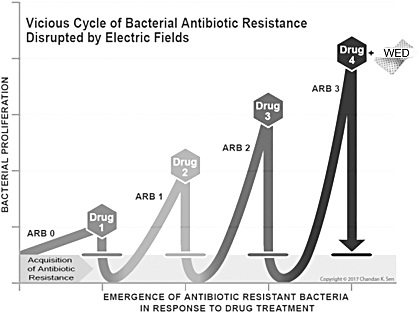
Bacterial genetic mutations alter functional pathways that are targeted by traditional antibiotic therapies, resulting in evolution of the following: (i) masked or decoy drug targets, (ii) drug-inactivating enzymes, and (iii) drug pumping mechanisms. Because much of the effort in clinical management of infections is still dependent on pharmaceutical options, each of these pathways may be viewed as a drug-inducible loop that when treated with other drug-based strategies, results in a futile cycle, forcing the evolution and persistence of even more resilient strains with multidrug resistance (MDR) properties (Fig. 6). Within the protected biofilm cocoons, gene exchange favors rapid transfer of such drug resistance traits. This poses a critical challenge in combating infection and warrants development of productive nonpharmacological or combinatorial strategies to fight biofilm infection. Because electroceutical therapy is not subject to the metabolic pathways of drug resistance, it has the potential to circumvent drug resistance.
In 1992, it was reported that weak (1.5 V/cm and 15 μA/cm2) electric fields (17, 19) could significantly enhance killing of biofilm bacteria by antibiotics. This bioelectric effect suggested a possible application of electrical therapeutics for antibiotic-resistant bacterial strains. WED, when tested in vitro in the context of an MDR strain of P. aeruginosa, attenuated the expression of MexR and MexT multidrug efflux pump regulators (6). Follow-up studies using a porcine wound model infected with a mixture of MDR P. aeruginosa and Acinetobacter baumannii strains showed that WED disrupted biofilm infection by these strains (8). Other groups have tested WED against several antibiotic-resistant strains in vitro and found that WED was inhibitory to almost all the strains tested (38, 40).
Electroceuticals against viral diseases
Electroceuticals could be a new antiviral strategy. Electrostatic forces are critical for the structure and function of viral particles and could be exploited to destabilize viruses. WED was recently shown to disrupt the infectivity of some viruses in vitro (e.g., coronavirus and lentivirus) (77). The zeta potential (electrostatic interactions in particle dispersions) determines viral particle stability (77). WED therefore was found to rapidly lower the zeta potential, possibly causing defects in viral particle stability and therefore lowering infectivity. This compelling observation provides an exciting opportunity for further exploration of the use of electroceuticals as antiviral strategies.
Electroceuticals to manage health care-acquired infections (hospital-associated infections)
Hospital-associated infections (HAIs), also called nosocomial infections, are a pressing public health threat, estimated by the Centers for Disease Control to affect 1 in 25 hospitalized patients on any given day. In addition to the morbidity and mortality rates associated with HAIs, there is also a heavy economic burden estimated at $28–$33 billion in excess costs. These HAIs include central line-associated bloodstream infections, catheter-associated urinary tract infections, surgical site infections (SSIs), and ventilator-associated pneumonia. Several routes of transmission of the infectious agent contribute to the persistence of this problem in hospital settings, including contact with contaminated surfaces such as hospital textiles (including bedding and drapery [curtains and/or privacy screens]) facial masks and scrubs, among others (21, 77). The increasing evidence of biofilms in catheters and central lines has necessitated development of more sophisticated methods of sterilization and modification of medical devices to make them uninhabitable for biofilm-forming organisms (67). Several lines of research have focused on coating surfaces of catheters and central lines with various polymers, silver ions, and other nanoparticles and even treatment with photodynamic therapy. However, despite these advances, the HAI problem still persists, indicating a need for more effective measures in eradicating the infectious agent. In this context, electroceutical-based surface modifications may be viewed as a viable next-generation solution. For instance, coating the inner lining of catheters or central lines with electrically conductive materials that generate mild electric fields could interfere with adhesion and survival of microbial pathogens. Similarly, patterning hospital privacy curtains or linens with such conductive materials could make these surfaces incompatible for establishment of biofilms and thereby drastically decrease the incidence of HAIs.
Conclusions
Bioelectricity has largely been the concern of mammalian electrophysiology, with the central focus being neuromuscular excitation. Bacterial electrophysiology is an emergent discipline. We now know that bacterial life, growth, and function rely on an intrinsic bioelectrical milieu, the perturbation of which could inhibit or kill these organisms. Weak electrical fields, otherwise safe for humans, can achieve such benefit, exemplifying the Arndt–Schulz rule (for every substance, small doses stimulate, moderate doses inhibit, and large doses kill). At the same strength that kills microbes, beneficial effects of such electroceuticals have been observed in improving human keratinocyte migration—a contributor to wound closure. The electric field may stimulate immune cell function as well. A deeper mechanistic understanding of how electroceuticals may influence microbes, hosts, and host–microbe interactions is likely to help inform the design of next-generation electroceuticals aimed at prevention and management of infection. This is an opportune moment in time as there is a surge of interest in electroceuticals in medicine (66). The electroceutical market, projected to reach $35.5 billion by 2025, is rapidly becoming a cynosure in the global market. Electroceuticals broadly encompass all bioelectronic medicines that employ electrical stimulation to affect and modify functions of the body. Brain stimulation therapies, electrical muscle stimulation, cardiac stimulation therapies, cochlear stimulation implants, and tumor-treating fields in cancer are currently used in medical practice. The World Health Organization reports that more than 50% of SSIs can be antibiotic resistant. Electroceuticals emerge as a serious alternative. Investment into advancing electroceutical management of surgical infection warrants serious consideration.
Footnotes
Author Disclosure Statement
C.K.S. discloses a financial competing interest as owner of stock options of Vomaris Innovation, Inc.
Funding Information
This work was partly supported by the National Institutes of Health NR015676 and DK 125835.