
Abstract
A selection of five proteinogenic amino acids—glycine, isoleucine, phenylalanine, tyrosine, and tryptophan—were studied in the mid-infrared and in the far-infrared with the purpose to facilitate the search and identification of these astrobiologically and astrochemically relevant molecules in space environments. The molar extinction coefficients (ɛ) of all mid- and far-infrared bands were determined as well as the integrated molar absorptivities (ψ). The mid-infrared spectra of the five selected amino acids were recorded also at three different temperatures from -180°C to ambient temperature to +200°C. We measured the wavelength shift of the infrared bands caused by temperature; and for the most relevant or temperature-sensitive infrared bands, a series of linear equations were determined relating wavelength position with temperature. Such equations may provide estimates of the temperature of these molecules once detected in astrophysical objects; and with the reported values of ɛ and ψ, it will be possible to estimate the relative abundance of these molecules in space environments.
1. Introduction
Proteinogenic and non-proteinogenic amino acids are known to be present in meteorites, especially in carbonaceous chondrites. An inventory of the amino acids found in meteorites can be found in the works of Commeyras et al. (2006), Martins and Sephton (2009), Martins (2011), Pizzarello and Shock (2017), and Koga and Naraoka (2017). It is well established that amino acids and other complex organic molecules that are prebiotic precursors of molecules and macromolecules of biological interest formed in the icy mantles of dust grains and in the interstellar medium before formation of the Solar System. These molecules were then trapped within asteroids, comets, and planetesimals, some of which were delivered to primordial Earth (Nuevo et al., 2007; Hudson et al., 2008; Kwok, 2012, 2016, 2019; Kaiser et al., 2013; Cataldo and Iglesias-Groth, 2017; Cataldo, 2018). Of course, thermal and aqueous alteration processes also play a key role in the final amino acid distribution and their enantiomeric excess found in carbonaceous chondrites (Elsila et al., 2016).
The experimental evidence of this scenario is becoming impressive through a number of experiments that involve irradiation of simulated icy mantles of dust grains of the interstellar medium (see for example Bernstein et al., 2002; Caro et al., 2002; Hudson et al., 2008; Öberg, 2016; Modica et al., 2018; Arumainayagam et al., 2019; Kobayashi, 2019; Ciaravella et al., 2019) and astrophysical observations (Kwok, 2019; Scibelli and Shirley, 2020). Furthermore, it has been shown that even elemental carbon in the form of fullerene or under other allotropic states can be the source of atomic carbon suitable for the synthesis of amino acids under appropriate high energy fields (Iglesias-Groth et al., 2013; Ursini et al., 2019; Krasnokutski et al., 2020).
A series of studies have shown that all the proteinogenic amino acids and a selection of non-proteinogenic amino acids have a radiation stability sufficient to survive 4.6 × 10⁹ years of irradiation due to radionuclide decay when buried at a certain depth in asteroids and comets and shielded from the direct action of cosmic rays (Cataldo et al., 2011a, 2011b, 2011c, 2013a, 2013b; Iglesias-Groth et al., 2011a; Cherubini et al., 2014). Thus, the scenario involving formation of amino acids in the interstellar medium, followed by their incorporation in planetesimals, asteroids, and comets in star-forming regions and late delivery to Earth, is further reinforced by these radiation chemistry experiments performed on amino acids, following a calculation of the radiation dose received from radionuclide decay calculated many years ago by the Nobel laureate Harold Urey (1955, 1956). Very recently, the possible formation of oligomers and peptides as well from amino acids under prebiotic conditions is receiving a great deal of attention (Ligterink et al., 2018; Kitadai and Nishiuchi, 2019).
Another fascinating topic with regard to prebiotic amino acids found in meteorites has to do with the enantiomeric excess systematically detected by many authors and pioneered by Pizzarello and Cronin (2000), among others. This topic has been comprehensively reviewed in the work of Meierhenrich (2008) and more recently the work of Garcia et al. (2019). It is thought that the enantiomeric excess in initially racemic amino acids is induced by circularly polarized light irradiation from astronomical sources that may selectively transform a racemic mixture into a scalemic mixture (Bailey et al., 1998). This scenario is strongly supported by a series of interesting experimental results on the irradiation of interstellar ice analogs with circularly polarized UV light (Meierhenrich, 2008; Garcia et al., 2019).
With all these premises, an increased effort has been underway for the search for amino acids in space by astrophysical observations, as briefly reviewed in the work of Iglesias-Groth and Cataldo (2018). Similar to the case of fullerenes (e.g., Cami et al., 2010; García-Hernández et al., 2012; Iglesias-Groth, 2019), amino acids could, in principle, be recognized in space through mid-infrared and also far-infrared spectroscopy, given that new and forthcoming orbiting telescopes will cover the far-infrared or terahertz spectral window with high spectral resolution and sensitivity. In particular, C60 fullerene was discovered in the young planetary nebula Tc 1 and then in many other sources by way of the Spitzer infrared orbiting telescope due to the peculiar four-band pattern in the infrared (García-Hernández et al., 2012).
The integrated molar absorptivity of fullerene and fulleranes measured in the laboratory (Iglesias-Groth et al., 2011b, 2012) was used for an estimation of the relative abundance of these molecules in certain planetary nebulae (Diaz-Luis et al., 2016). The C60 molecule was not detected in the gas phase but as adsorbed on a solid surface, probably carbon dust (Cami et al., 2010; García-Hernández et al., 2012). By analogy, it can be assumed that, if amino acids are embedded or generated within ices, they are more refractory than the other species; thus it could be that, as a young star forms or a comet dust sublimates, the refractory components such as amino acids are left behind and could be identified at ambient, or even moderately elevated, temperatures through infrared spectroscopy.
Consequently, a selection of amino acids was spectroscopically studied in the mid- and far-infrared (Iglesias-Groth and Cataldo, 2018). As shown in the works of Cami et al. (2010), García-Hernández et al. (2012), and others, the integrated molar absorptivity of at least the most relevant infrared bands is needed for a quantitative analysis of detected molecules in space. Thus, the integrated molar absorptivity has been determined in the spectra of fullerenes. The present work represents an extension of previous work on amino acid spectroscopy by Iglesias-Groth and Cataldo (2018), integrated molar absorptivity of fullerenes by Iglesias-Groth et al. (2011b), fulleranes by Iglesias-Groth et al. (2012), and endohedral fullerenes by García-Hernández et al. (2020). Here, we report measurements of integrated molar absorptivity both in the mid-infrared and far-infrared spectral regions for a series of selected amino acids: glycine, isoleucine, phenylalanine, tyrosine, and tryptophan.
It must be emphasized that our intention was to study all the proteinogenic amino acids and, in this work, focused on the above-mentioned five amino acids. While glycine is the simplest amino acid and was quite recently the subject of infrared studies in astrochemically relevant conditions (Maté et al., 2011; Souza-Corrêa et al., 2019), the integrated molar absorptivity of its mid- and far-infrared bands is still unknown. Glycine is the most interesting candidate to be sought in different astronomical objects. Isoleucine has been little studied in the infrared, but it was found in meteorite analyses and, hence, is another interesting candidate to be sought in different astronomical objects. Consequently, the infrared data of the present study are of a particular relevance. With regard to the aromatic amino acids phenylalanine, tyrosine, and tryptophan, it is true that they are seldom found in meteorite analyses, but we have included them in this work as a part of the planned systematic investigation in the mid- and the far-infrared of all the proteinogenic amino acids.
2. Experimental
2.1. Materials
All the amino acids used in the present work were purchased from Aldrich-Merck. The spectroscopy matrices cesium iodide, low-molecular-weight polyethylene, were also obtained from Aldrich-Merck.
2.2. Laboratory equipment
The mid-infrared spectra were recorded in transmittance on a Nicolet 6700 Fourier transform infrared (FT-IR) spectrometer from Thermo-Scientific with a resolution of 4 cm−1. The spectrometer could work at higher resolution, but 4 cm−1 is the optimal condition for samples in the solid state embedded in a matrix such as CsI, for instance. The low-temperature apparatus consisted of a variable temperature cell from the Specac model P/N 21525 equipped with heated KBr windows and a temperature-controlled sample holder, which is able to work in the range between +250°C and -180°C. The low temperature limit of -180°C was reached by using liquid nitrogen. The variable temperature cell was evacuated with a Buchi vacuum pump model V-710 equipped with four diaphragm heads and a three-stage vacuum creation process, which delivers 3.1 m3 h−1 and an absolute vacuum of 1 mbar.
The far-infrared spectra from 600 cm−1 down to 50 cm−1 were recorded with the Nicolet 6700 FT-IR spectrometer always at a resolution of 4 cm−1, after opportune adjustments of the optical bench (change of the beam splitter and the detector). Furthermore, the entire spectrometer optical bench and the sample compartment was continuously purged with completely dry air free of CO2 at 12 L/min. The CsI, or polyethylene pellet, used in the infrared spectra measurement was produced by a Silfradent double column press equipped with a pressure meter. Aldrich macro–micro pellet die was loaded with the pre-weighed CsI and amino acid, closed, and compressed in a double column press while evacuated at 2 mbar. The thickness and diameter of the pellets were measured with a Somet digital micrometer with a sensitivity of 0.01 mm.
Note that transitions with very high absorbance may be subject to deviations from the Beer-Lambert law, and those band strengths could be infraestimated.
2.3. Pellet preparation for the determination of the integrated molar absorptivity and the molar extinction coefficient in the mid-infrared
In a typical procedure, the selected amino acid (1.2 mg) was quickly mixed with 250.0 mg of CsI in an agate mortar, and the two components were finely ground and mixed together. The powder was transferred into the macro–micro pellet die and compressed at 6 tonnes cm−2 with a Silfradent press. The resulting pellet, with a measured thickness of 0.71 mm, was mounted into the sample holder of the Specac variable temperature cell and inserted into the cell. The cell was then evacuated with the aid of a Buchi pump to a vacuum of 0.1 torr and then heated gradually at +60°C in order to permit the humidity, which is eventually absorbed on the internal surfaces of the cell and in the KBr pellet, to evaporate. To reach below room temperature, liquid nitrogen was added cautiously and in small amounts in the cavity present inside the Specac cell. Such a cavity is connected with the sample holder and allows for cooling of the sample to the desired temperature. The temperature of the sample was monitored with thermocouples present in the cell. The lowest temperature reached with this apparatus was −180°C, while the highest temperature was +250°C. Heating is provided by the Joule effect supplied to the Specac cell by an external thermal control unit.
A more detailed description of the apparatus (photographs) used for the measurement can be found in the work of Cataldo and Iglesias-Groth (2010). The spectra were recorded in absorbance mode. The measurement of the intensity (height) of the absorption bands with automatic subtraction of the baseline was made through the OMNIC software from Thermo, which was dedicated to the FTIR spectrometer. Similarly, the integrated band intensity was also measured through the OMNIC software. The error level both in the determination of ɛ and ψ values is in the range of ±13%.
2.4. Pellet preparation for the determination of the integrated molar absorptivity and the molar extinction coefficient in the far-infrared
The selected amino acids (1.2 mg) were mixed in an agate mortar with polyethylene (220 mg), a matrix suitable for far-infrared spectroscopy. The pellet was compressed in the Silfradent press at 6 tonnes cm−2 inside the macro–micro pellet die. The resulting pellet was obtained with a measured diameter of 12.55 mm and a measured thickness of 0.73 mm. The far-infrared spectra were collected exclusively at room temperature in absorbance mode without the use of the Specac cell. Also, in the far-infrared the error estimation both in ɛ and ψ values is in the range of ±13%.
3. Results
3.1. On the molar extinction coefficients and integrated molar absorptivity of the mid-infrared bands of selected amino acids
Detailed accounts of the procedure for determination of the molar extinction coefficients (ɛ) and the integrated molar absorptivity (ψ) of the infrared bands can be found in the works of Cataldo and Iglesias-Groth (2010), Iglesias-Groth et al. (2011b, 2012), García-Hernández et al. (2020), all of which focus on fullerenes, fulleranes (the hydrogenated fullerenes), and endohedral fullerenes infrared spectroscopy. We note that our understanding of these parameters is important in astrochemistry for the quantitative estimation of the abundance of a given molecule once it has been identified qualitatively in a certain space environment through the infrared band pattern. A typical example of the application of the integrated molar absorptivity was reported in the work of Cami et al. (2010), where the ψ parameter of the fullerene infrared bands was employed in the evaluation of the relative abundance of C60 fullerene present in the young planetary nebula Tc 1. Similarly, Diaz-Luis et al. (2016) employed the ψ parameter determined for fullerenes and fulleranes (Iglesias-Groth et al., 2011b, 2012) for the estimation of the relative abundance of these molecules in a series of planetary nebulae.
Through the Lambert-Beer law, the molar extinction coefficient (or molar attenuation coefficient) is expressed as follows:
that is, for a selected wavelength λ or (wavenumber ν = λ−1) the corresponding absorbance A in the spectrum multiplied by the path length b of the matrix that contains the sample expressed in cm and the concentration of the sample under analysis in the matrix expressed in mol L−1, this gives the molar extinction coefficient ɛλ expressed in L mol−1 cm−1 (Cataldo and Iglesias-Groth, 2010; Iglesias-Groth et al., 2011b, 2012).
On the other hand, the integrated molar absorptivity ψ is derived by the integration of the absorbance over the entire absorption band:
The integrated absorptivity in an infrared spectrum that has wavenumbers in the abscissa has the dimensions of cm−1. If the path length b is also expressed in cm and the concentration is expressed in mol cm−3, then ψ has the dimensions of cm mol−1. By multiplying by a factor 10−5, it is possible to convert into the astrochemical practical value of km mol−1 (Cataldo and Iglesias-Groth, 2010; Iglesias-Groth et al., 2011b, 2012).
The mid-infrared spectra of proteinogenic α-amino acids and the assignment of the vibrations can be found in numerous works and reviews, for example those of Parker (1971) and Barth (2000). On the other hand, little is known about the molar extinction coefficients (ɛ) and, moreover, about the integrated molar absorptivity (ψ) of the amino acids’ infrared bands. Regarding the molar absorption coefficients of amino acids, a reference work is that of Wolpert and Hellwig (2006), where the ɛ values of all proteinogenic amino acids were determined in aqueous solutions. However, the ɛ and moreover ψ values of α-amino acids in the solid state, which are of more relevance in astrochemistry and astrobiology, have not yet been determined. In the present work, a selection of amino acids, starting from the simplest glycine and involving as well phenylalanine, tyrosine, isoleucine, and tryptophan, were studied in the solid state for determination of ɛ and ψ values of the most relevant infrared bands.
Tables 1–5 show the results of our determination of both ɛ and ψ values on glycine, isoleucine, phenylalanine, tyrosine, and tryptophan respectively in the solid state. The data are derived from the spectra reported in Figs. 1–6. Regarding the integrated molar absorptivity, Tables 1–5 report the integration ranges over which the ψ value of each relevant band was determined.
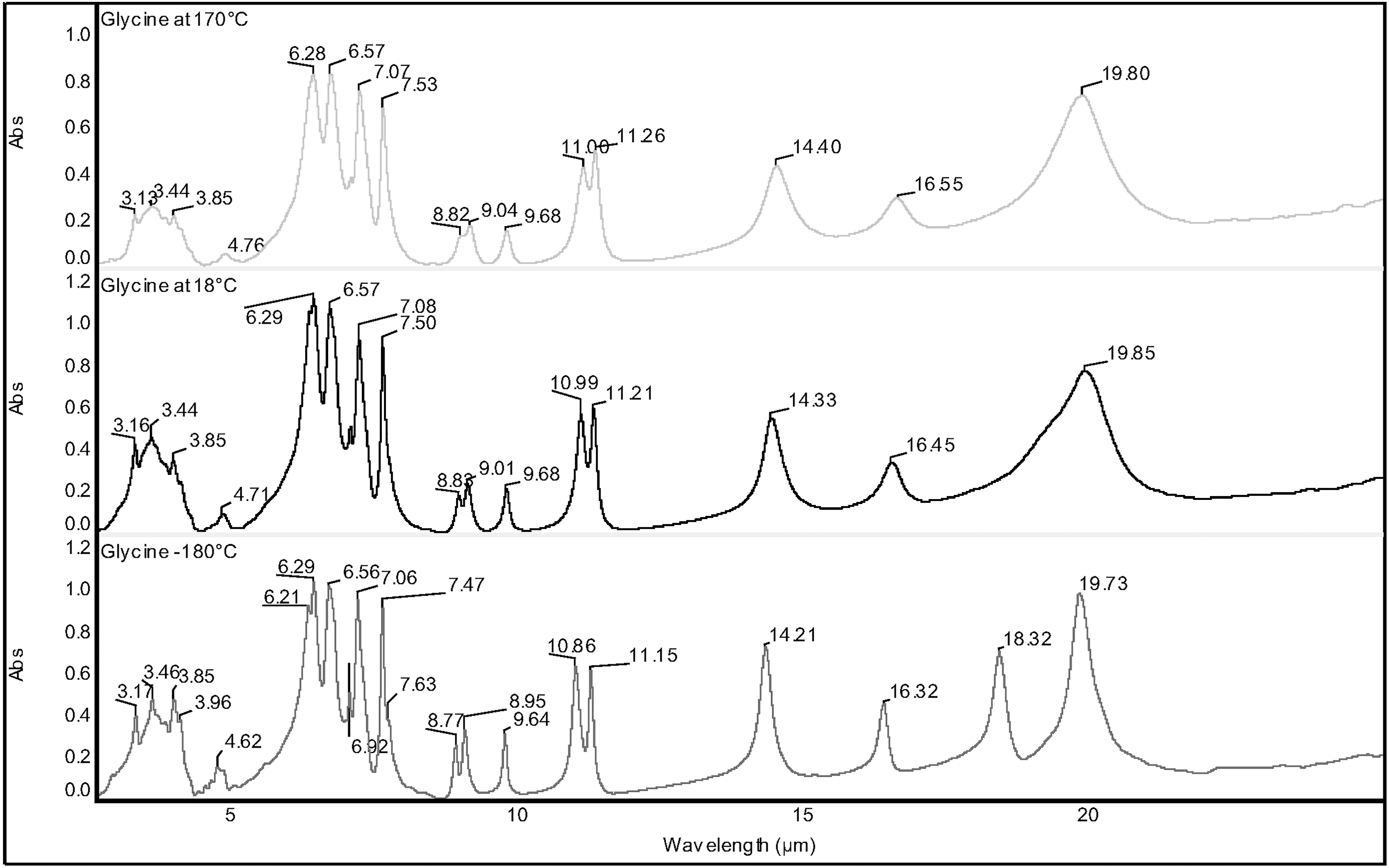
FT-IR spectra of glycine in CsI matrix recorded (from top to bottom) at +170°C, +18°C, and −180°C, respectively.
Mid- and Far-Infrared Bands of Glycine with Molar Extinction Coefficients and Integrated Molar Absorptivity
3.2. The molar extinction coefficients and integrated molar absorptivity of the far-infrared bands of selected amino acids
Currently, the far-infrared spectral range is preferably referred to as terahertz spectral range. To cover this low-frequency and low-energy spectral range, it is necessary to change the beam splitter of the FT-IR spectrometer. The standard beam splitter used to cover the mid-infrared spectral range is made in KBr, but to reach the far-infrared, the most common beam splitter is made by Mylar. In our Nicolet 6700 spectrometer, not only the beam splitter but a specific far-infrared detector as well should be mounted by substituting the standard detector for the mid-infrared. With these changes in the optical bench, our FT-IR spectrometer can cover the typical far-infrared spectral window comprised between 16.67 and 200 μm (600 and 50 cm−1). Furthermore, instead of CsI, which in any case may be satisfactory in the far-infrared spectral range, polyethylene was used as a pellet matrix. The ɛ and ψ values in the far-infrared of each selected amino acid are reported in bold italicized characters at the bottom of Tables 1–5 and are derived from Fig. 6. In all data reported in Tables 1–5, the tail of the mid-infrared bands overlaps with the head of the far-infrared spectral window so that certain infrared bands were measured two times with a mid-infrared spectrometer and sample configuration for Figs. 1–5 and with a far-infrared spectrometer and sample configuration for Fig. 6. It is possible to observe a fair agreement in the ɛ and ψ values as determined for different conditions, which helps ensure the accuracy the values determined.
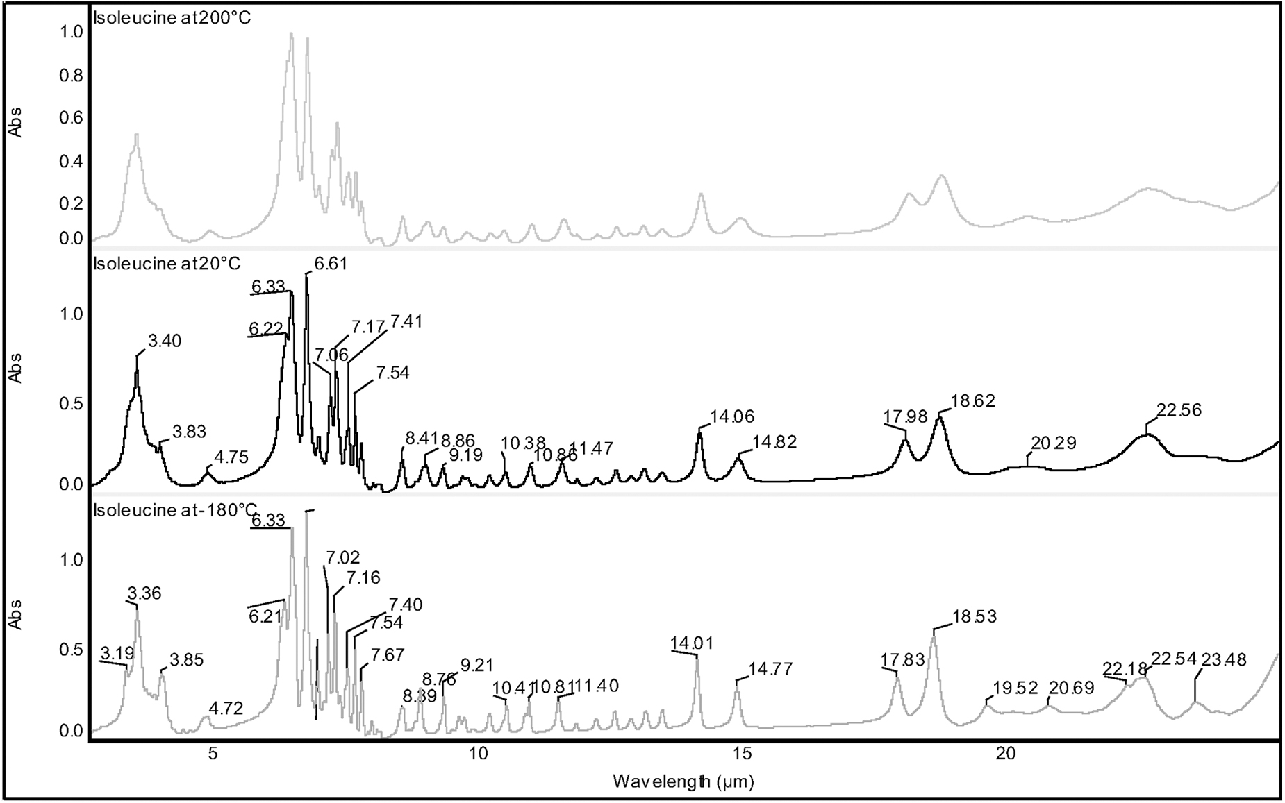
FT-IR spectra of isoleucine in CsI matrix recorded (from top to bottom) at +200°C, +20°C, and −180°C, respectively.
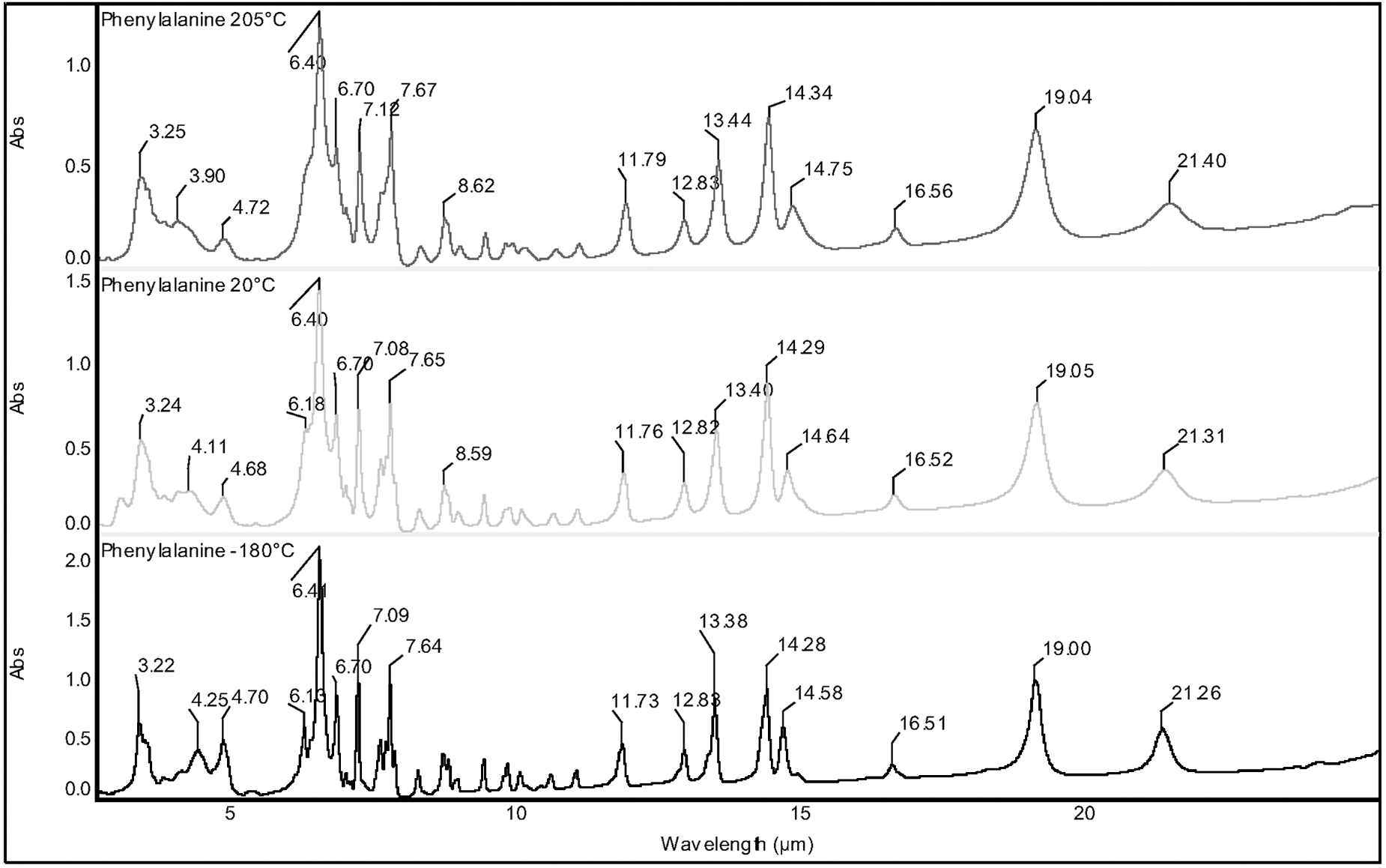
FT-IR spectra of phenylalanine in CsI matrix recorded (from top to bottom) at +205°C, +20°C, and −180°C, respectively.
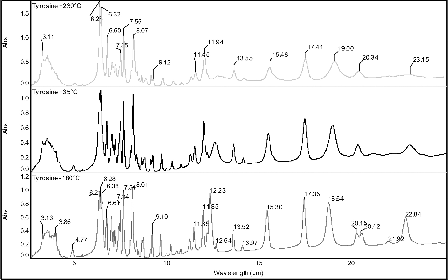
FT-IR spectra of tyrosine in CsI matrix recorded (from top to bottom) at +230°C, +35°C, and −180°C, respectively.
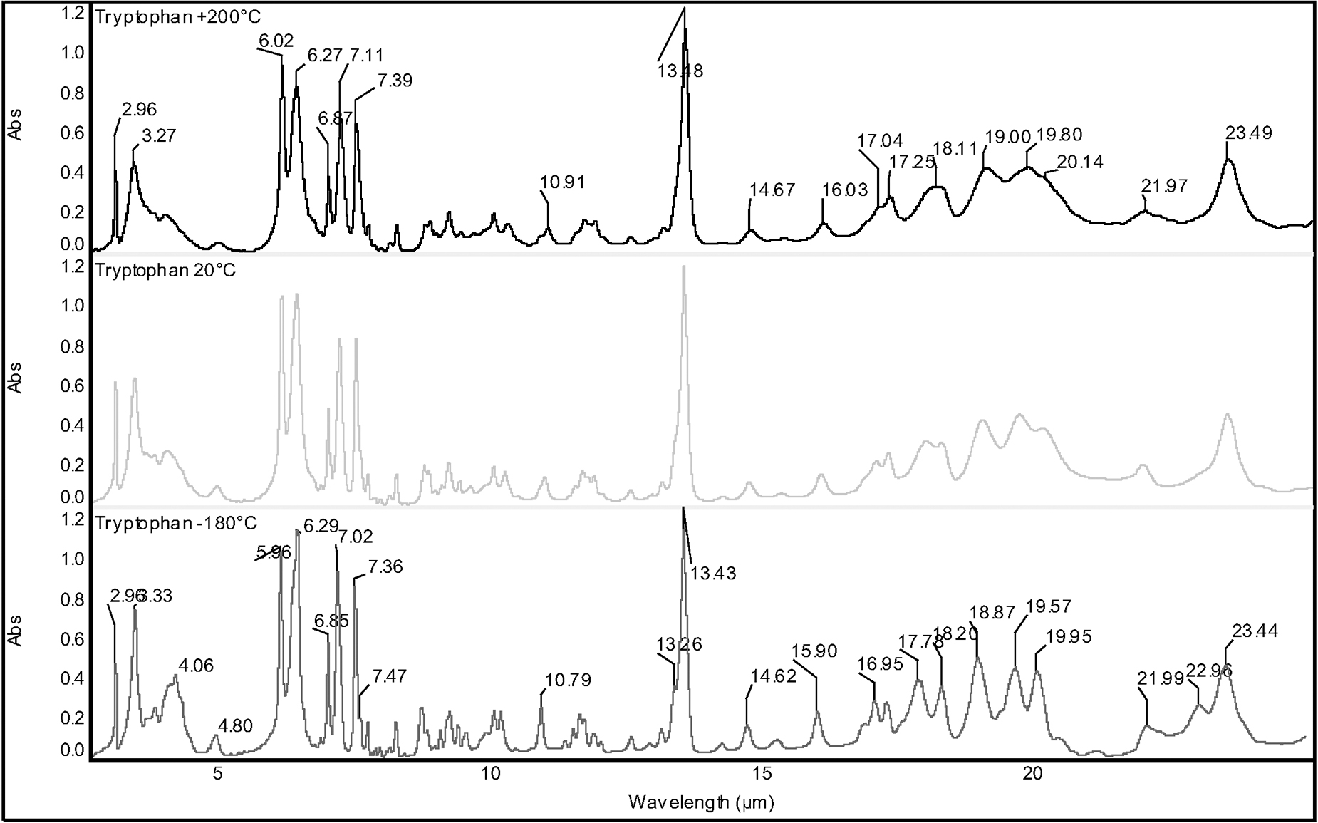
FT-IR spectra of tryptophan in CsI matrix recorded (from top to bottom) at +200°C, +20°C, and −180°C, respectively.
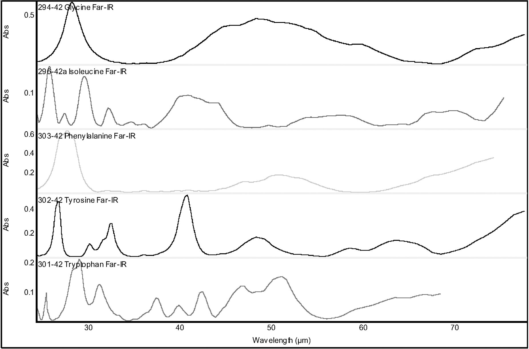
FT-IR spectra in the far-infrared range recorded at ambient temperature on CsI matrix; from top to bottom: glycine, isoleucine, phenylalanine, tyrosine, and tryptophan.
3.3. Infrared spectra from −180°C to room temperature to +200°C
The mid-infrared spectra of the selected amino acids were studied in a wide range of temperatures from -180°C (i.e., the liquid nitrogen temperature) to ambient temperature and the maximum permitted temperature before decomposition. Figure 1 shows the infrared spectra of glycine at −180°C, +18°C, and +170°C. The latter temperature was considered the highest reachable temperature without glycine decomposition. The low-temperature infrared spectroscopy of glycine was studied in the works of Chernobai et al. (2007) and Chesalov et al. (2008), and the spectral changes observed were found to be due to bond force constant dependence from temperature as well as polymorphism of glycine crystals. Chernobai et al. (2007) offered a complete analysis of the vibrational assignments and discussion of the crystalline state transition of glycine.
From an astrochemical perspective, it is necessary to know how the infrared spectrum of any given amino acid changes as a function of temperature. For example, Fig. 1 shows that the glycine infrared spectrum at −180°C allows the broad band at 19.85 μm at +18°C to resolve into two well-resolved bands at 19.73 and 18.32 μm. Furthermore, all the infrared bands of glycine appear narrower and sharper at low temperatures; and certain doublets, such as those at 10.86 and 11.15 μm, 8.77 and 8.95, or even 6.21 and 6.29 μm, appear much more resolved at −180°C. In the event that glycine (or any other amino acid) is conclusively discovered in space, a case could be made that the actual temperature of glycine (or any other amino acid) can be estimated from certain details of the detected infrared spectrum. Such details may indeed have to do with the fact that a band like that at 19.85 μm is resolved into two bands at very low temperature, each band width of the spectrum; the degree of resolution of selected doublets; and the relative intensity ratio as well as the possibility to detect certain weak bands such as those at 6.21, 6.92, and the shoulder at 7.63 μm. Another spectral tool with which to estimate the actual temperature of the detected glycine in space could be achieved through the use of linear equations that describe the shift of the band position as a function of temperature.
In general, the infrared bands are shifted to shorter wavelengths (and higher frequencies) at lower temperatures. This phenomenon called “temperature effect” is due to a combination of different effects that involve quantum mechanics and changes in bond length (and hence force constant) as reviewed in the work of Avram and Mateescu (1972). By studying the most sensitive infrared bands of glycine as function of temperature (Fig. 1), linear equations were derived and are reported in Table 1. By determining the exact position of the glycine infrared bands at 8.83, 10.99, 14.32, and 16.45 μm in an eventual infrared spectrum in space, it is possible to estimate the actual temperature of the molecules. Thus, for example, taking the following equation from Table 1 (with T absolute temperature):
then
The intercept in Eq. 3 is found at 8.743 μm, and it represents the position expected for this absorption band at nearly 0 K (Iglesias-Groth et al., 2012). Thus, for all equations that show wavelength dependence from a temperature reported in Table 1, it is possible to predict, from the intercept, the given band position at nearly 0 K.
The isoleucine mid-infrared spectra in a wide range of temperatures are reported in Fig. 2. The full analysis of the vibrational bands of isoleucine was reported, for example, in the work of Moorthi et al. (2014). Similar comments as already detailed for glycine spectra can be applied as well to isoleucine spectra, especially to that at −180°C, which provides the better-resolution spectrum. Table 2 also reports the linear equations determined for all the isoleucine infrared bands that show the higher temperature dependence or the infrared bands with the higher ɛ or ψ values, which should be those more easily detected in a space environment. These bands could be used to estimate the temperature of isoleucine in a space environment.
Mid- and Far-Infrared Bands of Isoleucine with Molar Extinction Coefficients and Integrated Molar Absorptivity
A full account of phenylalanine vibrational analysis can be found, for example, in the work of Kaczor et al. (2006). The relative infrared spectra of phenylalanine recorded at −180°C, +20°C, and +205°C are reported in Fig. 3. The infrared bands of phenylalanine are less sensitive to temperature and undergo smaller bathochromic shifts from low temperature to higher temperatures in comparison to the case of glycine and isoleucine, judging from the slopes of the linear equations that connect wavelengths with temperature (see Table 3).
Mid- and Far-Infrared Bands of Phenylalanine with Molar Extinction Coefficients and Integrated Molar Absorptivity
Tyrosine is molecularly a strict parent of phenylalanine, and the infrared spectroscopical review of these two molecules can be found in the works of Barth (2000, 2007). The infrared spectra of tyrosine at three different temperatures can be found in Fig. 4, while Table 4 also reports wavelength dependence from temperature of the most relevant infrared bands. The latter properties are completely equivalent to those measured in the case of phenylalanine.
Mid- and Far-Infrared Bands of Tyrosine with Molar Extinction Coefficients and Integrated Molar Absorptivity
Figure 5 shows the infrared spectra of tryptophan at −180°C, +20°C, and +200°C, and in this case the low-temperature spectrum is definitely richer and of better resolved infrared bands than the higher-temperature spectra. Table 5 shows that the temperature dependence of certain infrared bands of tryptophan is quite high. This is especially true in the case of the band at 13.43 μm, with the highest temperature dependence among the five amino acids studied in the present work. The vibrational analysis of tryptophan in the mid-infrared was again summarized by Barth (2000, 2007).
Mid- and Far-Infrared Bands of Tryptophan with Molar Extinction Coefficients and Integrated Molar Absorptivity
Figure 6 shows the far-infrared spectra of the selected amino acids recorded at ambient temperature. The main bands’ position is summarized at the bottom of Tables 1–5 in bold italicized characters. Due to limitations in our experimental setup, it was possible to collect only the far-infrared spectra at ambient temperature and not at the extreme temperatures as was the case in the mid-infrared spectral window.
4. Discussion
It is important to note here that the reported molar absorptivities were obtained under specific laboratory conditions and should be used with caution when attempting abundance determinations of amino acids in space. They could be useful to provide insight into the relative abundances of these species when observed under similar physical conditions in the same spectral regions. They are also valuable because they may allow for acquisition of the order-of-magnitude estimates for abundances. Precise abundance determinations will require that absorptivity values of amino acids are measured in laboratory experiments that resemble as much as possible the conditions of interstellar space.
It is well known that in aqueous solutions the position of the infrared bands of amino acids is greatly affected by the pH of the solution or, it could be said, the degree of dissociation and protonation of the amino acids. A similar destiny is also reserved for the ɛ values of the amino acids; that is, they are affected by the pH of the solution (Wolpert and Hellwig, 2006). Indeed, a direct comparison of the amino acids’ ɛ values determined in the solid state and in aqueous solution leads to large differences in some cases.
However, for glycine, the infrared bands at 9.67, 10.99, and 11.20 μm (1034, 910, and 893 cm−1) show the same ɛ values both in the solid state and in aqueous solution as reported in the work of Wolpert and Hellwig (2006). These bands are due to νCN and νCC stretching modes, which evidently are not too affected by the environment where the glycine is surrounded. Furthermore, the good match between the ɛ value determined in CsI matrix and in aqueous solution is another confirmation of the accuracy of the collected data. The good match in molar extinction coefficients as determined either in in the solid state (CsI) or in aqueous solution, as reported in the work of Wolpert and Hellwig (2006), can be observed in the case of isoleucine bands at 7.06, 8.87, 10.07, and 10.38 μm (1417, 1128, 993, and 964 cm−1); phenylalanine bands at 6.86 and 8.15 μm (1458 and 1226 cm−1); tyrosine bands at 6.22 and 9.10 μm (1608 and 1099 cm−1); as well as in the case of the tryptophan bands at 6.00, 8.12, and 17.00 μm (1666, 1231, and 588 cm−1). In all these cases, the same comments as expressed for glycine apply for the other amino acids. The infrared bands that display similar ɛ values in the solid state and in aqueous solutions are necessarily due to stretching or bending modes not directly affected by the interaction of the -COO- group and -NH2 moiety of amino acids with aqueous solutions.
As discussed in the work of Iglesias-Groth and Cataldo (2018), the determination of the mid- and far-infrared spectra of amino acids under various experimental conditions is of paramount importance for the future search for these molecules in space. The vibrations of amino acids in the far-infrared spectral range are essentially due to skeletal deformations, torsions, bending, rocking, and wagging modes. Furthermore, hydrogen bonding modes can also contribute in this spectral range.
New orbiting telescopes will be able to cover a significant fraction of the relevant spectral range of these transitions with much higher spectral resolution and sensitivity than before. This is the case, for instance, for the MIRI instrument on board the James Webb Space Telescope, which is well suited to the study and, ultimately, the unveiling of amino acid transitions in various astrophysical contexts, from nearby molecular clouds and reflection nebulae to protoplanetary disks and the interstellar medium of star-forming regions. Indeed, the far-infrared spectra of all the proteinogenic amino acids were recently determined and the far-infrared vibrations assigned and discussed in a broader perspective involving previous literature data (Iglesias-Groth and Cataldo, 2018).
In the case of far-infrared bands, very limited information is available on molar extinction coefficients (ɛ) and integrated molar absorptivities (ψ). It is very relevant to extend the present work to other amino acid species. If bands from various amino acids will ever be detected in the far-infrared in some astrophysical environment or object, their ɛ and ψ values will help complement the mid-infrared data, contribute to improve abundance estimates, and set more reliably constraints on amino acid relative abundances.
5. Conclusions
The results of this work constitute a series of key tools in the search for five selected amino acids (glycine, isoleucine, phenylalanine, tyrosine, and tryptophan) in space. First, the mid-infrared spectra of the five amino acids are reported, which were recorded at three different temperatures, namely, −180°C, +20°C, and +200°C. Furthermore, the far-infrared spectra of the selected amino acids are reported as well at +20°C. Both the integrated molar absorptivity (ψ) and the molar extinction coefficients (ɛ) of all mid- and far-infrared bands were determined and are reported.
With these data, it will not only be possible to search for, and eventually identify, the selected amino acids in different space environments, but this will also allow for the estimation of the local abundance of the eventually detected amino acids through the use of the ψ and ɛ values. The dependence on temperature of certain key bands (the most intense or the most temperature-dependent) of selected amino acids may allow for estimation of the actual temperature of the amino acids once they are discovered in a given astrophysical object.
ORCID
Franco Cataldo
Funding
We acknowledge the support of the Instituto de Astrofísica de Canarias and the Ministry of Science and Innovation of Spain via project ESP2015-69020-C2-1-R.
Footnotes
Abbreviation Used
Associate Editor: Sara Seager