
Abstract
Abstract
Introduction:
Human breastmilk is a dynamic, multifaceted biological fluid containing nutrients, bioactive substances, and growth factors. It is effective in supporting growth and development of an infant. As breastmilk has been found to possess mesenchymal stem cells, the importance of the components of breastmilk and their physiological roles is increasing day by day. The present study was intended to identify the secretions of growth factors, mainly vascular endothelial growth factor (VEGF) and hepatocyte growth factor (HGF), from human breastmilk mesenchymal stem cells under basal conditions of in vitro cell culture using synthetic media and human cord serum.
Materials and Methods:
The growth factors were analyzed with the enzyme-linked immunosorbent assay technique.
Results:
The cultured mesenchymal stem cells of breastmilk without serum revealed significant differences in secretions of the VEGF and HGF growth factors (8.55 ± 2.26402 pg/mL and 230.8 ± 45.9861 pg/mL, respectively) compared with mesenchymal stem cells of breastmilk with serum (21.31 ± 4.69 pg/mL and 2,404.42 ± 481.593 pg/mL, respectively).
Conclusions:
Results obtained from our study demonstrate that both VEGF and HGF are secreted in vitro by human breastmilk mesenchymal stem cells. The roles of VEGF and HGF in surfactant secretion, pulmonary maturation, and neonatal maturity have been well established. Thus, we emphasize that breastmilk-derived MSCs could be a potent therapeutic source in treating neonatal diseases. Besides, due to its immense potency, the study also emphasizes the importance of breastfeeding, which is promoted by organizations like the World Heatlh Organization and UNICEF.
Introduction
A
The mammary gland is a metabolically active, dynamic organ that undergoes significant changes during pregnancy, lactation, and transformation. 1 Breastmilk is the secretory product of this mammary gland in the postpartum period. It is contemplated to be a dynamic, multifaceted biological fluid containing nutrients, bioactive substances, and growth factors. It provides growth, development, immunity, and protection to the newborn from various gastrointestinal and respiratory infections. 2
The reappearance and remodeling of mammary gland tissue and its ability for expansion point out the existence of stem and progenitor cells in the mammary gland. Human breastmilk has been found to possess a putative mammary stem cell population with expression markers such as CK5, CK14, CK19, and nestin.3,4 In addition, it is also identified as harboring heterogeneous unique cellular components like mesenchymal stem cells (MSCs), hematopoietic stem cells, side population cells, and endothelial cells.3,5 As a result this breastmilk-derived stem cell population is believed to possess great potency for the treatment of a wide variety of neonatal diseases without any special intervention. Breastmilk has also been demonstrated to possess a subpopulation set of cells that showed positivity for markers of pluripotency such as Oct4, Sox2, and Nanog. 6
Much attention has been devoted to the physiological roles of growth factors in the growth, maturation, and maintenance of the neonatal gastrointestinal tract.7–9 Human breastmilk contains various growth factors that participate in various biological functions in infants. 10 Among them, vascular endothelial growth factor (VEGF), hepatocyte growth factor (HGF), and epidermal growth factor are of foremost importance. VEGF regulates angiogenesis and vasculogenesis. 11 HGF stimulates growth, motility, and morphogenesis in epithelial cells and other diverse cell types. 12 Besides, these growth factors play a role in surfactant secretion, pulmonary maturation, and neonatal maturity.13,14 In a recent study, the secretion of growth factors and cytokines by mesenchymal stem cells along with its immunomodulatory properties are well described. 15 Besides, from our previous study, we showed that the levels of growth factors are negligible in maternal serum but higher in milk supernatant. 16 We already know that the MSCs are being used in experimental animal models as well as in human trials to evaluate their therapeutic safety and efficacy in various diseases. 17 These cells after administration play a paracrine role through the secretions of cytokines and growth factors to stimulate the organ's resident progesterone. 18 With this background we aimed to study whether the MSCs from human breastmilk secrete VEGF and HGF in vitro in culture medium.
Materials and Methods
Subjects
The study was approved by the Institutional Ethics Committee of Charotar University (Changa, Gujarat, India). We collected breastmilk samples from 26 lactating women during postpartum days. They were healthy, without any associated medical disorder like diabetes, hypertension, or any other infectious diseases. The methods and purpose of the study were explained to the participants, and their informed written consent was obtained prior to enrollment.
Collection of breastmilk samples
The samples were collected every day from Day 0 until Day 7 postdelivery, usually in the morning. The samples were collected by a sterile technique in sterile 15-mL Falcon™ tubes (BD, Heidelberg, Germany) and transported to the laboratory for isolation of cells.
Isolation and culture
The breastmilk was diluted 1:2 with Dulbecco's modified Eagle's medium (DMEM) (Gibco, Invitrogen, Carlsbad, CA) containing antibiotic (2 μg/mL amphotericin B and 2 μg/mL ciprofloxacin) and centrifuged at 300 g for 15 minutes at room temperature. The supernatant was discarded, and the cell pellet was washed twice with sterile phosphate-buffered saline by centrifugation at 285 g for 10 minutes. The viability of the isolated cells was assessed by the trypan blue exclusion assay. The final cell pellet was seeded in a 35-mm2 treated tissue culture dish (BD, Franklin Lakes, NJ) using DMEM containing 10% heat-inactivated human umbilical cord blood serum and supplemented with penicillin (100 units/mL)/streptomycin (100 μg/mL) and incubated at 37°C under 5% CO2 and 95% humidity.
The medium was changed every 48 hours and replenished with DMEM supplemented with 10% human cord blood serum. At 80% confluency, cells were passaged using trypsin-EDTA.
Characterization of isolated cells using flow cytometry
Isolated cells were characterized with a BD FACSAria™ cell sorter using a 488-nm argon ion laser and 632-nm red laser for excitation. Fluorescence emission was collected using the corresponding detectors. About 1 × 10 5 cells were stained with saturating concentrations of fluorochrome-conjugated antibodies. The cells were incubated with allophycocyanin for CD105 and CD117, fluorescein isothiocyanate for CD44, and phycoerythrin for CD140b. The cells were incubated in the dark for 20 minutes at room temperature, washed three times with wash flow buffer, and resuspended in 500 μL of phosphate buffer solution. Data analysis and acquisition were then performed using FACSDiva™ software (BD). A minimum of 10,000 events was characterized and recorded.
Growth curve analysis
Growth curves were plotted to evaluate population growth characteristics of the isolated cells in DMEM supplemented with 10% human umbilical cord blood serum and 1% antibiotic/antimycotic solution, and characterization was performed on 12-well plates in duplicates, with a seeding density of 2 × 10⁵ cells per plate at Day 0. Cells were harvested and counted every day, until Day 10. Results were plotted on a log-linear scale, and population doubling time (PDT) was calculated using the following formula:
Analysis of growth factors
The concentrations of VEGF and HGF in the supernatant of cultured human breastmilk MSCs with cord serum and human breastmilk MSCs without cord serum were measured in triplicate using an enzyme-linked immunosorbent assay, in a blind fashion, according to the manufacturer's instructions (Quantikine® human HGF and VEGF immunoassays; R & D Systems, Minneapolis, MN). The kit has the characteristics of intra-assay precision (precision within an assay) where three samples of known concentration were tested 20 times on one plate to assess intra-assay precision. The kit also has inter-assay precision (precision between assays) where three samples of known concentrations were tested in 40 separate assays to assess inter-assay precision. These assays use the quantitative sandwich enzyme-linked immunosorbent assay technique. An antibody that was specific for each growth factor had been precoated onto a microplate. Standards and samples were pipetted into the wells, and the immobilized antibody bound to any growth factor present. After unbound substances were washed away, an enzyme-linked antibody specific for each growth factor was added to the wells. Following washing to remove unbound antibody–enzyme reagent, a substrate solution was added to the wells. Color developed in proportion to the amount of each growth factor bound in the initial step. The color development was stopped, and the color intensity was measured. The optical density was determined for each well using a microplate reader set to 450 nm.
Statistical analysis
The statistical analysis was done by means of the SPSS statistical software package (SPSS, Inc., Chicago, IL). Levels of VEGF and HGF were expressed as mean ± standard deviation values. Fisher's (F) test was applied to compare normally distributed variables. A probability (p) of ≤0.05 was considered significant.
Results
The clinical data of the study are given in Table 1.
Data are mean ± standard deviation values.
F, female; M, male.
The cultured cells showed initially more of an epithelial contour at primary culture (Fig. 1A) and, upon further passages, developed into fibroblastic-like morphology, representing the mesenchymal cell phenotype (Fig. 1B and C). The cultured cells were characterized using MSC markers such as CD105, CD44, CD117, and CD140b (platelet-derived growth factor receptor-beta) (Fig. 1D). From the results, we identified that MSC-specific markers are highly expressed in cultured breastmilk. With the fibroblastic morphology at later passage and the marker positivity, the mesenchymal cell phenotype is confirmed.

Breastmilk-derived cells observed at ×400 phase-contrast microscopy and flow cytometric data:
The growth curve analysis demonstrates the growth potency of the MSCs cultured from breastmilk (Fig. 2). The PDT was found to be 3.2 days.
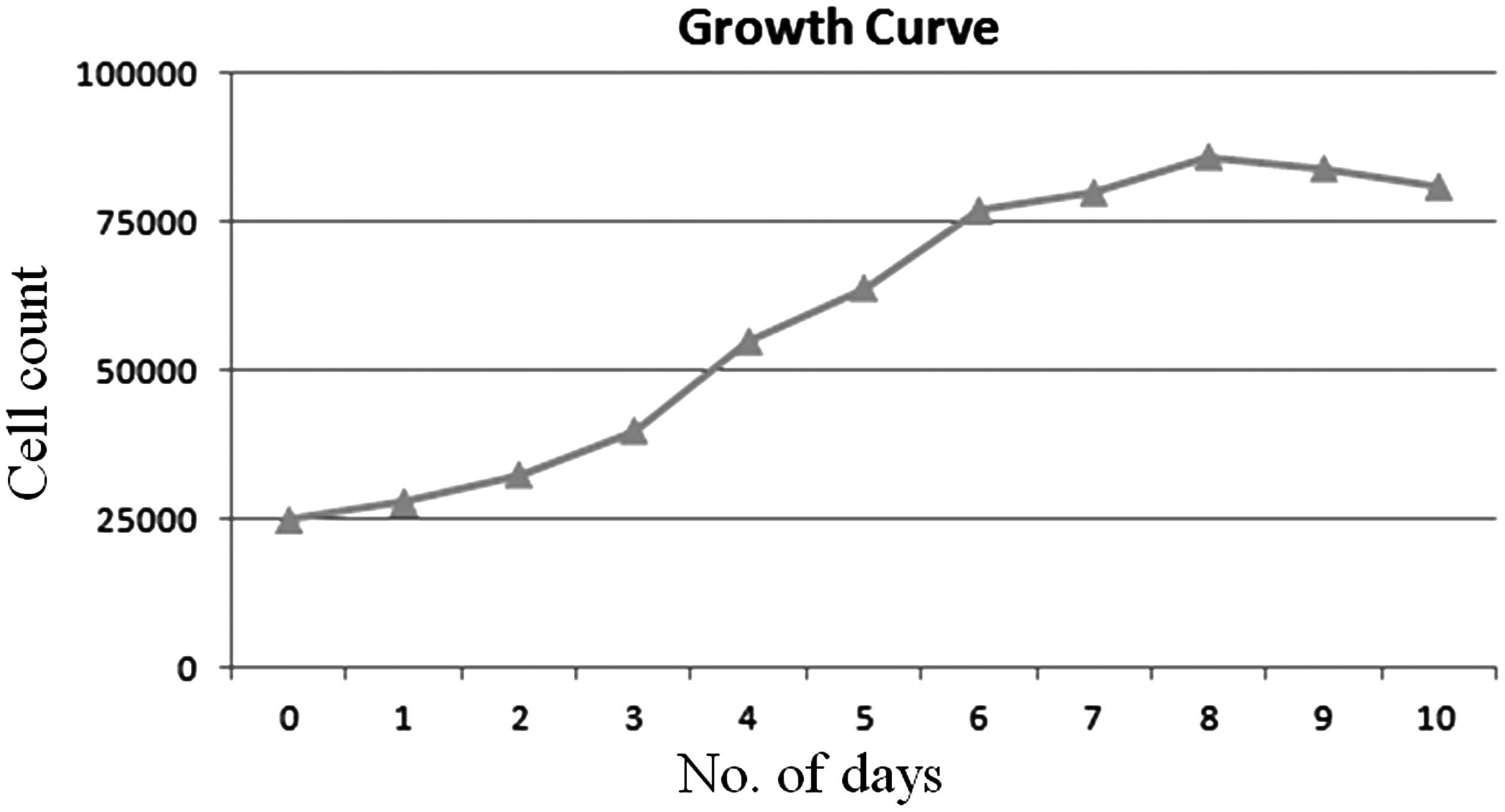
Growth curve analysis. The growth potency of the breastmilk-derived mesenchymal stem cells shows a decline in growth from Day 10.
We assessed the growth factor secretion by these breastmilk-derived MSCs with and without cord serum to check that the growth factors secreted are from MSCs and not influenced by cord serum. The VEGF levels secreted by MSCs with and without serum were 21.31 ± 4.69 pg/mL and 8.55 ± 2.26402 pg/mL, respectively. Similarly, the HGF levels secreted by MSCs with and without serum were 2,404.42 ± 481.593 pg/mL and 230.8 ± 45.9861 pg/mL, respectively (Table 2). Thus these findings indicate that these growth factors could be secreted from these MSCs without the influence of serum.
Data are mean ± standard deviation values. The p value indicates a significant difference exists between breastmilk MSCs with cord serum and breastmilk MSCs without cord serum.
HGF, hepatocyte growth factor; VEGF, vascular endothelial growth factor.
Discussion
The milk of each species is specific and has a unique composition that has evolved over millions of years to suit the infant for which it is intended.19,20 Breastmilk is undoubtedly a one of a kind natural source of nutrition for the human infant. In recent years study of the cellular components of breastmilk in the search for stem cells has gained consensus. The cellular fraction of human milk contains various types of cells, including MSCs, as demonstrated by various investigators.3–5
The present study is different from those studies reported above. It intends to identify the secretions of growth factors, mainly VEGF and HGF, from human breastmilk MSCs. The rationale of choosing these growth factors is due to itheir potency in growth and development of neonates.
VEGF and HGF stimulate growth, motility, morphogenesis, and development.11,12 Overall, they help in growth and development of the organ system of the neonate such as pulmonary development, gastrointestinal development, and so on.13,14 Kobata et al. 21 had demonstrated high levels of growth factors (epidermal growth factor, VEGF, and HGF) in human breastmilk. Siafakas et al. 22 have also shown high concentrations of VEGF in human breastmilk and its binding to specific receptors on the cells of intestinal epithelium. Our previous study interestingly demonstrated that the levels of VEGF and HGF in human breastmilk were severalfold higher than those observed in the umbilical cord blood. 16 The role of these growth factors in the development of an individual is well known. However, the source of these growth factors is uncertain, and whether the breastmilk-derived MSCs could secrete these growth factors has not yet been established.
The breastmilk-derived cells were cultured using synthetic medium (DMEM) and human cord blood serum under the basal conditions of in vitro cell culture. Here the cultured MSCs of breastmilk without serum revealed significant secretions of the VEGF and HGF growth factors (8.55 ± 2.26402 pg/mL and 230.8 ± 45.9861 pg/mL, respectively), compared with MSCs of breastmilk with serum (21.31 ± 4.69 pg/mL and 2,404.42 ± 481.593 pg/mL, respectively). The levels in serum alone were 16.632 ± 0.773 pg/mL and 2,581.6 ± 108.275 pg/mL, respectively. 16 These results indicate that MSCs are the source of the growth factors secreted under culture conditions. Besides, it is apparent that the MSCs solely secrete these growth factors without the influence of the serum.
The capacity to secrete protective biologically active factors designates MSCs among the most suitable tools for paracrine contribution to tissue regeneration. 23 MSCs have been found ubiquitously throughout the body and are routinely isolated from nearly all human tissues, as well as being mostly found in the perivascular niche. 24 They have been obtained from a wide variety of fetal and adult tissues: adipose, 25 placenta, 26 lung, 27 umbilical cord, 28 synovial membrane, 29 and dental pulp, 30 among many others. MSCs were first isolated from bone marrow. 31 The unique properties of MSCs make it an ideal source for its regenerative applications. Their abilities to migrate into injured tissues, engraft, and differentiate into many cell types are directly involved in promoting tissue regeneration processes. 32 The paracrine mediators initiate chemotactic signaling, which results in activation of resident stem cells and mobilization of circulating systemic stem cells.33,34 Clinical trials have already established use of MSCs in an autologous and allogenic manner, as MSCs possess immunomodulatory properties.35–37
Besides, the pluripotent property of these MSCs also plays a vital role for their application in treating a wide variety of diseases. The presence of markers of pluripotency is also identified in breastmilk. Hassiotou et al. 6 demonstrated the multilineage differentiation potential of these pluripotent cells in breastmilk and identified its differentiation ability to form cells of ectodermal, mesodermal, and endodermal origin, including bone, liver, and neuronal cells. This has also opened new avenues for the use of breastmilk as a noninvasive source of pluripotent cells for stem cell therapies.
The hypothesis that MSCs act through a paracrine mechanism by secreting growth factors is supported by our experiment that noted secretions of VEGF and HGF in vitro by breastmilk-derived MSCs. Thus, we recommend breastmilk-derived MSCs as a source of regenerative therapeutics in treating neonatal disorders. Besides, we also propose the concept that “breastfeeding is a natural source of stem cell therapy,” where the child receives the stem cells, growth factors, and other cellular components from the mother as a natural gift, favoring growth and development of the neonate.
Conclusions
In conclusion, we have begun to draw on possibilities of using human breastmilk as a promising source of stem cell therapeutics. This observation thus emphasizes the importance of breastfeeding, which is promoted by organizations like the World Health Organization and UNICEF. Further work on other growth factors and cellular components of breastmilk might shed light on its therapeutic applications. Besides, it paves way to store these cells to widen the network of clinical trials.
Footnotes
Acknowledgments
We thank all the lactating women for kindly providing breastmilk samples to us.
Disclosure Statement
P.M.K., I.S., A.B.N., J.S.P., and S.A.S. have no conflict of interest to declare.