
Abstract
Insulin-dependent diabetes mellitus is one of the leading causes of death world wide. Donor-derived pancreas and islet of Langerhans transplantation are potential cures, however, postmortem ischemia impacts islet quality. The murine βt3 cell line was used as a model to study apoptosis after hypothermic storage by comparing Unisol™ with Belzer's machine perfusion solution (BMPS) and the University of Wisconsin (UW) solution. The objective was to determine which of these solutions provided the best support for βt3 cells and which solution demonstrated the least amount of apoptotic activity. Several apoptosis markers were measured that included the translocation of phosphatidylserine, caspase activity, and the formation of DNA laddering. In addition, metabolic activity and membrane integrity were also measured. The results demonstrated that the three solutions behaved similarly during overnight cold storage at 4°C. However, Unisol was consistently better than UW solution and BMPS, demonstrating better cell viability and recovery, and lower levels of apoptotic activity when all three parameters were measured. These results demonstrated that apoptosis plays an important role in the survival of cells and tissues during cold storage. Development of solutions to help prevent or decrease the levels of apoptosis after cold storage will likely improve overall cell and tissue recovery and survival in a clinical setting.
Introduction
I
In the search for a cure for diabetes, researchers have sought ways to return normal pancreatic function to the body. The methods used have included whole pancreas transplants, human islet transplants, animal islet transplants, fetal tissue exchange, creation of artificial pancreas or beta cells, and transplantation of genetically engineered cells.1,2 All of these procedures have both positive and negative attributes. Pancreatic islet transplantation received a strong boost from the introduction of glucocorticoid-free immunosuppressive regimens. As a result, there is now a consensus that islet transplantation may be a viable option for the treatment of IDDM. The short-term success of the first glucocorticoid-free protocol3,4 and progress in modification of the protocol for longer term post-transplant islet function 5 stimulated our search for technologies that may help overcome the shortage of pancreata for islet isolation.
Procurement of heart beating donor pancreases for islet isolation and transplantation is in its infancy. Many pancreata suitable for islet isolation and transplantation are not procured due to concerns about postmortem ischemia. Postmortem ischemia during hypothermic transport on ice results in autolysis of the insulin-producing β cells in the islets, inadequate islet yields, and poor function. Current practice is to flush and transport the pancreas with University of Wisconsin (UW) solution on ice. We anticipate that better pancreas preservation may be achieved by perfusing the pancreas during hypothermic storage.6–8 Allogeneic kidneys have been shown to function better after perfusion in a large prospective, randomized multicenter study. 9 The long-term objective of our studies is the development of an optimized pancreas storage solution for hypothermic perfusion of the pancreas with preservation of the islets of Langerhans for transplantation.
Using cells isolated from the organ of interest provides a good model for evaluating how the whole organ will behave under hypothermic conditions. In addition, removal of the cells from hypothermic storage back into physiological conditions also emulates cell and organ behavior during rewarming, when reperfusion injury usually occurs. To this end, we used a murine β-cell line to study apoptosis after hypothermic storage. A comparison of the lead commercially available organ perfusion solution, Belzer's machine perfusion solution (BMPS), and the lead commercially available organ flush solution used for static storage of organs, UW solution,10–16 with a new proprietary solution, Unisol™, was performed.17–20 The objective was to determine which of these solutions provided the best support of βt3 cells by evaluating the degree of apoptosis after hypothermic storage. It is anticipated that these studies will translate to large mammal pancreas models and eventually human pancreas preservation.
Materials and Methods
Cell line
A murine cell line, βt3, derived from pancreatic islets was used in these studies. 12 The cells were maintained at 5% carbon dioxide at 37°C and cultured in Dulbecco's modified Eagle's medium (DMEM) supplemented with 15% horse serum, 4.5 g/L glucose, 2.5% fetal bovine serum, 100 U penicillin, 100 μg streptomycin, and 2 mM glutamine. Cells, 1 × 10⁵/well, were plated in 24-well microtiter plates and cultured overnight in supplemented DMEM before use in experiments.
Storage conditions
After plating the day before, the cells were washed with phosphate-buffered saline (PBS) and then stored in 0.5 mL/well of the intracellular formulation of Unisol,17–18 BMPS, 10 or UW solution 10 at 4°C for 16–24 hours. After cold storage, the cells were washed thrice with PBS and then incubated in supplemented DMEM for 1 hour at 37°C for recovery before cell viability and apoptosis assessment. 22
Measurement of cell viability
alamarBlue™ (Trek Diagnostics) was used to assess cell viability by measuring the oxidation/reduction reactions that take place within cells. alamarBlue was added directly to the plates containing cultured cells in culture medium and incubated for 3 hours at 37°C. Upon reduction, alamarBlue changes color and this color change can be measured and quantified. The culture plates were read using a Gemini EM fluorescent microplate reader (Molecular Dynamics) at an excitation wavelength of 544 nm and an emission wavelength of 590 nm. After each reading, the cells were washed twice with PBS and then placed back in tissue culture. Results shortly after rewarming (day 0) demonstrate cell viability and, after 1–2 days, decreases indicate cell death due to apoptosis, and increases over 1–4 days or longer reflect cell proliferation.
In addition to measuring the metabolic activity, the viability of the cells was also assessed by measuring their membrane integrity with the ViaCount assay and the Guava cell analysis system (Guava PCA-96). The ViaCount assay uses a permeable nuclear dye to stain live cells and an impermeant dye to stain dead or damaged cells. When the labeled cells are analyzed in the Guava PCA-96, the cell populations can be identified and quantified for an accurate live and dead cell count. After storage and recovery, the cells are washed with PBS and then gently removed from the well using trypsin. Approximately 50 μL of cells was mixed with 450 μL of ViaCount reagent and incubated at room temperature for 5 minutes before being analyzed in the Guava cell analysis system. Subsequent days poststorage were processed and analyzed in a similar manner.
Apoptosis assays
Using the Guava cell analysis system (Guava PCA-96), four separate assays examining early and late apoptotic events were performed. Nexin staining binds to phosphatidylserine that translocates to the outside of the membrane early in apoptosis. The caspase activity is measured in two ways. First, a tagged general caspase inhibitor will bind to activated caspases and the marker allows the relative caspase activity to be measured. In a second caspase assay, inhibitors specific for caspases 3 and 7 will be used. Caspase 3 and 7 perform the initial cleaving of cellular substrates, which promotes the morphological and biochemical states that are indicative of apoptosis.23,24 Finally, a modified version of the TUNEL assay was used to measure the DNA ladder. As the DNA fragments expose the 3′-hydroxyl ends, terminal deoxynucleotidyl transferase adds Br-dU to the ends, which can subsequently be labeled using tagged antiserum. Once labeled, ∼2000 cells were counted and the percent of the apoptotic cells counted could be determined.
Statistics
All experiments were repeated at least thrice with 6–12 replicates in each experiment. Statistical differences were assessed by analysis of variance (ANOVA) and post-tests were done using one-way ANOVA and Tukey's multiple comparison test. p-Values <0.05 were regarded as significant.
Results
These experiments were designed to compare Unisol, a proprietary solution made in our laboratory, with the leading organ storage solution, UW, and the leading machine perfusion solution, BMPS. To this end, the pancreatic cell line βt3 was used, because we were interested in developing storage strategies for pancreas for transplantation or the isolation of pancreatic islets. βt3 cells were plated in 24-well plates and left in one of the three solutions mentioned at 4°C for 16–24 hours. After storage, the cells were washed and left in a culture medium to recover before assessment of cell viability and apoptosis.
Cell viability
Cell viability was assessed using two parameters, the metabolic activity and membrane integrity. The metabolic activity was measured using the nontoxic indicator alamarBlue, and percent cell viability was calculated based on an untreated control whose metabolic activity was measured when treated cells were placed into cold storage (Fig. 1). alamarBlue is nontoxic, and hence, it can be used to measure the same cell cultures over several days, as was done for these experiments. The alamarBlue assay results reflect changes within the entire cell culture, while the ViaCount assay discussed below is based on a sampling of cells, not the entire cell culture. In both cases, untreated controls measured at the start of cold storage (time zero) were used to calculate viability for all the days poststorage that were measured.
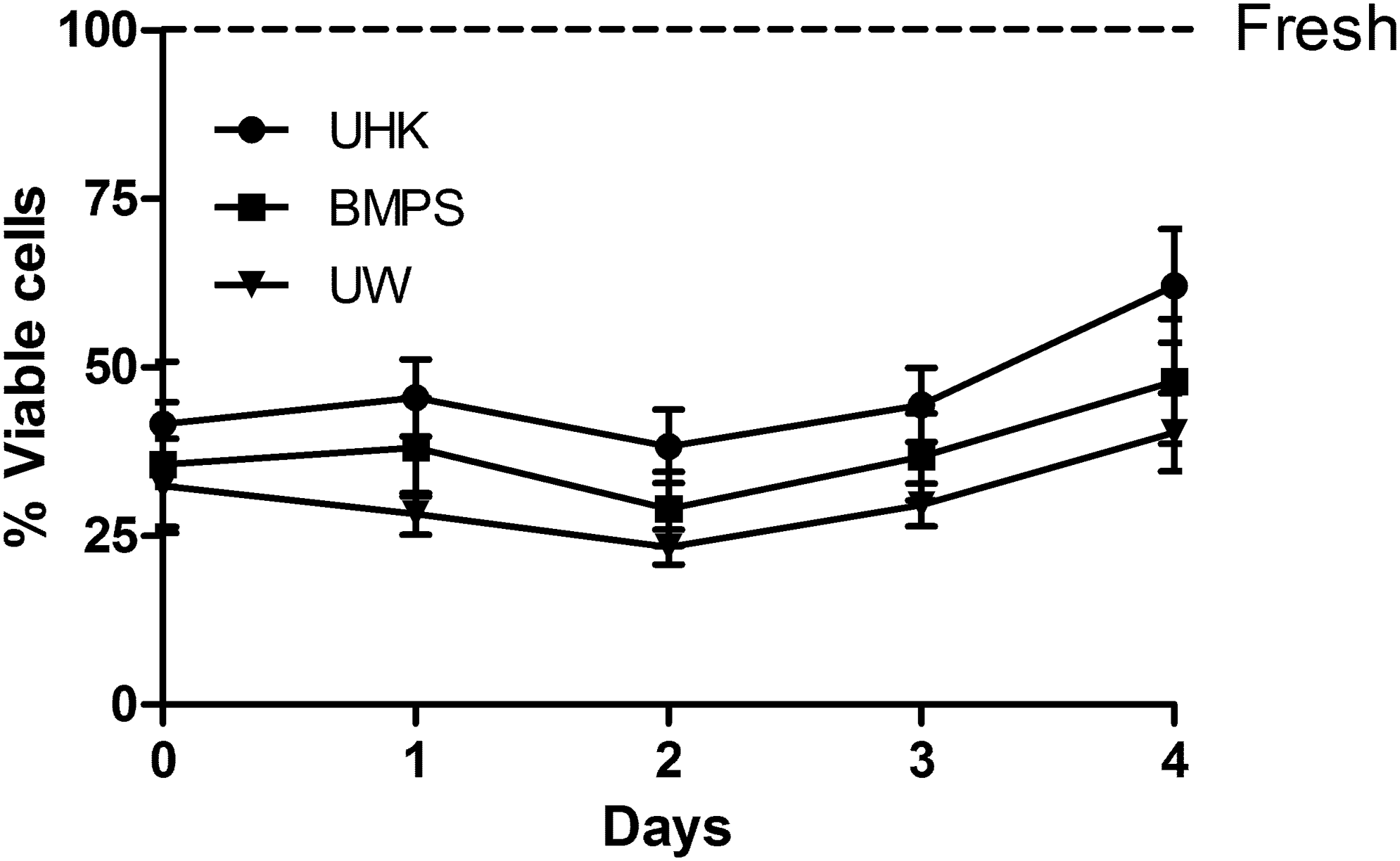
Cell viability of βt3 cells after storage at 4°C. βt3 cells were stored for ∼16–24 hours in Unisol™, BMPS, or UW. After a 1-hour recovery at 37°C, cell viability was measured every day for 4 days poststorage. Values represent the mean ± SEM for 12 replicates. Differences in viability were considered significant by one-way ANOVA, p < 0.05. ANOVA, analysis of variance; BMPS, Belzer's machine perfusion solution; UHK, Unisol™; UW, University of Wisconsin.
When the cells were removed from cold storage and their metabolic activity was measured, the initial cell viability was between 30% and 40% for all the solutions, with Unisol demonstrating the best viability at 40%. Over the next several days postrewarming, the viability of the cells fluctuated somewhat, but by day 4, cells stored in each of the solutions demonstrated that they had recovered and were starting to proliferate as observed by their increase in cell viability over initial values at day 0. Overall, Unisol demonstrated the best initial viability and recovery over the 4 days that the cells were assessed, showing a significant difference compared to the UW solution (p < 0.05).
Membrane integrity was also measured using the ViaCount assay and the Guava cell analysis system (Fig. 2). Although this assay has limitations, in that it can only count cells that are present in the sample at the time they are harvested, which can be variable across separate samples and over time in a treatment group, it was a good indicator of how the cells performed after storage. Fresh cells were evaluated at the time the experiment was started. Separate wells of cells that had been treated the same were evaluated for each solution immediately after rewarming and for several days postrewarming. Initial membrane integrity at day 0 showed significant differences between fresh untreated cells and each of the storage solutions used (Unisol, p < 0.05; BMPS and UW, p < 0.001). Significant differences were also noted between Unisol and the other two solutions (p < 0.001). Over the course of the next several days, the cells stored in each solution recovered quickly to values equivalent to fresh untreated cells with little difference in viability observed between each solution.
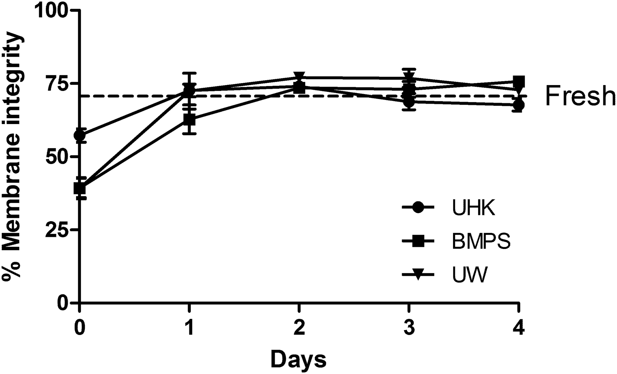
Membrane integrity after storage at 4°C. βt3 cells were stored as described in Figure 1. After recovery, the cells were stained and live cells with intact membranes were counted. A total of 2000 cells were counted using the Guava cell analysis system. Values represent the mean ± SEM for 12 replicates. Differences in viable cells were considered significant by one-way ANOVA.
Apoptosis
Nexin staining
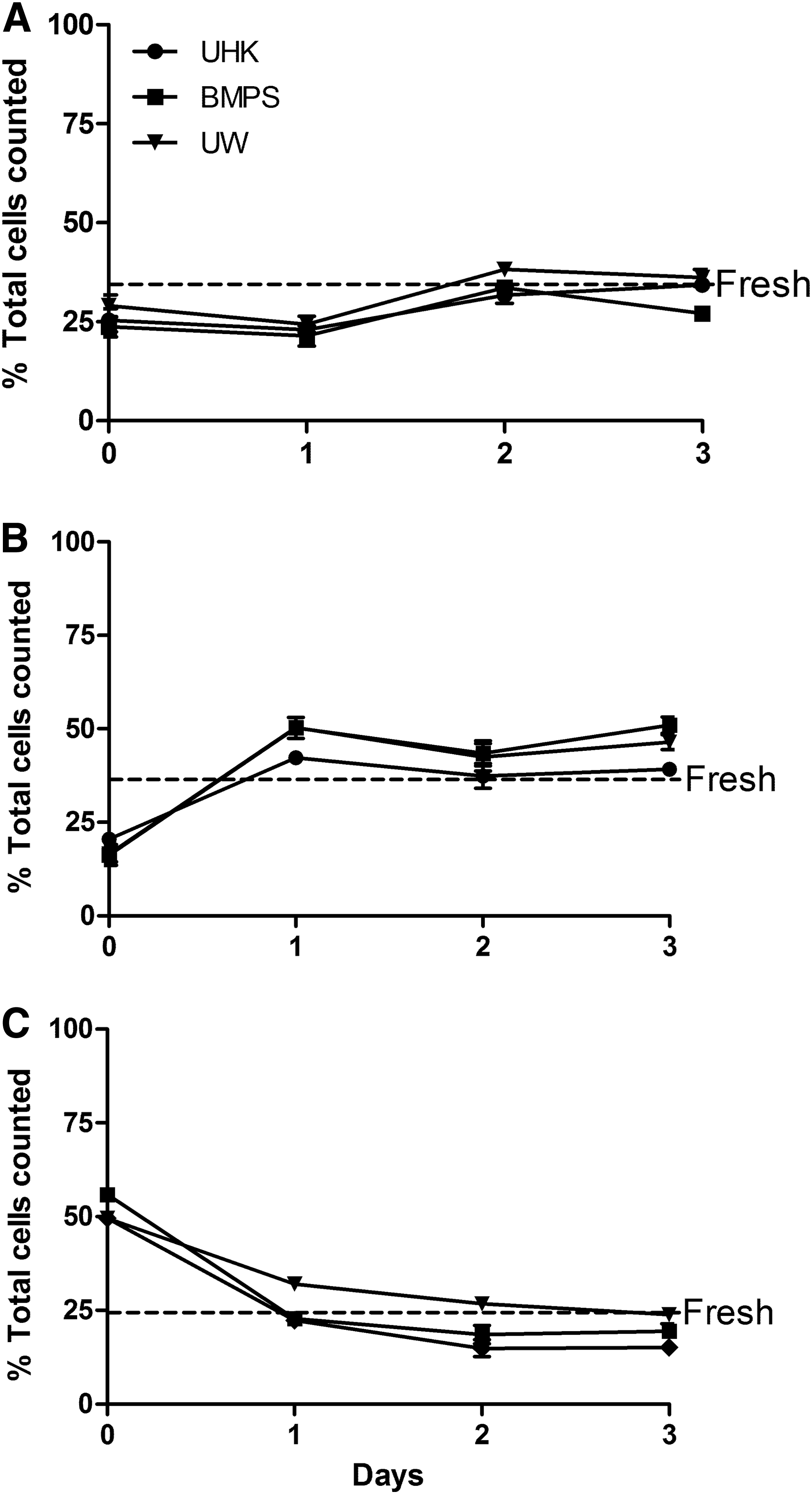
In this assay, the translocation of phosphatidylserine from inside the cell to outside, considered to be an early event in apoptosis, can be detected. The inclusion of the impermeant dye 7-AAD allows for the detection of dead cells so that live, dead, and apoptotic cells can be measured (Fig. 3). As described for measuring membrane integrity, several wells of cells were plated and left at 4°C for 16–24 hours for each solution assayed. Then, Nexin staining was performed on cells immediately after storage and for several days poststorage to assess the apoptotic activity. Nexin staining was also done on fresh untreated cells for comparison. Three graphs are presented in Figure 3, measuring live, dead, and apoptotic cells. The percentage of live cells in the three solutions was lower than fresh controls (∼50%–70%) with Unisol having the best live cell values followed closely by UW (p < 0.05) at day 0. The next day, Unisol had recovered close to fresh control values and was significantly better than UW or BMPS (p < 0.01). Live cell values for the three solutions improved somewhat over the next several days, approaching fresh control values, with Unisol showing the highest live cell value.
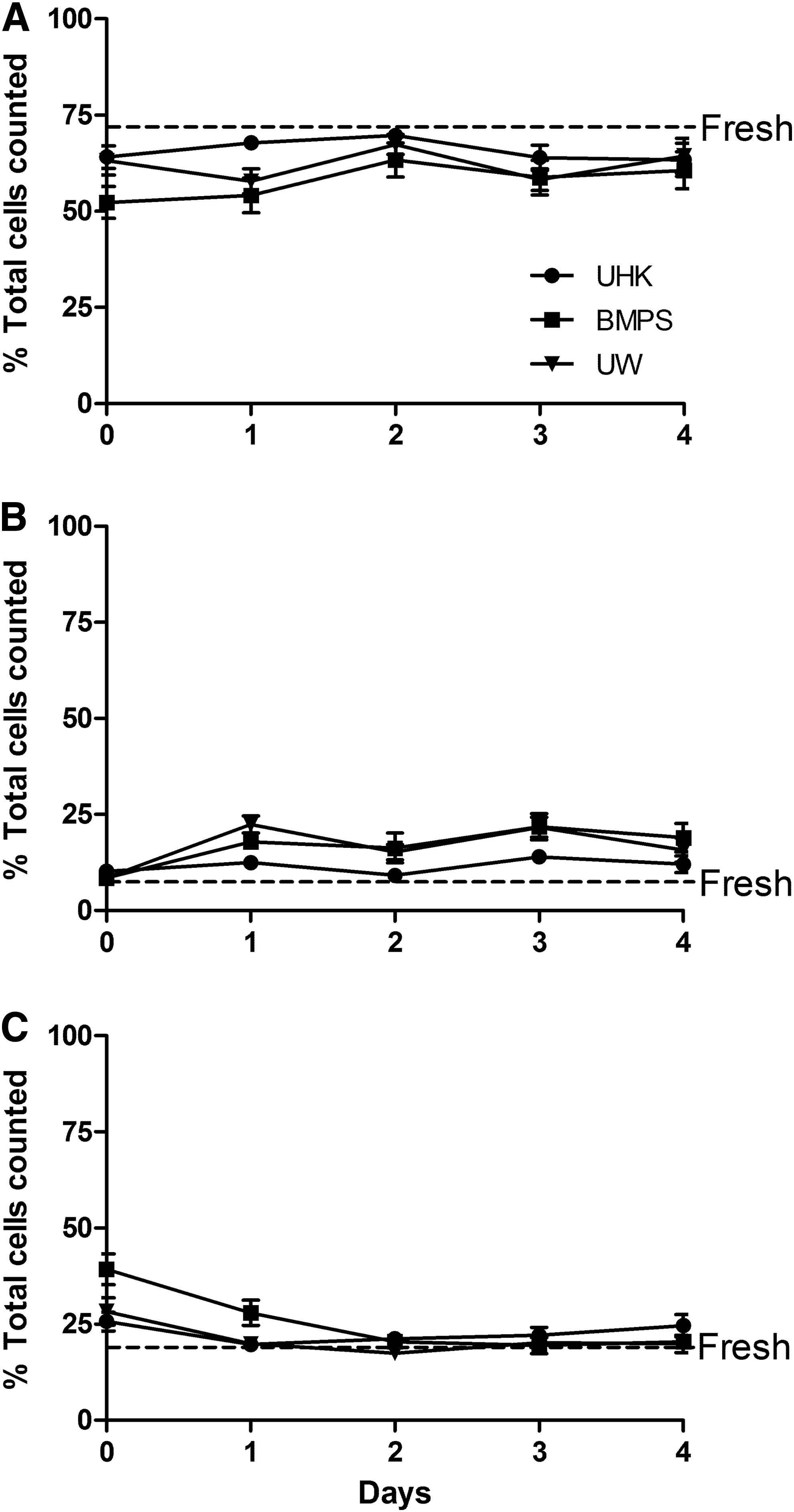
Measurement of apoptosis by nexin staining. βt3 cells were stored as described in Figure 1. The cells were stained and then 2000 cells were counted using the Guava cell analysis system. Live
An evaluation of the number of dead cells measured in each solution demonstrated that at day 0, BMPS had the highest level of dead cells, which was considered significant, compared to fresh control cells and Unisol (p < 0.05). There was still a significant difference in dead cells between BMPS and the other solutions at day 1 (p < 0.05). At later time points, all of the solutions expressed dead cell numbers that were equivalent to fresh cells.
For the apoptotic cells that were measured, the apoptotic activity was equivalent to fresh controls at day 0. However, at day 1 postrewarming, the number of apoptotic cells increases for all three solutions but is significant for BMPS and UW (p < 0.01). Unisol demonstrated the lowest apoptotic cell numbers of any of the three solutions and this was consistent at day 2 and 3 postrewarming (p < 0.05). Only at day 4 were equivalent apoptotic values between the solutions observed and these were still somewhat higher compared to fresh cells.
Caspase activity
Two measures of caspase activity were done. An overall survey of all caspase activity and then a more detailed assessment of the activities of caspases 3 and 7, the execution caspases that are the main initiators of the actual physical changes that take place within a cell undergoing apoptosis. A survey of the overall caspase activity evaluates live cells, dead cells, and cells that are considered to be at early to middle and late apoptotic stages (Fig. 4). Both assays work using labeled peptides that bind to caspases. Dead cells are also labeled using the impermeant dye, 7-AAD. Live cells are not labeled. Approximately 2000 events were counted. Caspase and dead cell counts are based on what has been labeled, while the live cell count is extrapolated from the unlabeled counts that comprise the 2000 events recorded. Things to consider are as follows: first, even a fresh, untreated cell population will have apoptosis occurring in it, so demonstration of caspase activity and dead cells in the fresh population is not surprising. Second, caspase activation does not necessarily mean that cell death must occur. A certain point within the apoptotic cascade must be reached before the cell is committed to apoptosis and cell death. Finally, due to the nature of the assay concerning what is labeled and what is not labeled, some cells that are labeled for caspase activity may also be considered live cells, even though they were not recorded as live in the assay.
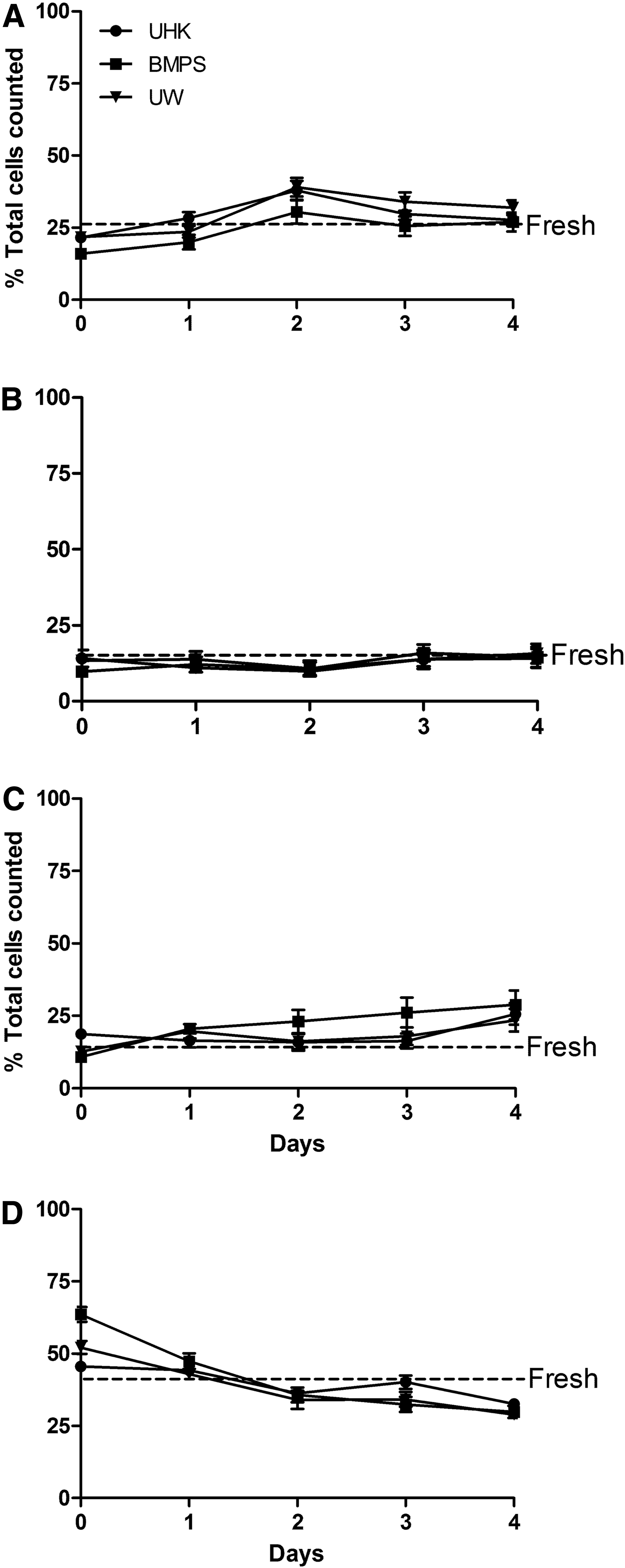
Measurement of apoptosis by general caspase staining. βt3 cells were stored as described in Figure 1. The cells were stained and then 2000 cells were counted using the Guava cell analysis system. Live
A comparison of live cells between the three solutions demonstrated that the number of live cells was considerably less than that of fresh controls (p < 0.01). BMPS also demonstrated significantly less live cells than either the UW or Unisol solutions (p < 0.01).
The three storage solutions also demonstrated significant differences compared with fresh controls when dead cells were measured; Unisol (p < 0.05), BMPS (p < 0.0001), UW (p < 0.001). Significant differences were also observed between the solutions. Unisol had the least number of dead cells (∼45%) measured, while BMPS had the most dead cells counted (∼63%).
Evaluating caspase activity, there was little difference between the storage solutions from the early to middle apoptotic events. A significant difference was observed between fresh cells and cells stored in BMPS (p < 0.01) at day 0; otherwise, all of the solutions were comparable to fresh cells. As for later apoptotic events, Unisol demonstrated the highest level of apoptotic activity at day 0, which then decreased to levels comparable to fresh cells for the remainder of the days tested. UW and BMPS demonstrated levels that were below fresh cells at day 0 but then increased over the 4 days that the cells were evaluated to levels that were greater than fresh cells.
Three and 7 caspase activity
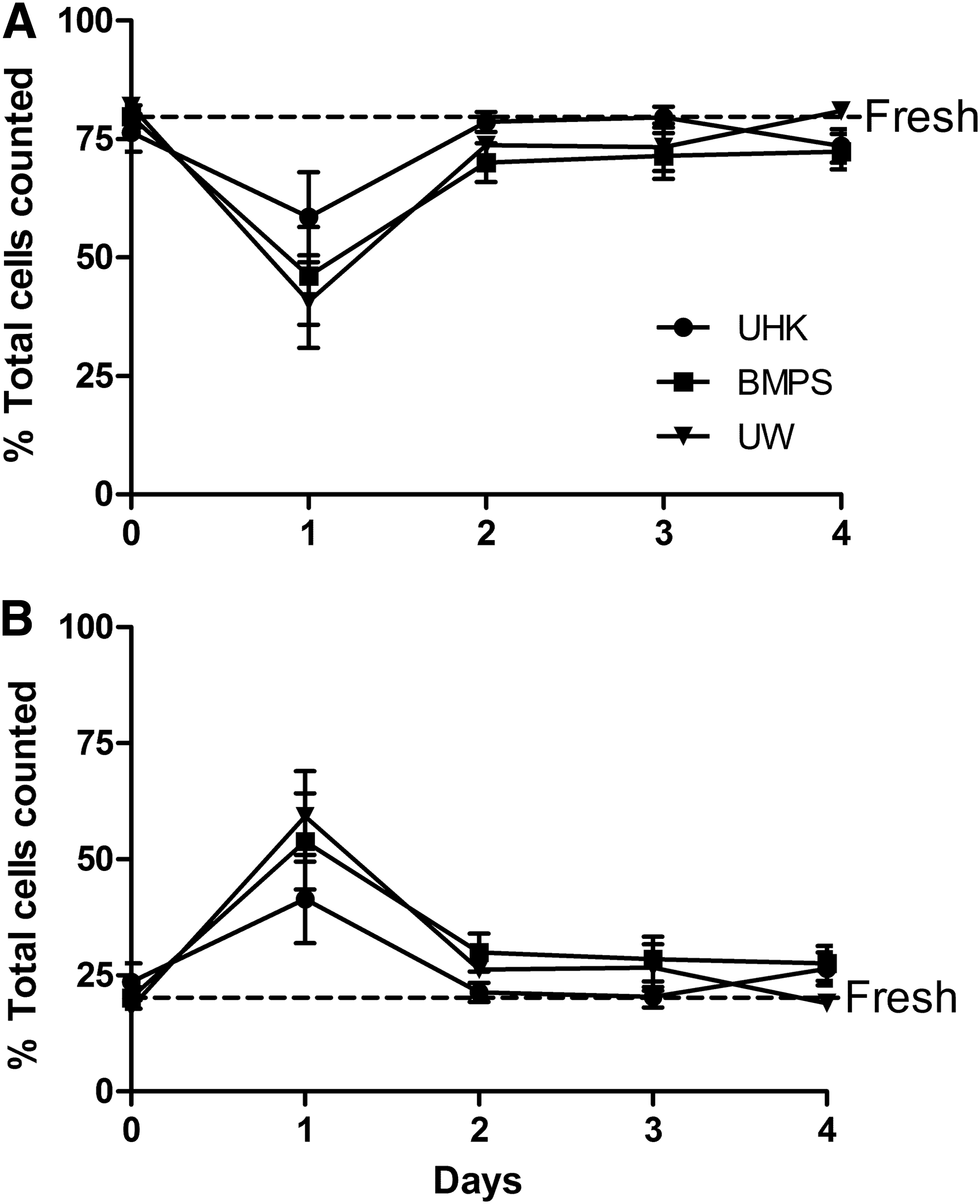
In a second series of experiments, a more directed look at caspase activity was performed. Specifically, the activity of caspases 3 and 7 was evaluated as they are considered the initiators of the morphological and biochemical changes that take place within a cell during apoptosis (Fig. 5). A comparison of live cells counted between each of the three solutions demonstrated numbers that were lower than fresh at day 0 and at day 1 with little if any differences observed between the solutions. At day 2, live cell counts were equivalent to fresh controls for all the solutions, which continued into day 3 for Unisol and UW. However, a drop in live cell numbers for BMPS was noted at day 3, which was significantly different compared with Unisol and UW (p < 0.01).
Measurement of apoptosis by caspase 3/7 staining. βt3 cells were stored as described in Figure 1. The cells were stained and then 2000 cells were counted using the Guava cell analysis system. Live
The number of dead cells counted for each of the solutions was significantly greater than the fresh control (p < 0.0001) at day 0, but these numbers rapidly declined. By day 1, cell counts for UW and BMPS were equivalent to fresh controls, while Unisol dead cell numbers declined a little more slowly but were equivalent to fresh by day 2.
Observation of the caspase 3 and 7 activity in these experiments showed that there was low caspase activity at day 0 for the three solutions tested compared to fresh controls. At day 1, however, caspase activity levels had increased above fresh control levels and remained there at days 2 and 3. Unisol demonstrated the lowest caspase activity on days 1–3, which was significant (p < 0.05) compared to UW and BMPS.
Tunel activity
In the final assay to measure apoptotic activity, the Tunel assay was performed, which measures the formation of the DNA ladder, a very distinctive characteristic of the apoptotic cascade. Only live cells and apoptotic cells are measured in this assay (Fig. 6). Each of the solutions demonstrated cell counts equivalent to fresh cells at day 0. Then, a large drop in live cells was measured with each solution at day 1, which then rebounded back to levels close to fresh cells by day 2 and beyond. Unisol live cell counts decreased the least at day 1 compared to UW and BMPS and its cell counts on days 2–4 were closer to fresh cells.
Measurement of apoptosis by TUNEL staining. βt3 cells were stored as described in Figure 1. The cells were stained and then 2000 cells were counted using the Guava cell analysis system. Live
Apoptotic cells for the three solutions demonstrated a corresponding response. At day 0, apoptotic cell levels were close to fresh control cells. At day 1, a large increase in apoptotic cell counts was observed that corresponded to a similar decrease in live cell counts for the same time period. Cell levels returned to values similar to fresh cells at day 2 and beyond. Again, Unisol demonstrated the least increase in apoptotic cell counts compared to UW and BMPS, and also had cell numbers that were more similar to fresh cells at days 2–4.
Discussion
The goal of this study was to evaluate the lead hypothermic storage solution, UW, the leading hypothermic perfusion solution, BMPS,10,11–16 and our own hypothermic storage solution, Unisol, to determine which solution provided the best conditions for cell survival during overnight storage at 4°C and which solutions demonstrated the least induction of apoptosis. The design of the experiments was such that the measurement of multiple points in the apoptotic cascade was made from the same set of cells and then measured for multiple days after storage. Included in these studies was the evaluation of two conventional markers of cell viability, metabolic activity and membrane integrity.
An overview of all the apoptosis and viability measures demonstrated that well-coordinated events were occurring as the cells were removed for cold storage. On the day the cells were removed from cold storage (day 0), many cells did not survive and viability was low as evidenced by low metabolic activity (Fig. 1) and compromised membrane integrity (Fig. 2). By day 1, metabolic activity and membrane integrity had improved, but an increase in apoptotic activity was also observed. This was evident in a decrease in live cells (Figs. 4 and 5), an increase in nexin staining (Fig. 3), an increase in caspase 3/7 activity (Fig. 5), and an increase in TUNEL staining (Fig. 6). Apoptosis does not just occur, but is an active cell death process that takes time to proceed and the results of the induced apoptosis became evident approximately 24 hours after the cells were removed from cold storage. By day 2, no more apoptosis was being induced as observed by the absence of change in apoptotic marker expression. The remaining cells were recovering and proliferating as shown by an increase in metabolic activity on days 3 and 4. The amount of measurable apoptosis activity also decreased to baseline levels on days 3 and 4. The only parameter that did not seem to change after day 0 is membrane integrity. Cold storage compromised the cell membrane. However, the remaining live cells quickly recovered to baseline values by day 1 and membrane integrity did not change over the remaining days of the study.
For each measurement of viability or apoptosis, there were not always significant differences between the three solutions. However, Unisol was consistently better than UW or BMPS for each parameter measured. In particular, the metabolic activity was better for Unisol over the other two solutions and the degree of apoptotic activity as measured by nexin staining, caspase 3/7 activity, and the TUNEL assay was lower for Unisol. The measurement of general caspase activity correlated with the other more specific apoptotic parameters that were tested, but because it was measuring all caspase activity, differences between the solutions were harder to observe.
The design of these hypothermic preservation solutions contains the following components: (1) to minimize cell and tissue swelling, (2) to maintain appropriate ionic balance, (3) to prevent a state of acidosis, (4) to remove or prevent the formation of free radicals, and (5) to provide substrates for the regeneration of high-energy compounds and stimulate recovery, upon rewarming and reperfusion.17–20,25–28 This base formulation of Unisol, which accounts for both biophysical and minimal biochemical requirements during low temperature storage, could then be used as a vehicle for the addition of other compounds that would facilitate survival, such as the addition of a caspase inhibitor like Q-VD-OPH. Unisol has also been evaluated for the preservation of a variety of cells, tissues, organs, and large mammals,19,22,29–35 but an analysis of apoptosis using Unisol has not previously been performed.
These results suggest that apoptosis may play a significant role in βt3 cell death after hypothermic storage at 4°C and in turn, apoptosis likely plays a role in pancreas and islet survival after cold storage as well. Further experiments have already begun evaluating the addition of potential supplements to Unisol, such as caspase inhibitors and other compounds that would relieve oxidative stress. 36 By using Unisol as the base solution, a cocktail of supplements specifically designed to preserve the pancreas and/or islets could be added that would facilitate cold storage and/or perfusion for future transplantation or clinical treatment. If successful, this strategy could be applied to other organs and tissues for transplantation. The analysis of the role of apoptosis in the survival of tissues and organs during cold storage provides avenues for better preservation and treatment with the outcome being better function upon reperfusion in patients.
Footnotes
Acknowledgment
This work was supported by Grant # DK076326-01 from the National Institute of Diabetes and Digestive and Kidney Diseases of the National Institutes of Health.
Author Disclosure Statement
No conflicting financial interests exist.