
Abstract
Background:
A functional artificial ovary is a promising strategy to recover fertility and restore endocrine function in cancer patients. The aim of this study is to optimize the follicle isolation protocol for cryopreserved human ovarian tissues.
Methods:
Each of the cryopreserved human ovarian cortex pieces (OCPs) from 10 patients was cut into two equal parts and randomly distributed into two treatment groups. Group 1: OCPs digested with Tumor Dissociation Enzyme (TDE); Group 2: OCPs digested with Liberase Dispase High (DH). The efficiency of both groups were evaluated in terms of yield, viability, morphology, and a short-term in vitro culture (IVC) in alginate scaffolds.
Results:
The TDE can isolate more primordial follicles and smaller diameter of follicles than Liberase DH. The TDE also enabled the isolation of more bright red follicles, higher percent of viable follicles, more morphologically normal follicles, and lower oxidative stress levels compared with Liberase DH. After eight days of IVC, follicles in the TDE group had a higher growth rate from Day 0 to Day 8, and higher viability on Day 8 than the Liberase DH Group.
Conclusion:
The TDE can be considered an alternative to Liberase DH, enables the isolation of a higher number of healthy follicles from human OCPs, and improves follicle survival after IVC in contrast to Liberase DH.
Introduction
Advances in cancer treatment have remarkably increased cancer patients' survival rates and prolonged patients' life expectancy. However, chemo/radiotherapy often leads to some irreversible side effects to cancer survivors: premature ovarian failure and infertility. 1 Cryobanking ovarian tissue supplies options to restore fertility for patients who have no sufficient time to stimulate the ovary and cryopreserve embryos or oocytes. 2
However, the re-implantation of cryopreserved ovarian tissue may reintroduce malignant cells and place them at risk of disease recurrence.3,4 Follicle surrounded by a layer of basement membrane can bar the blood capillaries, white blood cells, and nerve processes, as a physiological barrier protecting follicles from being contaminated by malignant cells of ovarian tissue. 5 Thus, embedding isolated preantral follicles in a bioengineered transplantable artificial ovary can be a safe, alternative treatment and may be regarded as an extension of cancer care. In this case, we need to establish a standard protocol for the follicle isolation and choose a scaffold to support these follicles' growth.
This possibility was demonstrated by Gosden and colleagues 6 that primordial follicles can be isolated from mice ovaries by enzymatic digestion and incubated in a collagen–gel matrix,6,7 or a plasma clot. 8 Subsequently, Roy and Treacy isolated intact preantral follicles from the human ovary. 9 So far, numerous mechanical methods, enzymatic methods, and their combination have been developed to isolate ovarian preantral follicles from rodents and human ovaries.10–12
The mechanical method combined with different types of collagenase (Ia, II, IX, XI) alone or together with bovine pancreatic deoxyribonuclease I (DNase I),12,13 which avoids sticky DNA ends, may isolate more follicles in comparison with mechanical methods, However, it may damage the basement membranes and affect the viability of follicles. 14
Liberase, a mixture of purified enzymes, has been reported as a powerful tool to improve the isolated follicles' quality compared with collagenase. 15 Liberase Dispase High (DH) research grade belongs to the group of Liberase Research Grade with highly purified Collagenase I and Collagenase II blended in a precise ratio and contains a high concentration of Dispase®, a non-clostridial neutral protease. Moreover, other researchers have shown that follicles isolated by Liberase DH had better morphology and higher developmental competence compared with collagenase. 16
Our prior study documented using “Tumor Dissociation Enzyme Reagent” (TDE, Innovative Diagnostic Systems Dr. Christian Sartori, Hamburg, Germany) to digest the cryopreserved ovarian tissue could yield better viability follicles than using Liberase TM. 17 Tumor Dissociation Enzyme (TDE) is a commercial enzyme cocktail from the DCS ATP-Chemo-sensitivity Assay Kit (DCS Innovative Diagnostik-Systeme, Hamburg, Germany) for gentle enzymatic digestion.18,19
Prior studies in our group reported that TDE contains highly purified collagenase and other enzymes that enable the digested suspensions to be not sticky, easy to isolate, and yield a higher number of healthy cells.17,20 However, we do not know the further influence on follicular growth and development after in vitro culture (IVC).
We hypothesized that the use of TDE may isolate more human follicles with proper morphology, vitality, and development potential after IVC. To test this, we apply TDE to digest our cryopreserved ovarian cortex in comparison with Liberase DH combined with DNase I. We analyzed the quantity and quality of isolated follicles in both groups, and culture isolated follicles in alginate gel for eight days to analyze the follicular growth in both groups.
Materials and Methods
All chemicals were purchased from Sigma (Sigma-Aldrich Chemie, GmbH, Schnelldorf, Germany), except where otherwise stated.
Collection, dissection, and distribution into treatment groups
The present study was approved by the Ethics Board of the Cologne University, Germany (applications 999, 184 and 13–147). Informed consent was obtained from 10 patients aged 18–35 years (23.8 ± 4.66). These patients were undergoing laparoscopic surgery within the FertiProtect program at the Department for Gynecology and Obstetrics of the University Hospital of Cologne. All ovarian tissues used for this research were cryopreserved and stored at the cryobank of the Maternal Hospital of Cologne University. According to the protocol approved in our department, 10% of ovarian tissue collected from patients was used for “patient-oriented research” to assess the tissues' viability for re-transplantation.
Ovarian tissue freezing and thawing
The cryopreservation of ovarian cortex pieces (OCPs) was performed according to our protocol, as previously published. 21 On freezing day, the OCPs were placed for 30 minutes at room temperature in basal medium. After this, the OCPs were placed into standard 5-mL cryovials (Thermo Fisher Scientific, Rochester, NY) and frozen by using IceCube 14S programmable freezer (Sy-Lab, Neupurkersdorf, Austria).
The thawing of frozen probes was performed as follows: The cryovial was held at room temperature in the air for 30 seconds with subsequent, direct immersion in a boiling water bath (100°C) for 60 seconds. The thawed OCPs were immediately transferred from the cryovials to a 10 mL thawing solution (basal medium containing 0.5-M sucrose). After stepwise dilution of the cryoprotectants, the OCPs were finally transferred to a basal medium for 10 minutes.
Isolation and collection of follicles
After warming, a small fragment of tissue from each patient was fixed, serving as controls; then, the OCPs were cut into two equal parts. Each part was weighted by using an analytical balance (Ohaus EP64C Explorer Pro Analytical Balance, Ohaus Corporation), and the volume of the piece was measured (Table 1). A total of 10 OCPs in each treatment group were evaluated.
Follicles Isolated from 10 Frozen Experimental Ovarian Tissues with Different Kinds of Enzymes
Group 1 – follicles isolated by TDE commercial enzyme cocktail.
Group 2 – follicles isolated by Liberase DH and DNase I.
The number of retrieved follicles independent from their maturity was significantly higher in group 1 in comparison with group 2 (p < 0.01).
TDE, Tumor Dissociation Enzyme.
Our research compared two enzyme-cocktails' influence on viability and ability to grow in vitro in a three-dimensional (3D) system of preantral follicles recovered from the cryopreserved ovarian cortex.
The tested enzyme cocktails were:
Group 1: TDE Reagent from the DCS ATP-Chemo-sensitivity Assay Kit (DCS Innovative Diagnostik-Systeme, Dr. Christian Sartori Labor, Hamburg, Germany), which was composed of various types of highly purified collagenase (personal communication with Dr. Christian Sartori Labor), is a commercial enzyme cocktail for enzymatic digestion of any solid tumors.18,19,22
Group 2: Liberase DH Research Grade is composed of collagenase I, II and the neutral protease Dispase (Sigma-Aldrich Chemie, GmbH). DNase I (bovine pancreatic deoxyribonuclease I), which is a DNA minor groove-interacting nuclease with a relatively low specificity (Sigma-Aldrich Chemie, GmbH),23,24 allows the isolate to be not too sticky, and easy to isolate.
For this research, 10 frozen OCPs immediately after warming were cut into two equal parts and randomly distributed in two treatment groups. The procedure of follicle isolation was performed, as previously described17,24 with some modification. Briefly, the OCPs were mechanically cut into ∼0.5 × 1 to 1 × 1 mm3 pieces. Then, the minced ovarian cortical tissue was suspended in basal medium containing 16% of TDE-enzymatic solution (Group 1) or in basal medium containing 0.28 Wünsch units/mL Liberase DH in basal medium plus 10 μg/mL DNase I (Group 2).
Both enzyme solutions were prepared on Leibovitz L-15 medium in combination with 50 μg/mL neutral red (NR) vitality dye, which shows a deep red color in living cells, allowing quick and rough visualization of isolated follicles. 25 The incubation time for both treatment groups was 75 minutes at 37°C on KS 260 basic shaker (IKA, Staufen, Germany) with 120 rotations/minute. Tissue digestion was terminated by the addition of 10 mL of cold Leibovitz L-15 medium supplemented with 20% fetal calf serum.
Follicles were manually picked up by using 125 μm V-denuded capillaries (Vitromed GmbH, Jena, Germany) under a stereomicroscope (Nikon, Düsseldorf, Germany). The follicle diameter was measured by the basement membrane surrounding the granulosa cell layer(s) that served as the follicular boundary. The stage of follicles was classified according to Gougeon and Fortune26,27 as follows: primordial follicle (<60 μm), oocytes surrounded by a single layer of flattened pre-granulosa cells; primary follicle (>60 μm to ≤75 μm), oocytes with a single layer of cuboidal granulosa cells; and secondary follicle (>75 μm to <200 μm), with at least two complete layers of granulosa cells.
Follicle viability assessment with fluorescence dye Calcein AM and ethidium homodimer-1
The stained cells isolated with NR follicles were subjected to fluorescent staining with Calcein AM and ethidium homodimer-1. 28 Briefly, the follicles were exposed to 2 μM of Calcein AM and 5 μM of ethidium homodimer-1 for 45 minutes at 37°C in the dark. Non-fluorescent cell-permeant Calcein AM enters the cell and is cleaved by esterase in living cells, giving an intense green fluorescence (excitation/emission, 495 nm/515 nm). Ethidium homodimer-1 enters cells with damaged membranes binding to nucleic acids of dead cells and shows a red fluorescence (excitation/emission, 528 nm/617 nm).
The viability of follicles was classified into four categories depending on the percentage of dead granulosa cells 15 : V1, live follicles with the oocytes and all granulose cells (GCs) viable; V2, minimally damaged follicles with <10% of dead GCs; V3, moderately damaged follicles with 10%–50% dead GCs; and V4, dead follicles with both the oocyte and/or >50% GCs dead (Fig. 1B–E).
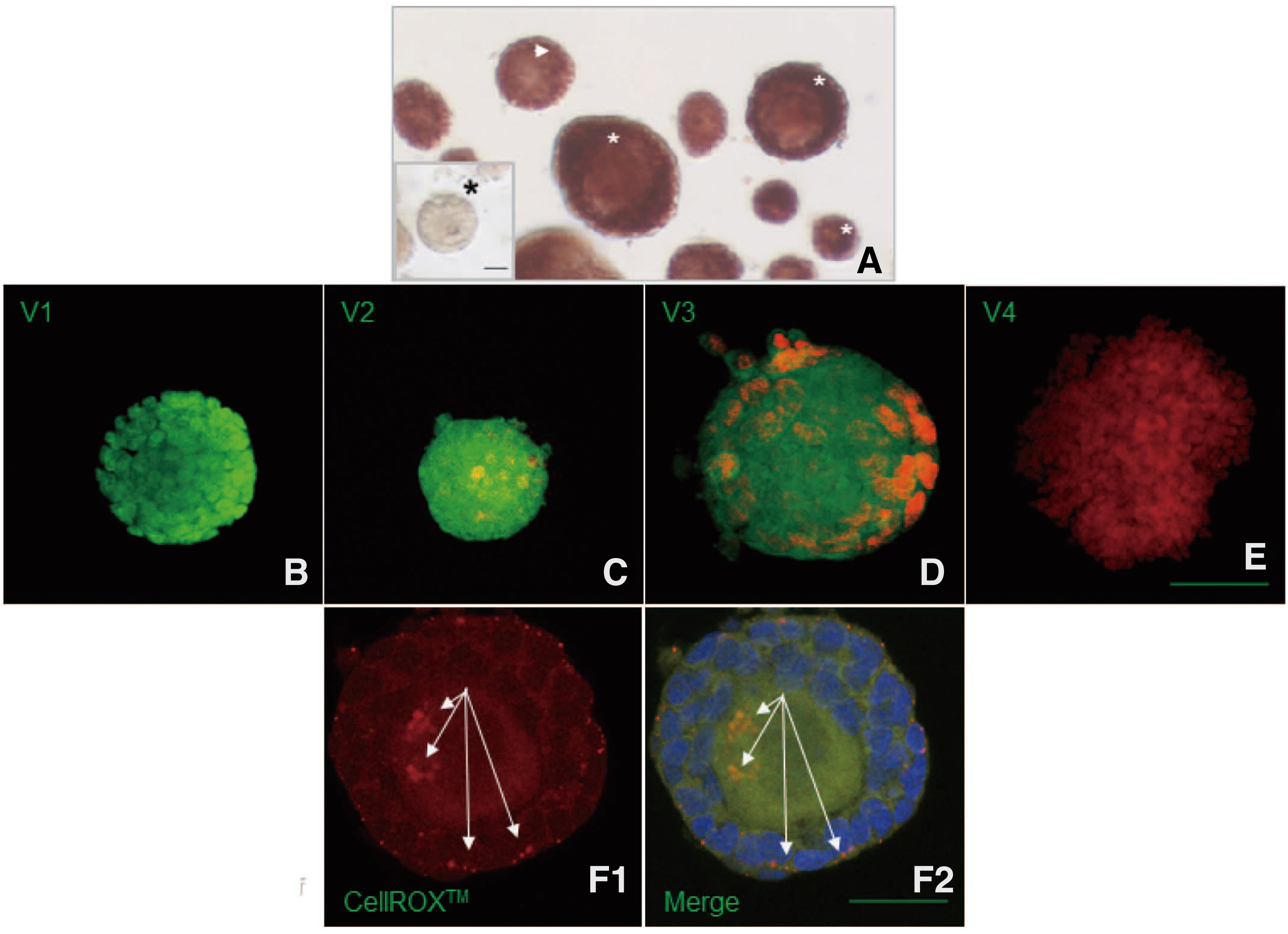
Example of isolated follicles treated with a neutral red viability dye and living/dead fluorochrome assay.
Viability assessment due to the measurement of oxidative stress level of isolated follicles
The isolated follicles were stained by the CellROX® Deep Red fluorogenic probe (Life Technologies), which was designed to reliably measure reactive oxygen species (ROS) in live cells. This dye detects cellular superoxide anions (O2˙−) and hydroxyl radicals (OH•), and it produces deep red fluorescence (excitation/emission, 640 nm/665 nm). The staining procedure was performed according to Korkmaz. 29 Briefly, the follicles were fixed in formalin, washed twice with DPBS, and incubated in CellROX Deep Red Reagent and anti-α-Tubulin antibody for 30 minutes at 37°C in the dark.
After incubation, follicles were covered by the Fluoromount-G mounting medium containing 4’,6-diamidino-2-phenylindole (DAPI, Fluoromount-G™ with DAPI; Thermo Fisher Scientific), and they were then observed under a confocal microscope (Olympus Fluoview FV 1000, Hamburg, Germany). The incubation of follicles with CellROX Deep Red Reagent shows a deep red fluorescence of ROS in cells (Fig. 1F). Semi-quantification of the oxidative level was calculated by using Fiji software (version 1.40; National Institutes of Health, Bethesda, MD), and the percentage of oxidative level (%) was calculated in these two digestion groups.
Follicle morphology assessment with fluorescence dye
The morphological evaluation of follicles was performed according to their morphology and the integrity of the granulosa cell layer 15 by staining with the anti-α-Tubulin antibody in combination with DAPI. The fluorescent-stain procedure was performed as follows: Isolated follicles were fixed and then stained with anti-α-Tubulin antibody for 30 minutes at 37°C in the dark. After incubation, follicles were covered by Fluoromount-G mounting medium containing DAPI and observed under a confocal microscope (Olympus Fluoview FV 1000).
According to staining results, the follicles were classified into four categories as follows: M1, spherical form with complete granulosa cell layer; M2, irregular form with complete granulosa cell layer; M3, irregular form with <10% granulosa cell loss; and M4, totally atypical form with 10%–50% granulosa cell loss (Fig. 2).

Example of morphological grading of isolated follicles using fluorescence dye. M1
Follicle embedding and IVC
Immediately after isolation, only well morphologically preserved brightly stained red follicles were collected for embedding in an alginate scaffold and cultured in vitro (Fig. 1A, white asterisk) at 37.5°C under 5% CO2 (Heraeus® HERAcell® CO2 Incubator, SelectScience) for up to 8 days (Fig. 3A). Every second day, half of the culture medium was replaced, and follicles were observed and photographed under a standard light microscope. After 8 days of IVC, follicles were lysed from the alginate scaffold and incubated with Calcein AM and ethidium homodimer-1 for viability evaluation.
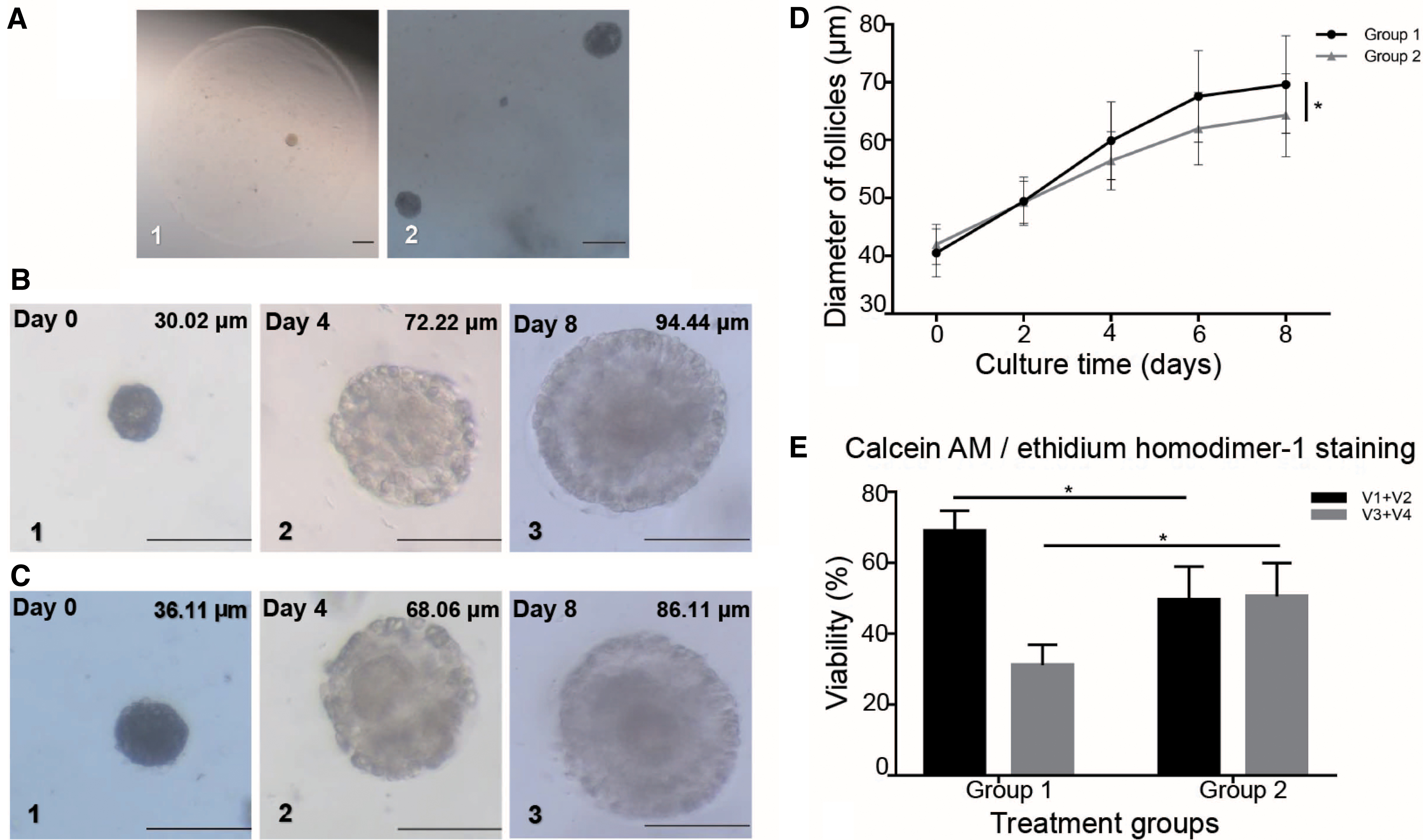
Growth and viability of isolated and embedded in alginate gel capsule follicles during in vitro culture depending on the type of enzymatic treatment of ovarian cortex.
Statistical analysis
The confocal images were processed and analyzed by using program Fiji software (version 1.40; National Institutes of Health, Bethesda, MD). Statistical Product and Service Solutions (SPSS) 19.0 software (Chicago, IL) was used for statistical analyses. A comparison was made between the TDE and Liberase DH groups with regard to the percentages of morphology, viability category, and after IVC. Data were statistically analyzed by the Kruskal–Wallis test. Results were expressed as mean ± standard deviation (SD). p < 0.05 was considered statistically significant. All experiments were repeated three times on different days.
Results
Retrieval rate of isolated follicles
After enzymatic digestion of the frozen ovarian cortex, a total of 1502 follicles in both treatment groups were recovered (790 follicles were isolated from Group 1, and 712 follicles were isolated from Group 2). Group 1 had more follicles recovered than Group 2 (79.00 ± 71.24 in Group 1 vs. 71.20 ± 67.71 in Group 2, respectively), (p < 0.05, Table 1). The diameter of isolated follicles in Group 1 was 44.41 ± 11.39 μm, and the diameter of isolated follicles in Group 2 was 45.76 ± 11.10 μm (Fig. 4B). The different maturity of isolated follicles in both groups is shown in Figure 4B. It was also demonstrated that the percentage of retrieved primordial follicles was significantly higher in the TDE group in comparison with Liberase DH group (Group 1: 80.54% ± 5.41% vs. Group 2: 77.73% ± 5.25%, p < 0.05) (Fig. 4B).
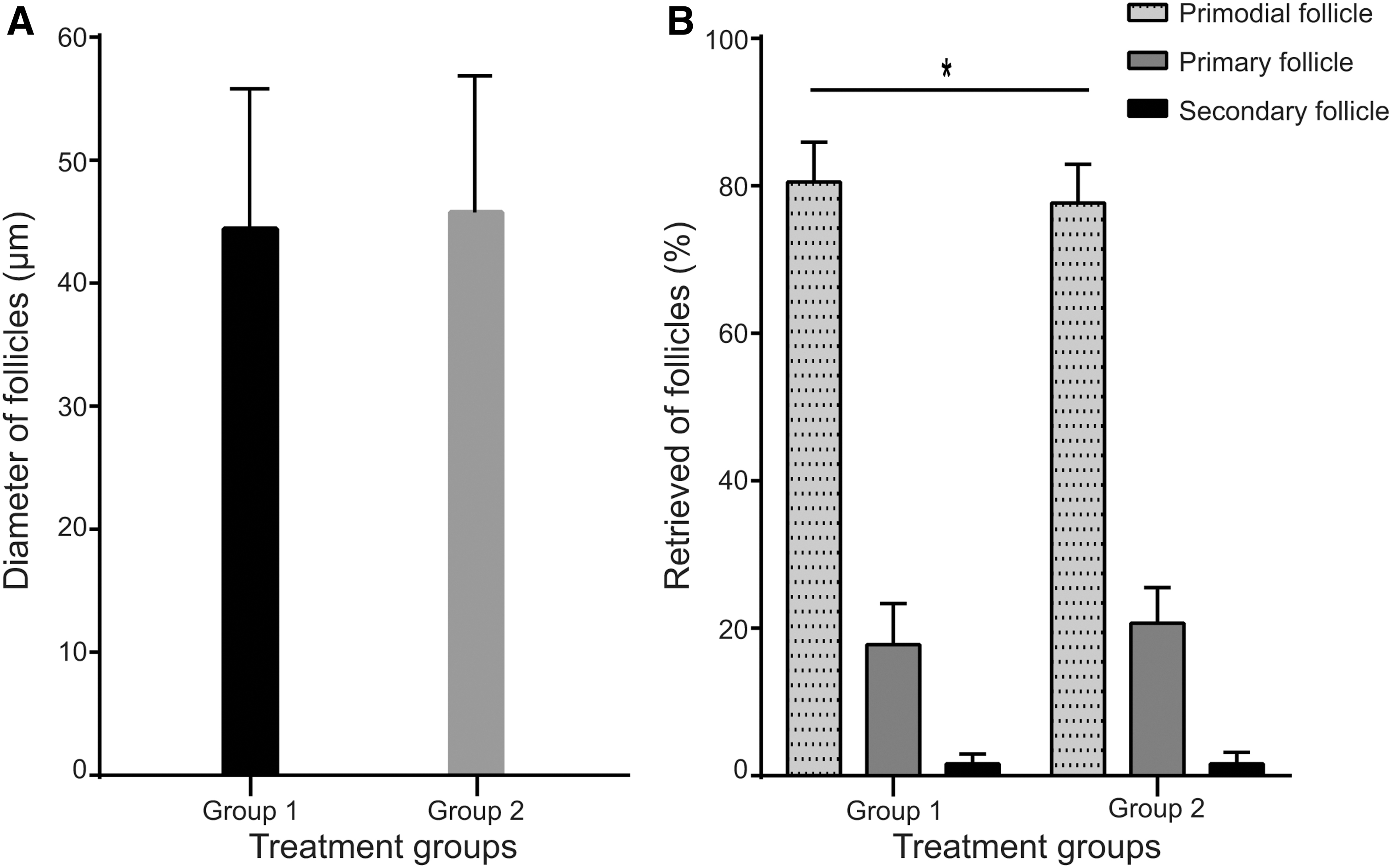
The distribution of follicles according to their diameter and maturity in each treatment group.
Viability assessment of retrieved follicles
NR dye
A total of 1502 follicles were collected and simultaneously stained with NR dye (Fig. 5A). The data on Figure 5A showed that the percentage of bright red staining follicles in Group 1 was 7% more than in Group 2 (p < 0.01), but the percentage of light red staining follicle in Group 1 was 5% less than in Group 2 (p < 0.01). However, the percentage of non-stained follicles was similar (p > 0.05) in both treatment groups.
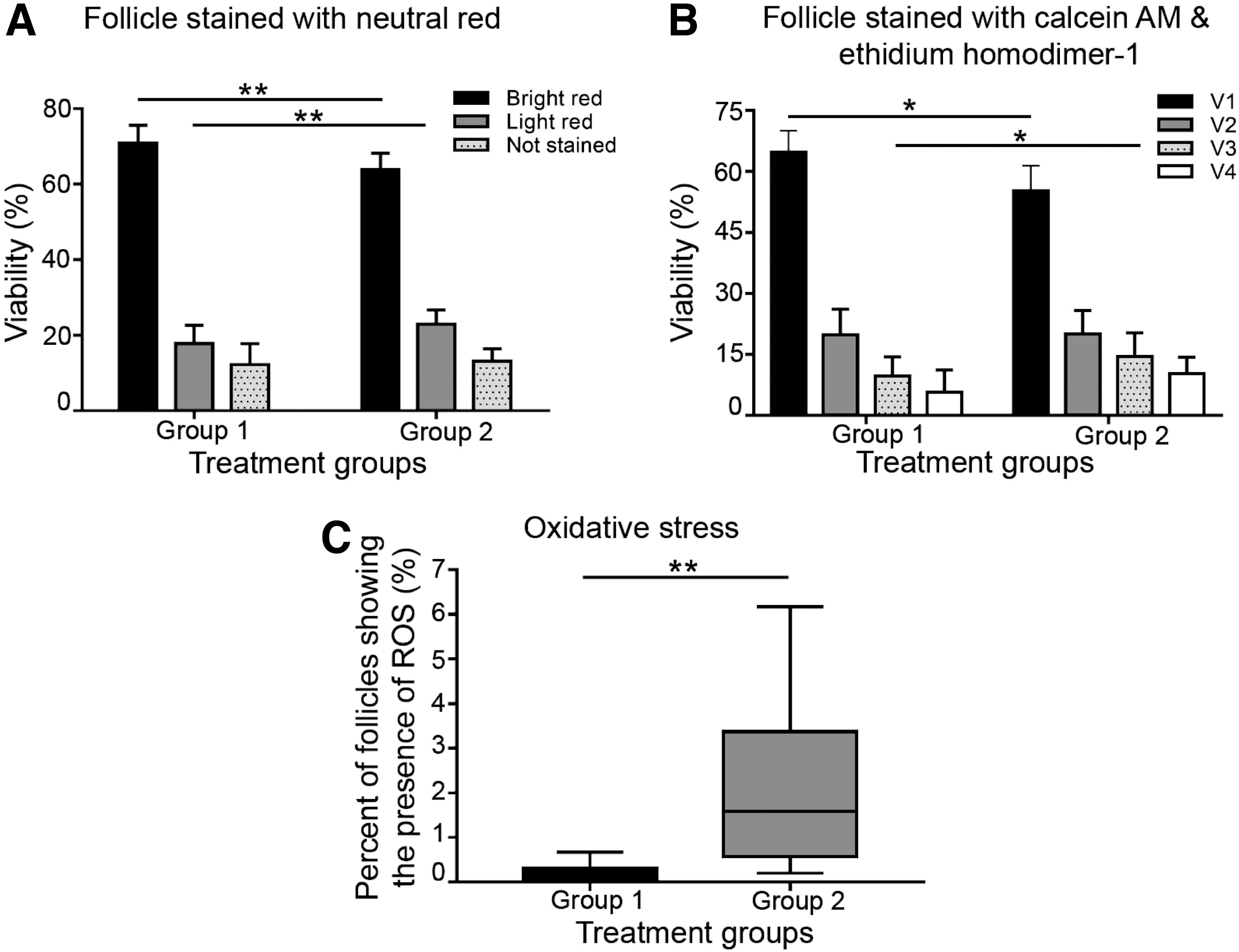
Viability assessment of isolated follicles depending on the type of enzymatic treatment of the ovarian cortex.
Calcein AM and ethidium homodimer-1
A total of 317 follicles were stained with Calcein AM and ethidium homodimer-1, as shown in Figure 5B. The data showed that Group 1 had more class V1 follicles isolated than Group 2 (60.81% ± 5.20% vs. 55.22% ± 6.15%, p < 0.05), but the percentage of class V3 follicles was less than Group 2 (9.67% ± 4.75% vs. 14.47% ± 5.88%, p < 0.05). However, the percentage of class V2 and class V4 follicles was not significantly different (p > 0.05) in both groups.
ROS assay
To measure the amount of ROS in live cells, a total of 20 isolated follicles in each treatment group were stained with CellROX. The data presented in Figure 5C show that generally the oxidative stress was very low in both treatment groups, but the percentage of oxidative level was significantly lower (p < 0.01) in Group 1 than in Group 2.
Morphology assessment
A total of 317 follicles were assessed for morphology by using fluorescence dyes anti-α-Tubulin antibody in combination with DAPI, as presented in Figure 6. It was shown that the percentage of morphologically normal follicles of class M1 was significantly higher (p < 0.05) in Group 1 than Group 2. However, the percentage of class M2, M3, and M4 follicles was similar (p > 0.05) between these treatment groups.
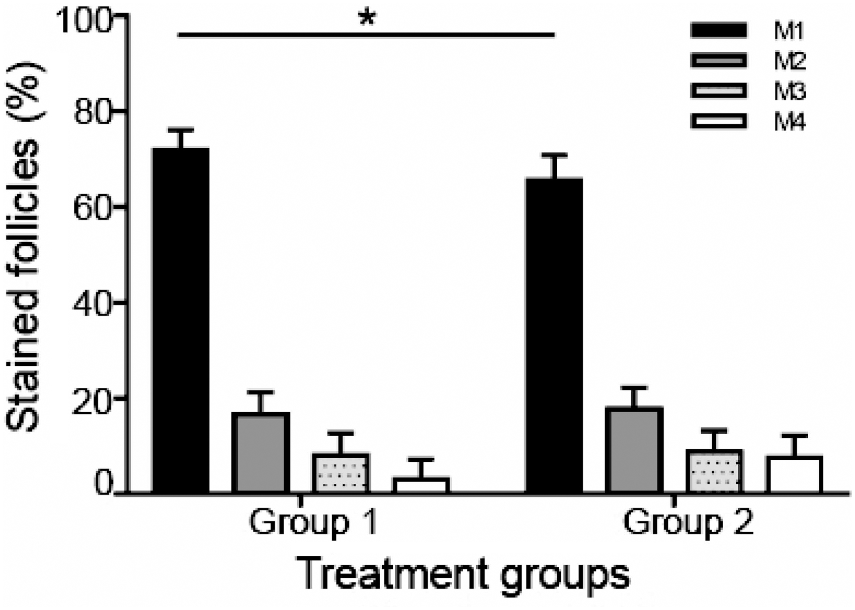
Morphology assessment of retrieved follicles depending on the type of enzymatic treatment of the ovarian cortex using nuclear stain DAPI with fluorescence contrasting of cytoskeletal α-tubulin. Significance of results is presented as mean ± SD; *p < 0.05.
IVC of follicles in 3D system using alginate scaffold
A total of 53 follicles in each treatment group were embedded in alginate beads (Fig. 3A) for IVC for up to eight days (Fig. 3B, C). It was shown that the diameter of the follicles on day 0 was similar in both groups (p > 0.05). However, after eight days of IVC, follicles in the TDE group had a higher growth rate from Day 0 to Day 8 than in the Liberase DH group (p < 0.05, Fig. 3D). We used Calcein AM and ethidium homodimer-1 for evaluating the viability of follicles after eight days of IVC (Fig. 3E). The data showed that the percentage of class V1 + V2 follicles, which represented viable follicles, was significantly higher (p < 0.05) in Group 1 compared with Group 2.
Discussion and Conclusion
Our results indicate that a new enzymatic digestion protocol using TDE, a commercial enzyme cocktail, is suitable for human follicle isolation as the first step to bioengineer a transplantable artificial ovary. The efficiency, safety, and productivity of the proposed protocol can be a suitable method for isolating many intact and viable follicles with good growth potential. The goal of this research was to replace Liberase DH as an enzyme digestion method to maximize the number and survival of isolated human preantral follicles, as the first step in constructing artificial ovaries for female cancer patients, preventing disease recurrence and for clinical application.
The ovarian tissue freezing and thawing protocol in our previous studies has shown promising results in viability and development and resulted in live births after the auto-seeding of cryopreserved ovarian tissue.20,21 Therefore, we are confident to use cryopreserved tissue. To assess the enzyme TDE, we compared it with Liberase DH combined with DNase I, which is widely used as the gold standard for human follicle digestion.24,30,31
Total amount of isolated follicles
In our study, TDE isolated more follicles than Liberase DH (p < 0.05, Table 1), especially in primordial follicles. This was similar to a recent study by Schmidt et al., which showed that the number of follicles isolated by TDE digestion is also significantly higher than using Liberase TM. 17 By comparison, Lierman et al. showed that no statistical differences were found between Liberase TM and Liberase DH, regarding the number and type of isolated follicles. 32 In our data, the follicles' diameter in Group 2 (Liberase DH) is 45.76 ± 11.10 μm, similar to the results obtained by Mouloungui et al. (diameter of follicles isolated by Liberase DH was 45.2 ± 8.2 μm). 33
As the ovarian cortex holds a vastly dense and fibrous structure, it is crucial to use a suitable enzymatic digestion protocol to maximize the yield of isolated follicles from the neighboring tissue. Liberase DH Research Grade contains highly purified Collagenase I and Collagenase II blended in a precise ratio to each other and contains a high concentration of Dispase. The TDE not only contains highly purified collagenase, but it also includes other enzymes: protease, dispase, or DNase, which allows deeper digestion, and obtains a greater number of isolated follicles than Liberase DH which is widely used for follicle isolation.
Quality analysis of the isolated follicles in our project was based on follicle viability assessment by NR dyes, vital fluorescent dyes, ROS assays, and general follicle morphology assessment by using fluorescence dyes.
The advantages of NR dye are that the dye can be reconstituted in physiological medium, is nontoxic, can be easily visualized under standard light microscopy, and has no long-term deleterious effects on the follicles. 34 The NR is a dye that readily diffuses through the cell membrane and concentrates in the lysosomes of viable cells, 25 and shows a deep red color (Fig. 1A). It is, thus, easy to distinguish between viable, damaged, or dead cells. 35 Our study showed that the percentage of bright red follicles in Group 1 was significantly higher (p < 0.01) than Group 2, which is similar to the observations of intense red-stained follicles in the suspension of cryopreserved ovarian tissues made by Schmidt et al. 17
Follicles stained by Calcein AM and ethidium homodimer-1 in our data have shown that Group 1 isolated more viable follicles than Group 2 (Fig. 5B). Liberase DH Research grade contains highly purified Collagenase I and Collagenase II, whereas endotoxin is one of the detrimental components of the collagenase extract, and it may reduce the vitality of follicles. 36 Indeed, for Liberase DH/DNase treatment, the percentage of class V1 follicles in our study was in accordance with previous reports: 65% (digested from cryopreserved ovarian cortex) 32 and 61% (digested from fresh ovarian cortex). 15
Other observations by Dolmans et al. 15 showed that early atresia was frequently found in follicles isolated by Liberase. This may infer that Liberase may alter the viability of isolated follicles by inducing apoptosis. 37
The ROS play a vital role as mediators of various intracellular signaling pathways 38 regulating apoptosis and necrosis. 39 Oxidative stress is the result of an imbalance in the production and elimination of the intracellular (ROS). However, a high level of oxidative stress can damage follicles 40 and affect their IVC. 41 A high concentration of oxidative stress may also enter mitochondrial membrane pores, emit cytochrome, trigger caspases, followed by apoptosis, 42 and affect oocyte maturation. 43 In our results, the percent of oxidative stress level was significantly lower (p < 0.01) in Group 1 than in Group 2.
Collectively, our results suggest that ROS generation was triggered during Liberase isolation procedures. Tirmenstein et al. found that Liberase containing highly purified collagenase can induce oxidative stress after isolation of hepatocytes, leading to a loss of cytochrome enzyme activity. 44 Liberase, which contains very low levels of endotoxin, still produced nitrite. The enzyme nitric oxide synthase (NOS) converts L-arginine to Nitric oxide (NO). 45 Once NOS is expressed, extremely high levels of NO can be produced. 44 The NO that is produced by NOS can react with a superoxide anion to form the ROS. 46
Similar observations were made by Förstermann and Kleinert, 47 where endotoxin has been shown to induce NOS in cells, leading to an increase of ROS. We can infer that Liberase contains very low levels of endotoxin, and it may induce NOS; a low level of NOS can produce high levels of NO; and NO can react with superoxide anion to form ROS. Therefore, we can deduce that TDE may well preserve ROS balance and reduce oxidative stress.
Our results showed that the percentage of class M1 follicles was significantly higher (p < 0.05) in Group 1 compared with Group 2. This means that using TDE digestion can preserve the morphology and granulosa cell layer of follicles. Laminin and collagen IV are essential components of the basement membrane of human primordial follicles. 48 Liberase DH presents a high concentration of dispase, which does not cleave laminin, but it can cleave fibronectin and collagen IV, 49 and damage the junction and membrane of the follicle.
That may explain our results of damaged follicles or extruded oocytes in the Liberase DH group. This is in accordance with previous investigations by Dolmans et al. 15 and Oktay et al. 50 that suggested that enzymatic isolation using Liberase may induce slight morphofunctional changes in the follicular cell compartment, such as alteration in shape or extensive disruption to the follicular wall.
The culture of isolated preantral follicles has several potential applications, including IVC and grafting or bioengineering of a transplantable artificial ovary or for molecular biology analyses. Therefore, ensuring good viability, preserving structure, and maintaining follicular development is essential before attempting such approaches in humans. Observations in preliminary studies reported that follicle isolation using Liberase were degenerated in IVC. 12 Thus, to examine the effect of different types of enzymic digestion on the follicle, we isolated follicles from cryopreserved ovarian cortex embedded in a 3D alginate scaffold for IVC for 8 days.
After 8 days of IVC in our study, follicles isolated from both TDE and Liberase DH treatments maintained good viability and preserved 3D structure, and they resulted in an increase in size in both groups. Our results are similar to those in follicles obtained by Yin et al., 23 with 7 days of IVC. Such an increase in follicle diameter after IVC may be due to some products generated during ovarian tissue cryopreservation: hypoxia, increasing of intracellular Ca2+, osmotic disruption of cellular membranes, generation of ROS, and lipid peroxidation.
These products generated by cryopreserved or ovarian cortex cut by a tissue sectioning and follicles isolated by enzyme may interrupt ovarian Hippo signaling and activate follicles in IVC. 51 Also, our results noted that follicles in the TDE group had a greater growth rate from Day 0 to Day 8 after eight days of IVC. Liberase, which contains very low levels of endotoxin, produces nitrite, 46 can induce oxidative stress in follicles after enzymatic isolation, 44 induce apoptosis IVC, 36 and strongly delay and impair grafting outcomes. 37 These results probably reflect that Liberase may weaken the initial health status of isolated follicles or a lack of factors essential to sustaining their survival and growth. 52 Hence, TDE can preserve follicles during digestion better than Liberase without impairing their ability to survive and grow in vitro.
To our knowledge, this study was the first to provide insight into the relationships between morphology, viability, cell intactness, oxidative stress, and diameter of growing preantral follicles between TDE and Liberase DH digestion. Gap junction formation based on the ability of the oocyte to communicate with granulosa cells and signal for cell division between oocytes and granulosa cells may explain the differences in growth rate in both groups. Our findings support the hypothesis that using TDE digestion can preserve granulosa cell–oocyte contact, which is important for cell proliferation in early stages.
Our study had several limitations: First, it did not evaluate the potential of isolated follicle contamination by cancer cells; Second, it did not examine the RNA expression levels of each oocyte and granulosa cell from a single follicle. Additional studies are needed to confirm the isolated follicle, with the risk of cancer cells reseeding by enzymatic digestion and isolated human ovarian follicles' RNA levels.
Conclusion
In conclusion, this study using TDE to isolate follicles can allow us to obtain a high number of viable follicles from the human ovarian cortex in contrast with the earlier described Liberase DH method, which is the initial and crucial step to construct an artificial ovary for future clinical applications. Our isolation technique will enable a foundation, as research is increasingly focusing on preantral folliculogenesis.
Footnotes
Acknowledgments
The authors thank all personnel involved in the clinical activities in fertility preservation for their passionate work. The women who donated their ovarian tissue for research are also greatly appreciated.
Ethics Approval and Consent to Participate
This study was approved by the Ethics Boards of University Cologne (applications 999,184 and 13 − 147). Written informed consent was obtained from all the participating couples.
Author Disclosure Statement
The authors declare that they have no competing interests.
Funding Information
No funding was received for this article.