
Abstract
Cyclic RGD (Arg-Gly-Asp) peptides radiolabeled with 68Ga have great potential for the early tumor detection and noninvasive monitoring of tumor metastasis and therapeutic response. Herein, the preparation of 68Ga-labeled DOTA-E[c(RGDfK)]2 (DOTA=1,4,7,10-tetraazacylododecane-1,4,7,10-tetracetic acid; E=Glutamic acid; R=Arginine; G=Glycine; D=Aspartic acid; f=phenyl alanine; K=lysine) using 68Ga directly eluted from a nanoceria–polyacrylonitrile (CeO2-PAN)-based 68Ge/68Ga generator developed in-house was reported. The 68Ga complex of DOTA-E[c(RGDfK)]2 was synthesized with >98% radiochemical purity by incubating 20 μg of the conjugate with 68GaCl3 (74–111 MBq) in acetate buffer (pH 3.5–4.0) at 90°C for 10 minutes. The complex exhibited excellent in vitro stability in 0.1 M EDTA solution at room temperature upto 1 hour studied (radiochemical purity: 98.0%). The biological efficacy of the radiolabeled conjugate was studied in C57/BL6 mice bearing melanoma tumors. The results of the biodistribution studies revealed significant tumor uptake (4.14±0.54%ID/g) within 10 minutes postinjection (p.i.), which increased further to 4.61±0.31%ID/g at 30 minutes p.i. The tumor-to-blood ratio was found to increase from 1.75±0.42 at 10 minutes p.i. to 2.25±0.20 at 60 minutes p.i., whereas the tumor-to-liver and tumor-to-muscle ratio between the same time points increased from 2.71±0.76 to 3.31±0.84 and 5.37±1.08 to 8.97±1.32, respectively. The study successfully demonstrated the preparation of 68Ga-DOTA-E[c(RGDfK)]2 as a potential positron-emission tomography radiotracer for possible use in tumor imaging by using 68Ga eluted from a reliable, easy-to-handle 68Ge/68Ga generator developed in-house, without any postelution purification of 68Ga.
Introduction
Tumor angiogenesis, which involves the formation of new blood vessels from pre-existing vasculature, is well recognized as an essential mechanism for tumor growth and metastasis. 1 –4 The angiogenic process depends on vascular endothelial cell migration and invasion, and is regulated by integrins, which are cell adhesion receptors. The function of integrins during tumor angiogenesis has been studied most extensively for integrin αvβ3, which is overexpressed on activated endothelial cells and different types of tumor. 5 –7 Therefore, integrin αvβ3 has been identified as an interesting molecular target for the early diagnosis as well as therapy of rapidly growing and metastatic tumors. 6 –11
Synthetic peptides containing the arginine–glycine–aspartic acid (RGD) tripeptide sequence can specifically bind to integrin αvβ3. 7,12 –14 In the past few years, significant progress has been made on the development of radiolabeled cyclic RGD peptides targeting integrin αvβ3 in various tumor models. The RGD peptide derivatives labeled with positron-emitting radionuclides are of particular interest due to the high sensitivity and excellent resolution of the positron-emission tomography (PET). 6,15 –20 Among the RGD radiotracers, [ 18 F]-AH111585 and [ 18 F]Galacto-RGD have undergone clinical investigations for noninvasive visualization of different types of malignancies in cancer patients. 20 –23 However, the relatively low tumor uptake, high cost, and lack of automated preparation modules for the 18 F-labeled monomeric RGD peptides present significant challenges for the widespread clinical application of these agents.
The introduction of 68Ga into clinical practice provided a significant breakthrough in the ongoing developments in functional and metabolic imaging using PET, which is independent of the availability of a cyclotron. 24,2568Ga decays by positron emission (89%) having a maximum energy 1.92 MeV with a short half-life of 68 minutes, which is compatible with the pharmacokinetics of many peptides and other small molecules. Moreover, 68Ga can be eluted from an in-house 68Ge/68Ga generator (68Ge, T1/2=270.8 days) that renders its availability independent of an onsite cyclotron. Although several 68Ge/68Ga generators have been reported in the past three decades, none of them are amenable for direct application in a clinical context. The 68Ga solution eluted from these generators is generally contaminated with residuals of matrix materials and other cations. The presence of these chemical impurities in the 68Ga solution is a major obstacle in radiolabeling receptor-specific biomolecules with appreciably high yield and specific activity. 26,27 Moreover, 68Ga is eluted with a relatively low radioactive concentration and may contain significant amounts of long-lived 68Ge as a radionuclidic impurity. Additionally, the commercial generators demonstrate a deteriorating performance in terms of elution yield and purity of 68Ga on repeated elutions over a prolonged period of time, usually 3–6 months. 28,29 The 68Ga eluate availed from these generators can only be used for radiopharmaceutical applications after tedious multiple postelution processing steps. 26,30,31 Although, automated systems for postelution purification of 68Ga eluate have been developed, the limited shelf-life of the generators and the high cost involved in the operation of the complex automation systems escalate the production cost of 68Ga and renders it cost ineffective compared to the most widely used diagnostic radionuclide, 99mTc. To circumvent these limitations, our group had earlier reported the development of a novel 68Ge/68Ga generator using a nano-ceria-polyacrylonitrile (CeO2-PAN) composite as the sorbent material. This was probably the first-reported generator system from which 68Ga could be availed with a appreciably high radioactive concentration and purity suitable for direct preparation of radiopharmaceuticals over a prolonged period of 1 year. 27 The present article describes the utilization of 68Ga directly availed from this novel generator toward preparation of a 68Ga-labeled RGD peptide derivative in high yield and purity for possible use as a potential PET radiotracer for tumor imaging. It is already well documented that with increase in peptide multiplicity, there is an increase in radiotracer tumor uptake and retention of radiolabeled RGD peptide derivatives. 7,16 –19,32 –42 This trend is observed from derivatives having monomer-to-tetramer RGD derivatives. 16 –19,32,33,36 –38,40 –42 However, a steady increase of uptake in other nontarget organs, for example, the liver, intestine, and kidneys, is also reported as the number of RGD molecule increase. A comparison of biological behavior of radiolabeled RGD monomer, dimer, and tetramer revealed that dimers exhibit rapid tumor localization and significant tumor retention with excellent target/nontarget ratios and is the most suitable candidate for tumor imaging. 7,16,32,36,38,41 Keeping this in mind, the cyclic RGD peptide dimer E[c(RGDfK)]2 (E=Glutamic acid, f=phenyl alanine, K=lysine) coupled to DOTA chelator (Fig. 1) is chosen as the targeting biomolecule for the present studies.
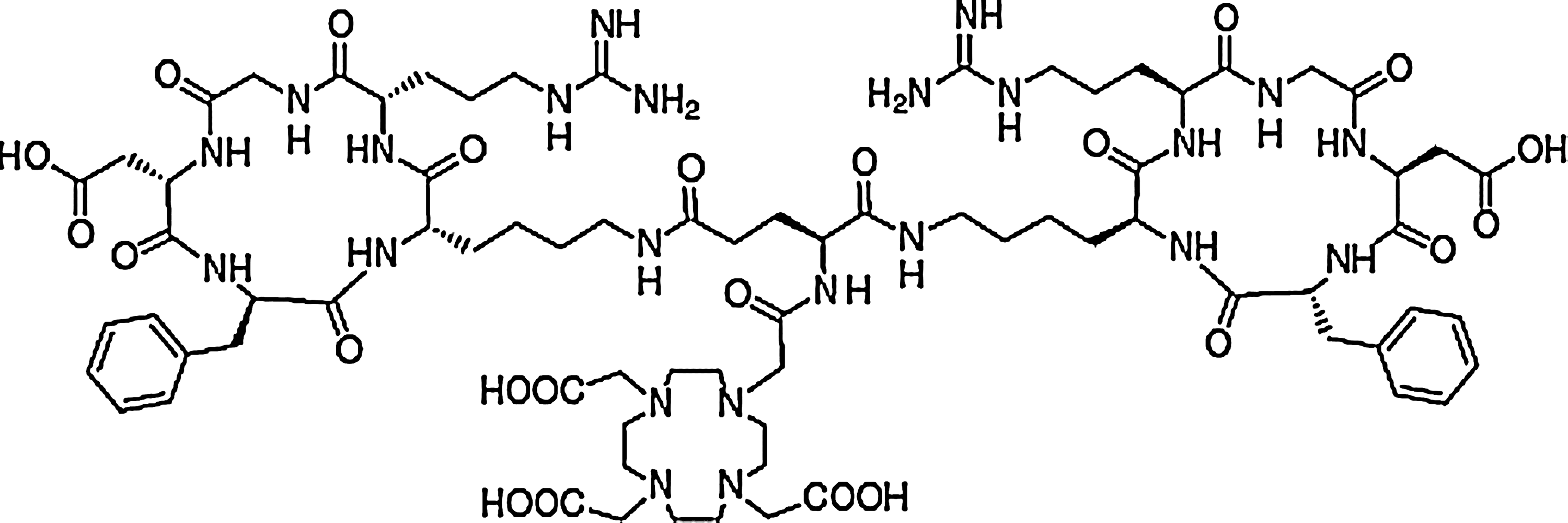
Structure DOTA-E[c(RGDfK)]2.
Materials and Methods
The RGD peptide conjugate, viz. DOTA-E[c(RGDfK)]2 (DOTA-RGD2) (Fig. 1), was custom synthesized by Ms. ABX Advanced Biochemical Compounds. All other chemicals used in the experiments were of AR grade and supplied by reputed chemical manufacturers.
Radioactivity assay of 68Ga activity was carried out by high-resolution gamma ray spectrometry using an HPGe detector (EGG Ortec/Canberra detector) coupled to a 4K multichannel analyzer system, measuring the 1077 keV γ-photopeak. Radionuclidic purity of 68Ga was also ascertained by the same system. A 152Eu reference source (Amersham, Inc.) was used for energy as well as efficiency calibration of the detector. All other radioactivity measurements were carried out by using a well-type NaI(Tl) scintillation counter (Electronics Corporation of India Limited) after adjusting the baseline at 450 keV and keeping a window of 100 keV, thereby utilizing the 511 keV annihilation gamma photon of 68Ga, unless mentioned otherwise.
Whatman 3-MM chromatography paper (UK) was used for paper chromatography studies. The high-performance liquid chromatography (HPLC) system equipped with a PU 1575 UV/VIS detector was obtained from JASCO (PU 1580). A well-type NaI(Tl) scintillation detector was coupled to the system for radioactivity measurements in the eluate. All the solvents used for HPLC analyses were of HPLC grade and purchased from reputed local manufacturers, degassed, and filtered before use.
Female C57/BL6 mice (6–8-week old) bearing melanoma tumors were used for evaluating the tumor avidity of the 68Ga-labeled agent. The melanoma cell line (ATCC-CRL-6475™), used for inoculation of the tumors, was purchased from the National Center for Cell Sciences. The animals were bred and reared in the laboratory animal facility of our Institute under standard management practice. Radioactive counting associated with the animal studies were carried out using a flat-type Nay(Tl) scintillation counter (Electronics Corporation of India Limited) by keeping the baseline and windows at 450 and 100 keV, respectively. All the animal experiments reported in the article were carried out in strict compliance with the relevant national laws relating to the conduct of animal experimentation.
68Ge/68Ga generator system
68Ga was obtained form a 740 MBq (20 mCi) 68Ge/68Ga generator developed in-house using a CeO2-PAN composite sorbent as the column matrix. 2768Ga activity was eluted from the generator with 2 mL of 0.01 M HCl solution and directly used for radiolabeling. The level of radionuclidic impurities and chemical contaminants present in the 68Ga solution was estimated using the reported procedures. 27
Preparation of 68Ga-DOTA-E[c(RGDfK)]2
For the preparation of the 68Ga-DOTA-RGD2 complex, 68GaCl3 solution (74–111 MBq 68Ga) was added to a solution of DOTA-RGD2 conjugate in 0.1 M ammonium acetate buffer (pH∼5.0) (containing 20 μg of peptide conjugate) in a clean glass vial. The resultant mixture was incubated at 90°C for 10 minutes in a boiling water bath after adjusting the pH to 3.5–4.0.
Various parameters, such as ligand concentration, incubation time, and temperature, were varied to determine the optimized protocol for obtaining the maximum complexation yield.
Quality control techniques
The radiolabeling yield and radiochemical purity were determined by paper chromatography (PC) and HPLC techniques.
Paper chromatography
Whatman 3-MM chromatography paper was used for PC. Five-microliter portions of the test solutions were applied at 1.5 cm from the lower end of the strips. The strips were developed in 50% aqueous acetonitrile, dried, cut into segments of 1 cm each, and the radioactivity associated with each segment was measured in an NaI(Tl) detector.
High-performance liquid chromatography
HPLC of the 68Ga-labeled RGD peptide conjugate was carried out using a dual pump HPLC unit with a C-18 reversed-phase HiQ-Sil (5 μM, 25 cm×0.46 cm) column. The elution was monitored by detecting both UV signals at 270 nm as well as radioactivity signal using an NaI(Tl) detector. Water (A) and acetonitrile (B) mixtures with 0.1% trifluoroacetic acid were used as the mobile phase, and the following gradient elution technique was adopted for the separation: 0–4 minutes 95% A, 4–15 minutes 95% A to 5% A, 15–20 minutes 5% A, 20–25 minutes 5% A to 95% A, and 25–30 minutes 95% A. The flow rate was maintained at 1 mL/min.
Optimization studies
Various reaction parameters such as concentration of peptide conjugate, pH of the reaction mixture, reaction time, and temperature were varied to achieve the maximum complexation yield of 68Ga-DOTA-RGD2. To determine the optimum ligand concentration required for obtaining maximum complexation, the amount of DOTA-RGD2 was varied from 5 to 40 μg, keeping the reaction volume constant. The effect of pH of the reaction mixture on complexation yield was studied by varying the pH of the reaction mixture within a range of 2–8 and determining the complexation yield at each pH. To ascertain the optimum reaction time and temperature, reactions were carried out by incubating the reaction mixture at room temperature and 90°C for different time periods (5, 10, 15, and 30 minutes) at an already optimized ligand concentration and pH. The results of all the complexation studies were reported as an average of three independent experiments.
Determination of octanol/water partition coefficients
The octanol/water partition coefficient of 68Ga-labeled DOTA-RGD2 was determined using the following protocol. A 25-μL aliquot of the radiolabeled conjugate prepared under optimized conditions was diluted to 1-mL volume using physiological saline. To this solution, 1 mL of n-octanol was added, and the mixture stirred vigorously for ∼10 minutes. Subsequently, the mixture was centrifuged at a speed of 3000 rpm for 5 minutes. Aliquots were withdrawn from both water and n-octanol layer and counted in an NaI(Tl) counter. The Log-P value was determined from these data and reported as an average of three independent measurements.
In vitro stability of 68Ga-DOTA-RGD2
The in vitro stability of the 68Ga-DOTA-RGD2 was ascertained by EDTA challenge using a 100-μL aliquot of the radiolabeled conjugate that was added to 1 mL of 0.1 M EDTA solution (pH ∼6.5) and the mixture stored at room temperature. The radiochemical purity was determined after 1 hour of storage by employing the standard quality control techniques described earlier.
In vivo stability of 68Ga-DOTA-RGD2
The in vivo stability of the 68Ga-DOTA-RGD2 complex was assessed by HPLC analysis of urine collected 30 minutes postadministration of the radiolabeled conjugate in normal C57/BL6 mice. Each mouse was administered with ∼3.7 MBq (∼100 μCi) of the radiolabeled conjugate through the tail vein. The urine samples were collected at 30 minutes postinjection (p.i) by manual void and were mixed with equal volume of 50% acetonitrile aqueous solution. The mixture was centrifuged at 5000 rpm 5 minutes. The supernatant was collected and passed through a 0.22-μm Millipore filter. The filtrate was analyzed by HPLC following the method described earlier.
Biodistribution studies
The biological behavior of the radiotracer prepared was studied in C57/BL6 mice bearing melanoma tumors. Melanoma tumors were developed by injecting ∼1×10⁶ melanoma cells (ATCC-CRL-6475™) suspended in 200 μL of PBS subcutaneously into the right thigh of each C57/BL6 mouse weighing 20–25 g. The animals were observed for visibility of tumors and subsequently allowed to grow for about 2 weeks to attain a tumor mass of 0.2–0.4 g. The radiotracer (∼100 μL, 3.7–5.5 MBq [100–150 μCi]) was injected into each animal through a lateral tail vein. The animals were sacrificed by cardiac puncture postanesthesia at 10, 30, and 60 minutes p.i. Four animals were used in each time point. Various organs, tissues, and tumors were excised after sacrifice, washed with physiological saline, dried, and the radioactivity associated with each organ and tissue was determined using a flat-type NaI(Tl) counter. The weight of each organ and tumor was also determined by using an analytical balance. The percent injected activity (%ID) in various organs, tissues, and tumor was calculated from the above data and expressed as percentage injected activity per gram (%ID/g) of organ/tissue. The activity excreted was indirectly determined from the difference between total ID and the %ID accounted for in all the organs. The total uptake in blood, bone, and muscles was calculated by assuming that 7%, 10%, and 40% of the body weight are constituted by these organs/tissue, respectively. 43,44
Saturation studies were also performed to determine whether the uptake of the radiotracer in melanoma tumor is receptor mediated. For this, four C57/BL6 mice bearing melanoma tumors were used, and each animal was administered with ∼3.7 MBq (100 μCi) of the radiotracer along with 500 μg (20–25 mg/Kg of body weight) of E[c(RGDfK)]2 (RGD2). Such a high dose of peptide was used to ensure that all the integrin αvβ3 receptors are blocked. The animals were sacrificed at 30 minutes p.i. %ID/g of organ/tissue was determined following the procedure mentioned above. The uptakes in different organs/tissue and tumor were compared to those obtained in the absence of excess RGD2.
Results and Discussion
68GaCl3
68Ga activity was obtained from the 68Ge/68Ga generator developed in-house with excellent purity and in adequate radioactive concentration (∼370 MBq/mL) suitable for radiopharmaceutical applications. 27 The amount of 68Ge impurity in 68Ga was <20 Bq (<105% of the total 68Ga activity) in all the elutions. The chemical impurities present in the 68Ga eluate in the form of Ce, Fe, and Mn ions were <0.1 mg/L (0.1 ppm), as ascertained by ICP-AES analyses of the decayed samples. Thus, the radionuclidic and chemical purity of 68Ga obtained from the generator was comparable to that obtained from commercial generators. 27 It could be noted that while the eluate 68Ga from commercial generators were subjected to multiple purification steps to obtain clinical-grade 68Ga, 26,28,29 the developed generator provides 68Ga of similar purity in a single step. Moreover, unlike the commercial generators, the elution performance of this generator remained consistently good over a prolonged period of time. 27
Characterization of 68Ga-labeled DOTA-RGD2 conjugate
The 68Ga-labeled DOTA-RGD2 conjugate was characterized by PC and HPLC. In PC using 50% aqueous acetonitrile as the eluting solvent, the activity corresponding to the 68Ga-DOTA-RGD2 complex moved toward the solvent front with a Rf of 0.8–0.9, whereas uncomplexed 68Ga remained at the point of spotting (Rf=0) under identical conditions.
The 68Ga-labeled DOTA-RGD2 complex was also characterized by HPLC, and a typical radio-HPLC pattern of the 68Ga-DOTA-RGD2 conjugate is shown in Figure 2. It was observed that the radiolabeled conjugate exhibited a retention time of 15.6 minutes. On the other hand, uncomplexed 68GaCl3 was eluted in the void volume. The extent of complexation and the radiochemical purity of the complex could therefore be determined from the HPLC.
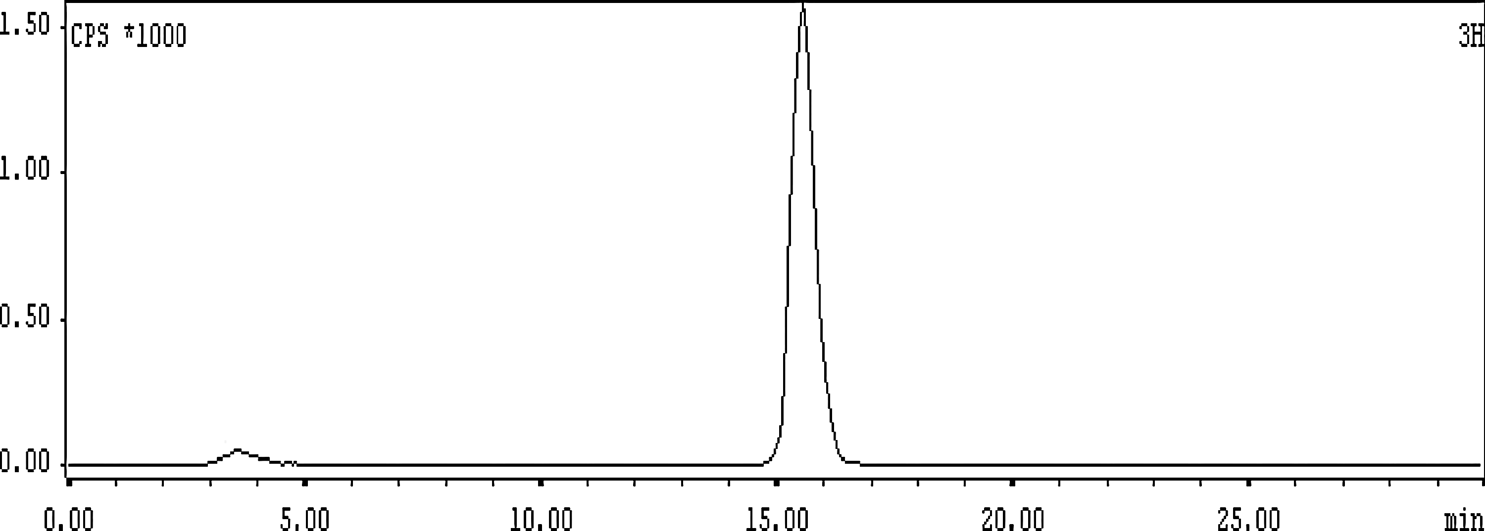
A typical radio-HPLC pattern of 68Ga- DOTA-E[c(RGDfK)]2 complex. HPLC, high-performance liquid chromatography.
Optimization studies
Studies on the effect of ligand concentration on complexation yield showed that a minimum of 20 μg of DOTA-RGD2 was required to obtain a complexation yield of 98.5%±0.3% (n=3). The effect of the amount of peptide conjugate on the complexation yield of 68Ga-DOTA-RGD2 is shown in Figure 3. Studies on the effect of pH of the reaction mixture on the complexation yield within a pH range of 2–8 indicated that the yield was maximum at pH ∼3.5–4.0.
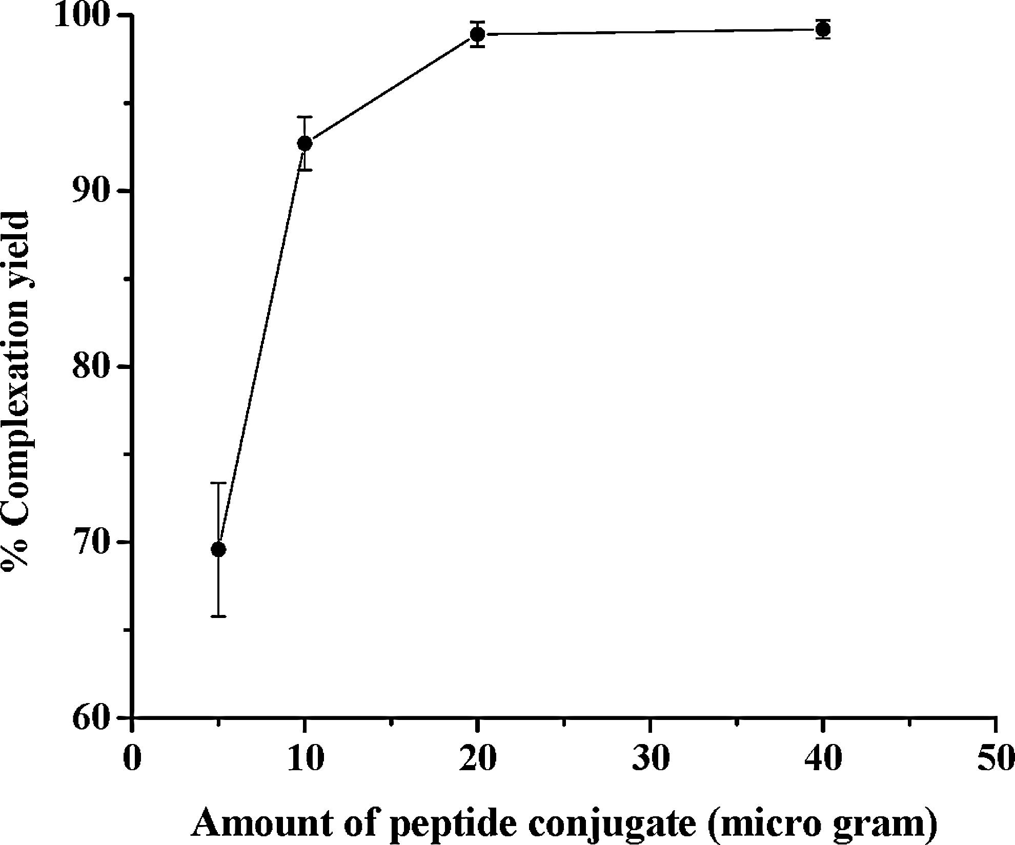
The effect of the amount of peptide conjugate on the complexation yield of 68Ga-DOTA-E[c(RGDfK)]2.
It was observed that a 98.5%±0.3% (n=3) complexation yield was achieved within 10 minutes when the reaction mixture was incubated at 90°C. On the other hand, a maximum of 80.3%±1.2% (n=3) complexation could be achieved when the reaction mixture was incubated for 30 minutes at room temperature. Therefore, 10-minute incubation of 20 μg DOTA-RGD2 with 68GaCl3 solution at pH ∼4 at 90°C was considered as the optimum concentration for radiolabeling.
Log P and stability of 68Ga-DOTA-RGD2
The Log P value of the 68Ga-DOTA-RGD2 complex was found to be −2.56±0.04 (n=3), indicating that the complex is highly hydrophilic in nature. The in vitro stability of the 68Ga-DOTA-RGD2 complex was studied by EDTA challenge, whereby the radiolabeled conjugate was stored in 0.1 M EDTA solution (pH ∼6.5) at room temperature. It was observed that the radiolabeled conjugate retained its radiochemical purity to the extent of >95% after 2 hours of storage in 0.1 M EDTA solution at room temperature. In vivo stability study carried out by HPLC analysis of urine collected 30 minutes postadministration of the radiolabeled conjugate into normal C57/BL6 mice showed that the complex retained its radiochemical purity to the extent of >95% in the urine sample. Figure 4 shows the radio-HPLC pattern of the urine sample.
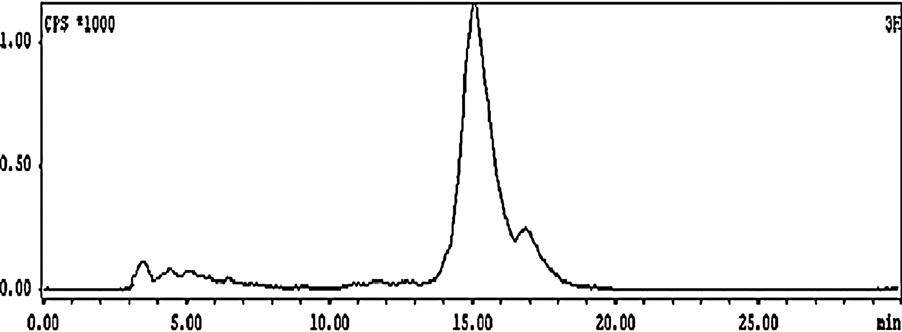
Radio-HPLC pattern of urine sample collected 30 minutes postadministration of 68Ga- DOTA-E[c(RGDfK)]2 complex into normal C57/BL6 mice.
Biodistribution studies
The uptake of the 68Ga-DOTA-RGD2 complex in the different organs/tissues of C57/BL6 mice bearing melanoma tumors expressed as %ID/g of organ/tissue at different p.i. times is shown in Table 1. The results of the biodistribution studies revealed a significant tumor uptake within 10 minutes p.i. (4.14±0.54%ID/g). The activity accumulated in the tumor was observed to increase further (4.61±0.31%ID/g) at 30 minutes p.i. Initial accumulation of activity was observed in various nontarget organs, viz. the liver, GIT, kidney, and lungs. However, with the progress of time, the uptake in nontarget organs was observed to gradually reduce. The tumor/organ ratio of the radiotracer at different time points p.i. for the major organs/tissues, viz. blood, liver, kidney, and muscle, is shown in Figure 5. The tumor-to-blood ratio was observed to increase from 1.75±0.42 at 10 minutes p.i. to 2.25±0.20 at 60 minutes p.i., whereas the tumor-to-liver and tumor-to-muscle ratio increased from 2.71±0.76 to 3.31±0.84 and from 5.37±1.08 to 8.97±1.32, respectively, between the same time points. The radiolabeled conjugate exhibited predominant urinary excretion, as more than 80% of the ID was observed to be cleared via the renal pathway within 60 minutes p.i.
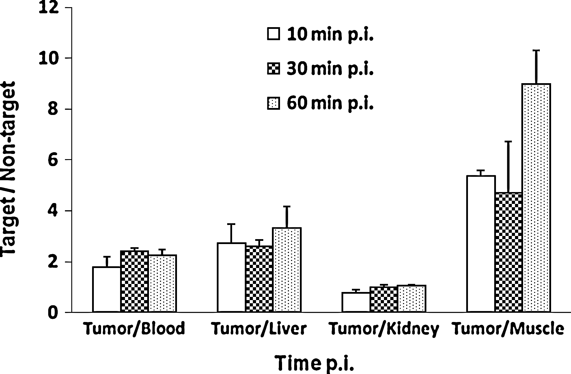
The tumor/background ratio of 68Ga-DOTA-RGD2 complex at different time points postinjection for the major organs/tissues in C57/BL6 mice bearing melanoma tumors.
Figures in the parentheses represent standard deviations.
Since the 68Ga-DOTA-E[c(RGDfK)]2 radiotracer is designed for imaging tumor by PET, the tumor uptake obtained in biodistribution studies is highlighted in bold font.
At every time point, 4 animals have been used.
Excretion has been calculated by subtracting the activity accounted in all the organs from the total activity injected.
Figure 6 compares the uptake of 68Ga-DOTA-RGD2 in various organ/tissues of C57/BL6 mice bearing melanoma tumors at 30 minutes p.i. in the absence/presence of excess RGD2. Clearly, coinjection of excess RGD2 almost completely blocked the tumor uptake of 68Ga-DOTA-RGD2 (0.39±0.14%ID/g with RGD2 vs. 3.80±0.55%ID/g without RGD2 at 30 minutes p.i.). Its uptake in normal organs was also significantly blocked by coinjection of excess RGD2 as shown in Figure 6. The blockage of radiotracer uptake observed strongly suggests that the tumor localization of the radiotracer is indeed receptor mediated. The partial blockage of uptake in the heart, lungs, liver, and spleen indicates that the accumulation of 68Ga-DOTA-RGD2 in these organs is mostly integrin αvβ3-mediated.
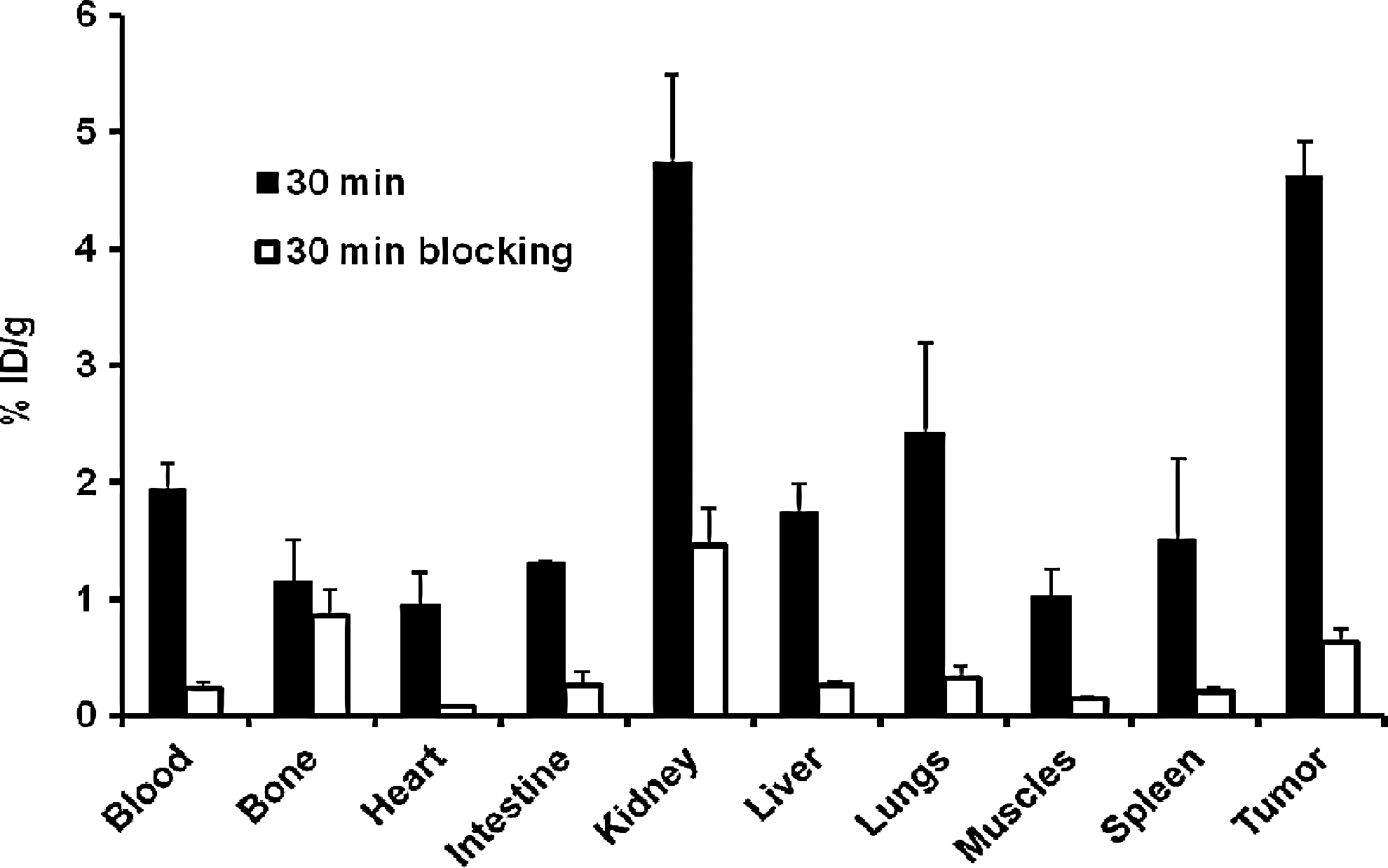
Comparison of 30-minute organ uptake of 68Ga-DOTA-RGD2 in C57/BL6 mice bearing melanoma tumors in the absence/presence of excess RGD2.
Conclusions
The present work describes the preparation of the 68Ga-labeled cyclic RGD peptide conjugate DOTA-E[c(RGDfK)]2 and the preliminary biological studies of the radiolabeled conjugate for possible use in in-vivo tumor imaging. 68Ga was obtained from a novel Nanoceria-based 68Ge/68Ga generator developed in-house. The 68GaCl3 activity eluted from the generator was used for radiolabeling without further purification. Radiolabeling of DOTA-E[c(RGDfK)]2 with 68Ga was achieved with an excellent radiochemical yield and purity (98.5%±0.3%) using 20 μg of peptide conjugate within 10 minutes at 90°C. The radiolabeled conjugate exhibited adequately high stability at room temperature. Biodistribution studies carried out in C57/BL6 mice bearing melanoma tumors revealed rapid and significant tumor uptake (4.14±0.54 and 4.61±0.31 IA/g at 10 minutes and 30 minutes p.i., respectively) with a satisfactory tumor-to-background ratio. These preliminary biological studies indicate the potential of the developed agent for possible use in detection of tumor by PET imaging, although further studies are warranted in animal models to determine the actual potential of the agent. However, the availability of a reliable and easy-to-handle 68Ge/68Ga generator like the one used in the present study would facilitate more research on new 68Ga radiopharmaceuticals for PET imaging.
Footnotes
Acknowledgments
The authors acknowledge Dr. Sharmila Banerjee, Radiopharmaceuticals Division, Bhabha Atomic Research Centre, for her keen interest and support. The sincere help received from the staff members of the Animal House Facility of Bhabha Atomic Research Centre during the course of animal experimentations is also gratefully acknowledged.
Disclosure Statement
The authors have neither received any outside funding nor any grants from any external agencies in support of this study. Our institutions do not have a financial relationship with any commercial entity that has an interest in the subject matter or materials discussed in this manuscript. None of the authors in this manuscript have any conflict of interest, financial, or otherwise in the publication of this material.