
Abstract
Background:
Pancreatic cancer (PaC) is a highly malignant gastrointestinal tumor with invasive and metastatic characteristics. Interleukin-6 (IL-6), a negative prognostic marker, contributes to PaC progression. However, the mechanism of IL-6 in PaC is not yet fully understood.
Methods:
miR-455-5p levels were first tested by reverse transcription-quantitative polymerase chain reaction (RT-qPCR) in PaC tissues or cells. Subsequently, PaC cell-related functions were identified through CCK-8, Transwell, and Western blotting. Changes in miR-455-5p and IGF-1R expression were confirmed using RT-qPCR and Western blotting. miR-455-5p methylation was assessed by bisulfite sequencing PCR.
Results:
The authors discovered that miR-455-5p was expressed at low levels in PaC tissues and cells, and miR-455-5p expression was observably reduced by IL-6 in PaC cells. In addition, IL-6 dramatically induces miR-455-5p methylation in PaC cells. Functionally, the data revealed that IL-6 could facilitate the malignant properties of PaC cells, including proliferation, epithelial–mesenchymal transition, and metastasis. The authors found that miR-455-5p could suppress the progression of PaC cells by downregulating IGF-1R in PaC cells. Mechanistically, IL-6 downregulated miR-455-5p and upregulated IGF-1R, and miR-455-5p reduced IGF-1R expression through targeted binding.
Conclusions:
The authors demonstrated that the miR-455-5p/IGF-1R axis is necessary for the induction of IL-6 in PaC progression. The results here may provide a theoretical basis for the application of the IL-6/miR-455-5p/IGF-1R axis in the clinical therapy of PaC.
Introduction
Pancreatic cancer (PaC) is a highly aggressive gastrointestinal system tumor. 1 The reasons for the high mortality of PaC include the protective effect of pancreatic location on tumor discovery, invasiveness, drug resistance, and reduction in the capacity of patients to withstand the positive treatment. 2 Currently, therapies for PaC include surgical resection, radiotherapy, immunotherapy, and chemotherapy. Although each treatment has its limitations, 3 surgery is the preferred therapeutic regimen for PaC. 4 However, since PaC is insidious, most patients cannot be treated surgically at the onset, 5 and surgery is still associated with higher postoperative morbidity. Chemotherapy, as a traditional method, has multiple disadvantages, such as low bioavailability, large side-effects, and induction of multiple drug resistances in the tumor. 6 Therefore, in-depth exploration of PaC pathogenesis is of key scientific value and practical significance for seeking effective clinical therapeutic methods.
Interleukin-6 (IL-6) is a pleiotropic cytokine secreted by multiple cells, including mesenchymal cells, fibroblasts, macrophages, endothelial cells, and tumor cells. 7 IL-6 has a crucial pro-inflammatory effect. 8 It is reported that IL-6 is highly expressed in many cancers, including lung, 9 gastric, 10 and ovarian cancers. 11 IL-6 has also been shown to accelerate cell growth, angiogenesis, invasion, and metastasis, and is associated with poor patient prognosis. 12,13 Multiple studies have also shown that IL-6 can affect PaC pathogenesis, which might be a latent therapeutic opportunity. 14,15 However, the mechanism of IL-6 in the PaC process has not been clearly elucidated. Further understanding of the complexity of the IL-6 pathway and the precise background of IL-6 inhibition or activation might further improve the clinical efficacy in PaC patients.
IL-6 has been reported to alter the malignant progression of multiple cancers through microRNA (miRNA) regulation. 16,17 miRNAs, as the key factors in gene networks, have a great influence on biological processes, including cell cycle, proliferation, death, and inflammation. 18 –20 Studies have revealed that miR-455-5p has low expression in various cancer tissues and has a tumor suppressive effect. 21,22 In addition, studies testified that miR-455-5p could prevent proliferation and trigger apoptosis of prostate cancer cells 23 ; miR-455-5p could block viability and metastasis, and induce apoptosis in cervical cancer cells through S1PR1, 24 whereas the impact of miR-455-5p in PaC progression is largely uncertain. The relationship between IL-6 and miR-455-5p is unclear. IL-6 has not been reported to be able to mediate miR-455-5p to affect PaC progression.
In this study, the authors evaluated the influence of IL-6 on miR-455-5p expression and PaC progression. In addition, they investigated the possible regulatory mechanisms of miR-455-5p in PaC. Confirming the current research might provide a new experimental basis for PaC targeted therapy.
Materials and Methods
Clinical specimens
Thirty PaC surgical specimens were collected from the Chongqing University Cancer Hospital. Each specimen was taken from PaC and paracarcinoma tissues. All specimens were confirmed by histopathologists and were immediately stored in liquid nitrogen after isolation. None of the patients received chemotherapy or radiotherapy before surgery. Permission was acquired from the ethics committee of Chongqing University Cancer Hospital. The participants provided written informed consent.
Cell culture
Human pancreatic cells (HPC-Y5 and BNCC101270) were obtained from Beina Company (China), and 293T, PANC-1 (CRL-1469), BxPC3 (CRL-1687), HPAF-II (CRL-1997), and SW1990 (CRL-2172) were obtained from ATCC (Manassas, VA). 293T, HPC-Y5, and PANC-1 cells were grown in Dulbecco minimum essential medium (Gibco), HPAF-II cells were maintained in Eagle's minimal essential medium (Gibco), BxPC3 cells were cultured in RPMI 1640 (Gibco), and SW1990 cells were cultured in Leibovitz's L-15 (Gibco). All media were supplemented with 10% fetal bovine serum (FBS; Gibco), and all cells were placed in an incubator at 37°C and 5% CO2.
Cell treatment
PANC-1 and BxPC3 cells (1 × 10⁵ cells/well) were uniformly seeded in 6-well plates and treated with 0, 1, 2, 4, and 8 ng/mL IL-6 (Sigma-Aldrich). IGF-1R plasmids and empty vectors were obtained from HanBio Biotechnology (Shanghai, China). Negative control and miR-455-5p mimics were obtained from GenePharma (Shanghai, China). PaC cells were also transfected with IGF-1R plasmids or miR-455-5p mimics using Lipofectamine 3000 (Invitrogen) for 48 h according to the manufacturer's instructions.
Reverse transcription-quantitative polymerase chain reaction
Total RNAs were isolated from cells treated in the previous step using TRIzol reagent (Invitrogen). After quality testing, the harvested RNAs (1.5 μg) were reverse-transcribed into cDNAs using the BestarTM qPCR RT kit (DBI; 2220). The cDNAs were then subjected to quantitative polymerase chain reaction (qPCR) with SYBR Green Master (Roche). The PCR cycling condition was 95°C, 2 min; 95°C, 20 s; 58°C, 20 s; 72°C, 20 s; 40 cycles. The data were calculated by the 2−ΔΔCt method. Table 1 presents the primer sequences.
The Sequences of Primers in Reverse Transcription-Quantitative Polymerase Chain Reaction
Western blot
The PaC cells were collected and increased with 1 mL phenylmethanesulfonyl fluoride solution (100 mmol/L) and 10 μL cocktail for protein extraction. The extracted proteins were quantified. Next, 5 × loading buffer and protein (40 μg) were mixed at 4 × 1, boiled for 10 min, and sodium dodecyl sulfate polyacrylamide gel electrophoresis was conducted after restoration to room temperature. The gel was then transferred to a polyvinylidene fluoride membrane (Millipore) and filter paper to form a “sandwich” structure. The membranes were then sealed and exposed to primary antibodies at 4°C overnight and secondary antibodies (Abcam; 1:1000) at 37°C for 1 h. The fluorescein substrate was uniformly added to the membrane surface for 5 min, and the protein was developed.
Bisulfite sequencing PCR
Genomic DNAs from the PaC cells were extracted using a DNA extraction kit (Applied Biosystems). After DNA purity and concentration were determined, DNA was modified with bisulfite according to the instructions of the methylation kit (Zymo Research). The reaction conditions were 95°C for 4 min, 35 cycles (94°C for 30 s, 55–60°C for 25 s, 72°C for 40 s), and 72°C for 6 min. The SssI enzyme was used as the positive control. PCR products were electrophoresed, and the results were observed based on the electrophoretic map.
CCK-8
PaC cells (2 × 10⁴ cells/well) were uniformly seeded into 96-well plates overnight. At 0, 24, and 48 h, 20 μL of CCK-8 solution (Dojindo, Japan) was disposed of in the wells. After incubation for 3 h at 37°C, the optical density was measured at 450 nm.
Transwell
The medium (600 μL) containing 10% FBS was added to the lower chamber of a transwell chambers (Millipore). Cell invasion was assessed in Transwell chambers, which were pre-paved with diluted Matrigel (1:8, 50 μL). The processed PaC cells (200 μL, 2 × 10⁵ cells/well) were added to the upper chamber and incubated at 37°C for 48 h. Then, the fluid in the upper chamber was discarded, and wet cotton swabs were applied to remove cells that had not penetrated the membrane. Cells that passed through the membrane were fixed and stained with 0.1% crystal violet. After cleaning, the cells were counted under a light microscope (Nikon, Japan) in random five fields, photographed, and statistically plotted.
Dual-luciferase reporter gene assay
The miR-445-5p and IGF-1R 3′-UTR latent binding sites were predicted using a biological information software (miRanda and TargetScan). After the IGF-1R gene was amplified by PCR, the wild-type (WT)-IGF-1R plasmids were constructed using the pisCheck 2 vector. IGF-1R mutants were constructed from the WT-IGF-1R plasmids using a point mutation kit. The authors co-transfected 293 T cells (1 × 10⁵ cells/well) in 24-well plates with miR-445-5p mimics and WT-IGF-1R or Mut-IGF-1R using Lipofectamine 3000 (Invitrogen). After 48 h of transfection, luciferase activity was tested using the dual-luciferase reporter assay (Promega).
RNA pull-down assay
The Pierce Magnetic RNA-Protein Pull-Down Kit was acquired from Thermo Fisher Scientific (Waltham, MA) for the RNA pull-down assay. Cell extracts were mixed with biotin-labeled RNA probes for IGF-1R 3′-UTR fragments (WT and Mut) and magnetic beads. The samples were analyzed using quantitative reverse transcription-polymerase chain reaction (qRT-PCR).
Statistical analysis
Measurement data are presented as the mean ± standard deviation from three independent experiments. The statistical results were analyzed using SPSS23.0 (SPSS, Chicago, IL). For data with homogeneous variance in a normal distribution, a two-tailed Student's t-test (two groups) or one-way analysis of variance (ANOVA; three or more groups) was used. The paired t-test was used to compare the miRNA and IGF-1R levels in pancreatic and paracancerous tissues, and the chi-squared test was used to analyze the correlation between clinical features and mRNA-455 expression levels. Statistical significance was set at p < 0.05.
Results
Downregulation of miR-455-5p in PaC
To confirm the miR-455-5p expression pattern in PaC, the authors first analyzed the changes in miR-455-5p expression in PaC tissues. As shown in Figure 1A, miR-455-5p was observably reduced in PaC tissues compared with paracarcinoma tissues. The authors also found that, relative to HPC-Y5 cells, miR-455-5p expression was lower in PaC cells (PANC-1, HPAF-II, BxPC3, and SW1990), especially in HPAF-II and SW1990 cells (Fig. 1B). In short, these data indicated that miR-455-5p downregulation might play a role in PaC progression, and the authors also selected two cell lines (PANC-1 and BxPC3) for the research.
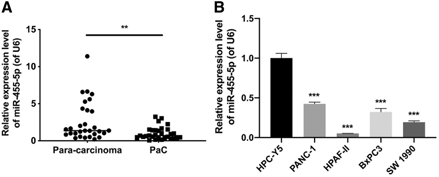
miR-455-5p was downregulated in PaC tissues and cells.
Relationship between miR-455-5p expression and clinicopathological features of PaC
Table 1 summarizes the association between miR-455-5p expression and clinicopathological variables in patients with PaC. Low miR-455 expression in PaC tissues was strongly correlated with tumor size, TNM classification, distant metastasis, and lymphatic invasion. However, no significant correlation was found between miR-455-5p expression and patient age and gender (Table 2).
Relationship Between miR-455-5p and Clinicopathological in 30 Patients with Pancreatic Cancer
IL-6 induced the methylation of miR-455-5p in PaC cells
Next, the authors verified the miR-455-5p expression and methylation in the PaC cells. These two strains of cells were treated with 0, 1, 2, 4, and 8 ng/mL IL-6. RT-qPCR results indicated that adding IL-6 could cause a striking miR-455-5p downregulation in PaC cells, and miR-455-5p downregulation expression also gradually decreased with an increase in IL-6 concentration (Fig. 2A). In addition, the bisulfite sequencing PCR results showed that IL-6 could contribute to a remarkable elevation in miR-455-5p methylation in PaC cells (Fig. 2B). Overall, these findings indicated that IL-6 downregulates miR-455-5p and enhances miR-455-5p methylation in PaC cells.
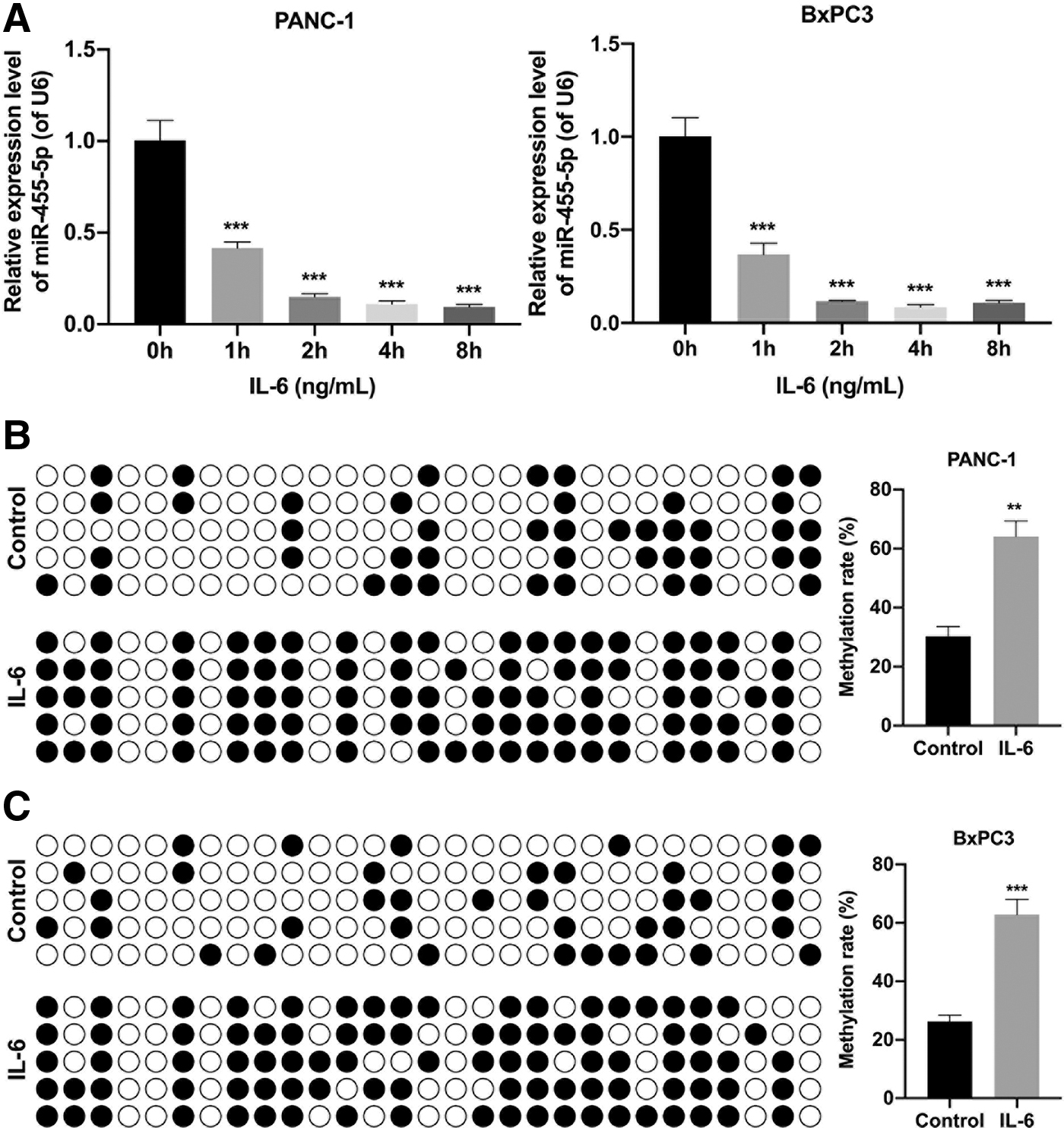
IL-6 induced miR-455-5p methylation in PANC-1 and BxPC3 cells.
IL-6 accelerated growth, EMT, and metastasis of PaC cells
The authors also investigated the influence of IL-6 on the malignant features of PaC cells. CCK-8 data showed that the cell proliferative capacity was significantly increased in the IL-6 group compared with that in the control group (Fig. 3A). Transwell assays showed that IL-6 could also prevent PaC cell migration and invasion (Fig. 3B). Meanwhile, the authors discovered that IL-6 could also generate noteworthy N-cadherin upregulation, Slug, and Twist and E-cadherin downregulation in PaC cells (Fig. 3C). The authors confirmed that IL-6 could increase the degree of malignancy of PaC cells.
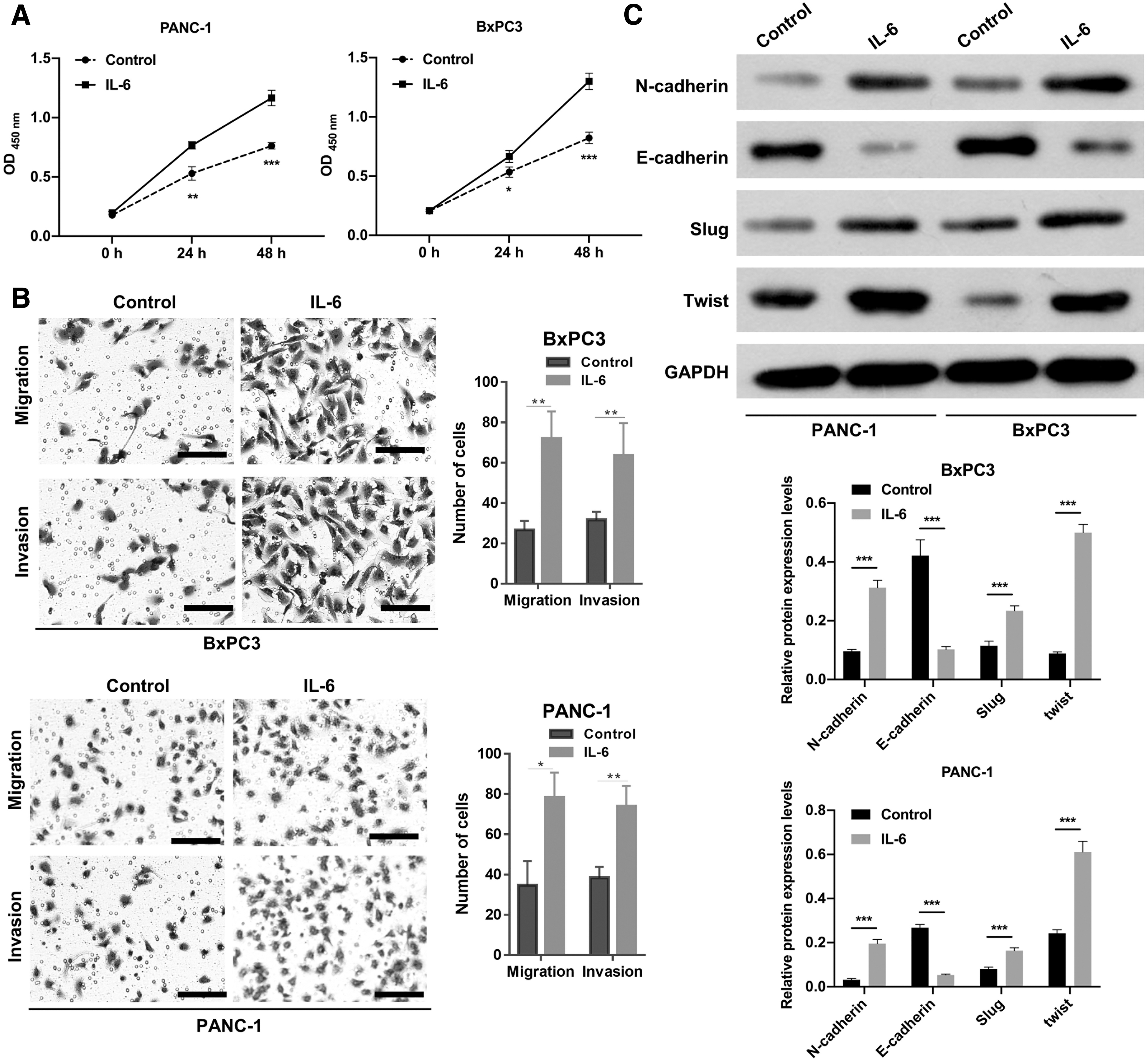
IL-6 accelerated growth, EMT, migration, and invasion of PaC cells.
IL-6 upregulated IGF-1R and miR-455-5p downregulated IGF-1R by targeting binding in PaC cells
Mechanistically, the authors further validated the downstream genes that miR-455-5p may regulate in PaC cells. First, the results revealed that IL-6 notably increases IGF-1R expression in PaC cells (Fig. 4A). Second, Western blot data indicated that miR-455-5p overexpression dramatically lowered IGF-1R expression in PaC cells (Fig. 4B). In addition, the authors found that miR-455-5p overexpression notably weakened the luciferase activity of WT-IGF-1R but did not change that of Mut-IGF-1R in 293T cells (Fig. 4C). miRNA-455-5p could bind to the 3′-UTR of IGF-1R (Fig. 4D). RNA pull-down assay results indicated that miRNA-455-5p was pulled down by Bio-WT-3′ UTR fragments of IGF-1R in PaC cells (Fig. 4E). To further verify the IGF-1R expression level in PaC tumors, the authors detected higher IGF-1R expression in PaC samples than in paracarcinoma samples using qRT-PCR (Fig. 4F
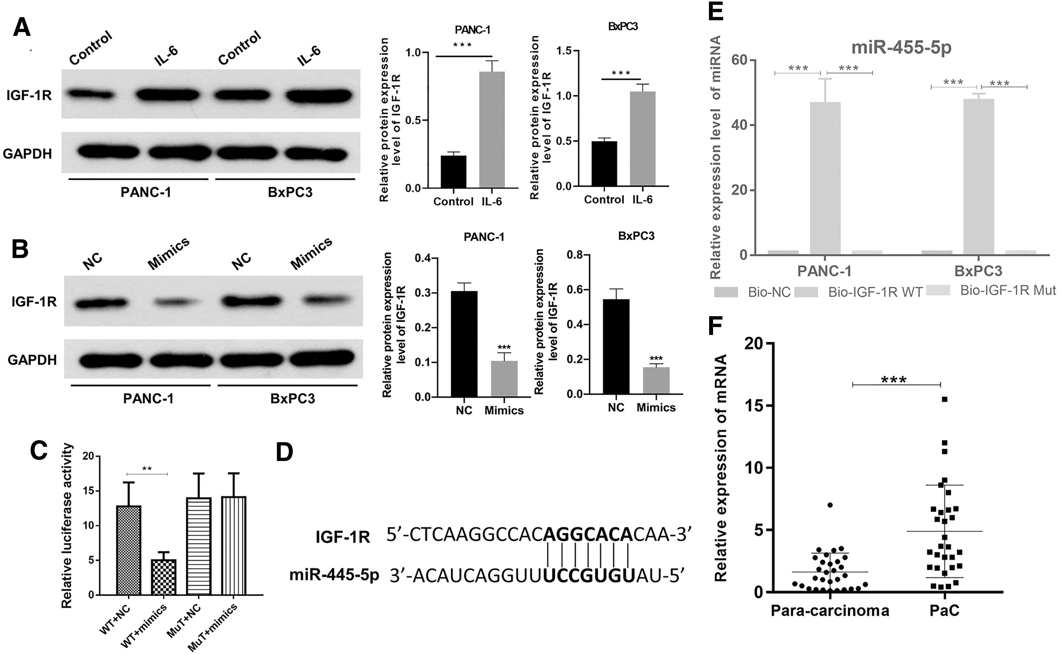
IL-6 upregulated IGF-1R, and miR-455-5p downregulated IGF-1R by targeting binding in PaC cells.
miR-455-5p overexpression suppressed PaC progression by IGF-1R
Based on IL-6 acceleration in the PaC process and the regulatory relationship between IL-6, miR-455-5p, and IGF-1R, the authors further verified whether miR-455-5p could influence PaC progression by changing IGF-1R expression. The CCK-8 results showed that IGF-1R overexpression markedly strengthened the proliferative activity of PaC cells, which could also be prominently restrained by miR-455-5p overexpression (Fig. 5A).
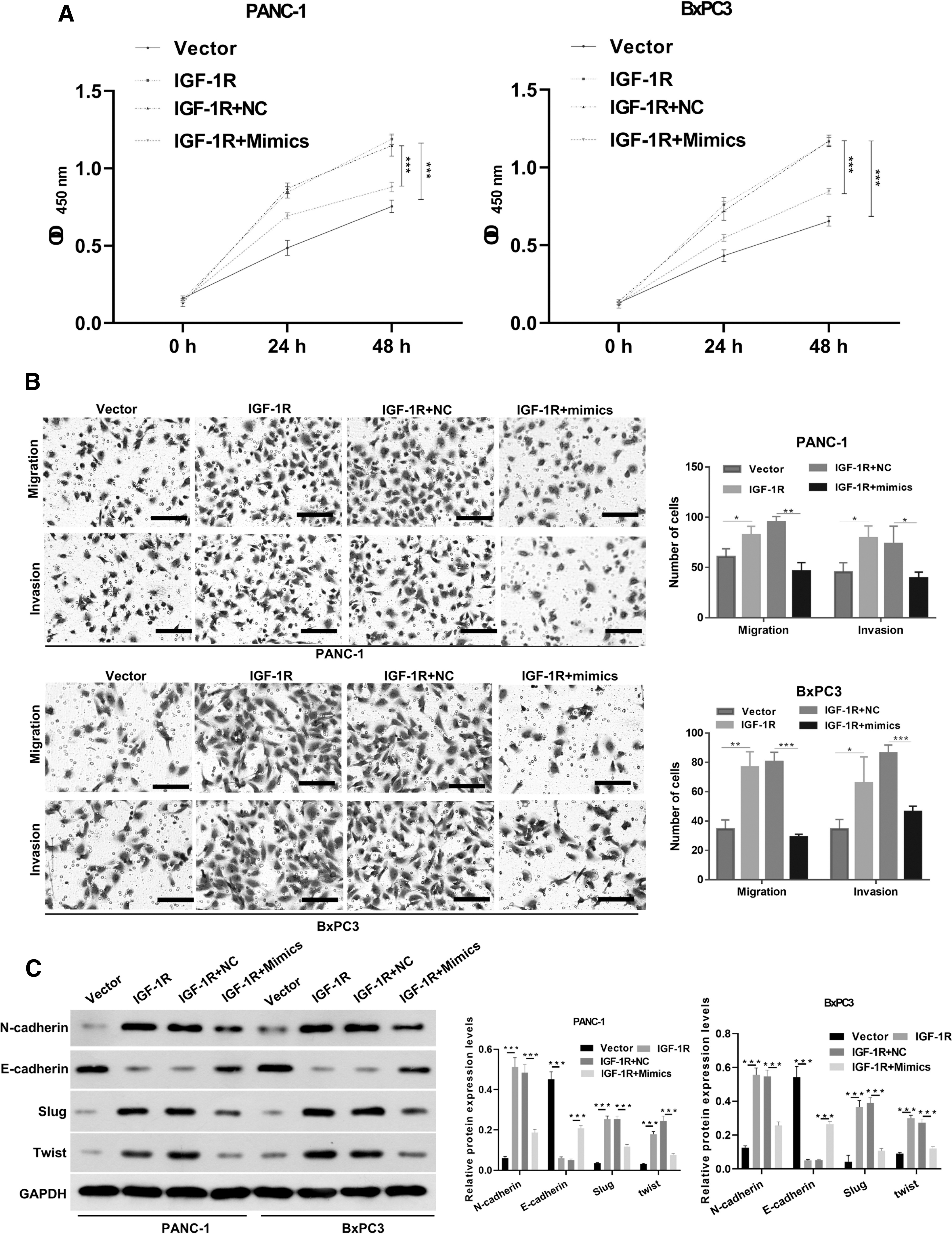
miR-455-5p overexpression suppressed PaC progression by IGF-1R. PANC-1 and BxPC3 cells were co-transfected with IGF-1R plasmids and miR-455-5p mimics.
In addition, the data indicated that IGF-1R overexpression dramatically boosted the migratory and invasive capabilities of PaC cells, which were also significantly repressed by miR-455-5p (Fig. 5B). Similarly, N-cadherin, Slug, and Twist levels were prominently increased, and E-cadherin levels were significantly diminished in the IGF-1R overexpression group relative to that in the vector group, while the expression changes of these four proteins mediated by IGF-1R could also be signally reversed by miR-455-5p overexpression (Fig. 5C). Thus, the data confirmed that miR-455-5p could inhibit the proliferation and metastasis of PaC cells by IGF-1R.
Discussion
PaC, as a highly aggressive gastrointestinal malignancy, has a high degree of malignancy. 25 Currently, PaC has the following characteristics: early diagnosis rate is not high, inconspicuous, high surgical mortality, easy metastasis, and low survival rate. 26 Molecular therapy, as a research hotspot, might provide a novel direction for the clinical therapeutics of PaC. 27 miRNAs, as endogenous and highly conserved ncRNAs, can alter the post-transcriptional modification of target genes by acting on 3′-UTR, thereby causing gene degradation. 19,20,28
Furthermore, miRNAs can affect cell proliferation, metastasis, and other processes, thus presenting carcinogenic or anticancer effects. 29,30 Based on literature reports, miR-455-5p could play a carcinogenic role in certain cancers (breast cancer and colon cancer), 31,32 and has tumor suppressive effect in certain cancers (gastric cancer and prostate cancer). 22,23 This is the first study to discover that miR-455-5p was significantly underexpressed in PaC tissues and cells, proving that miR-455-5p might play an inhibitory action in PaC progression.
The tumor microenvironment is the main location where tumor cells can communicate with the outside world, and it is key for the development of the tumor process. 33 There are numerous cytokines in the tumor microenvironment, which contain antitumor and pro-tumor categories in line with their functions. 34 Among them is IL-6, which participates in the regulation of immune response, inflammation, and other biological processes. 12 Moreover, IL-6 is also a key factor in cancer progression, including PaC. 15,35 In this study, the authors presented that IL-6 could memorably downregulate miR-455-5p in PaC cells, indicating that miR-455-5p inhibition in PaC progression might be mediated in part by IL-6. However, the mechanism by which IL-6 downregulates miR-455-5p in PaC is unclear.
In recent years, epigenetic regulation has been discovered to play a crucial role in tumor pathogenesis, especially DNA methylation and abnormal miRNA expression. 36 Research certified that abnormal methylation in or near CpG islands of numerous miRNAs is a key mechanism leading to decreased expression of certain tumor suppressive miRNAs. 37 In this study, the authors further demonstrated that IL-6 could prominently induce miR-455-5p methylation in PaC cells. This also suggests that DNA methylation might be the regulatory mechanism of the abnormally reduced miR-455-5p expression in PaC.
Invasion and metastasis are the characteristics of PaC development. 38 In clinical practice, most patients will have an invasion of surrounding tissues and organs and distant metastasis. 39 Tumor metastasis is a complex multipathway dynamic process. 40 Tumor cells first acquire the capacity to migrate, invade the extracellular matrix and basement membrane, enter the blood circulation, evade the surveillance and clearance of the immune system, spread to distant tissues and organs, and eventually form metastatic tumors. 39 An early key step in this cascade is epithelial–mesenchymal transformation (EMT). 41
EMT-associated regulatory factors comprise Snail, ZEB, and TWIST families. 42 Slug belongs to the Snail family, which can trigger the EMT by competitively combining with the E-cadherin. 43 In this study, the authors discovered that IL-6 could upregulate N-cadherin, Slug, and Twist, and downregulate E-cadherin in PaC cells, indicating that IL-6 could induce EMT of PaC cells. In addition, they verified that IL-6 could promote PaC cell growth and metastasis. Thus, they confirmed that IL-6 accelerated the malignant activity of PaC cells. Moreover, the data further revealed that miR-455-5p downregulation was key to the IL-6-induced PaC deterioration.
miR-455-5p was reported to suppress cancer cell growth and invasion by targeting IGF-1R. 44 This study confirmed that IGF-1R is a target gene of miR-455-5p in PaC cells. IGF is a peptide hormone that mainly affects cell proliferation and differentiation. 45 IGF-1R is the main functional IGF receptor, and the proliferative role of IGF is mainly realized through IGF-1R. 46 Furthermore, research also testified that IGF-1R has pro-proliferative, pro-metastatic, and antiapoptotic functions. 47 Moreover, IGF-1R has been proven to be key in PaC progression. 48 Also, the results further denoted that miR-455-5p also could block the malignant processes of PaC cells by IGF-1R.
Conclusions
This study provides a novel and latent regulatory loop (IL-6/miR-455-5p/IGF-1R) in PaC progression, and blockage of this pathway might also be a therapeutic strategy for PaC therapy.
Footnotes
Acknowledgment
This study was supported by Chongqing University Cancer Hospital, School of Medicine, Chongqing University.
Authors' Contributions
Y.Z., H.H., and B.S. designed the study, analyzed the data, and wrote the article. Y.Z., H.H., and L.H. performed the experiments. All the authors approved the final article.
Disclosure Statement
There are no existing financial conflicts.
Funding Information
No funding was received for this article.