
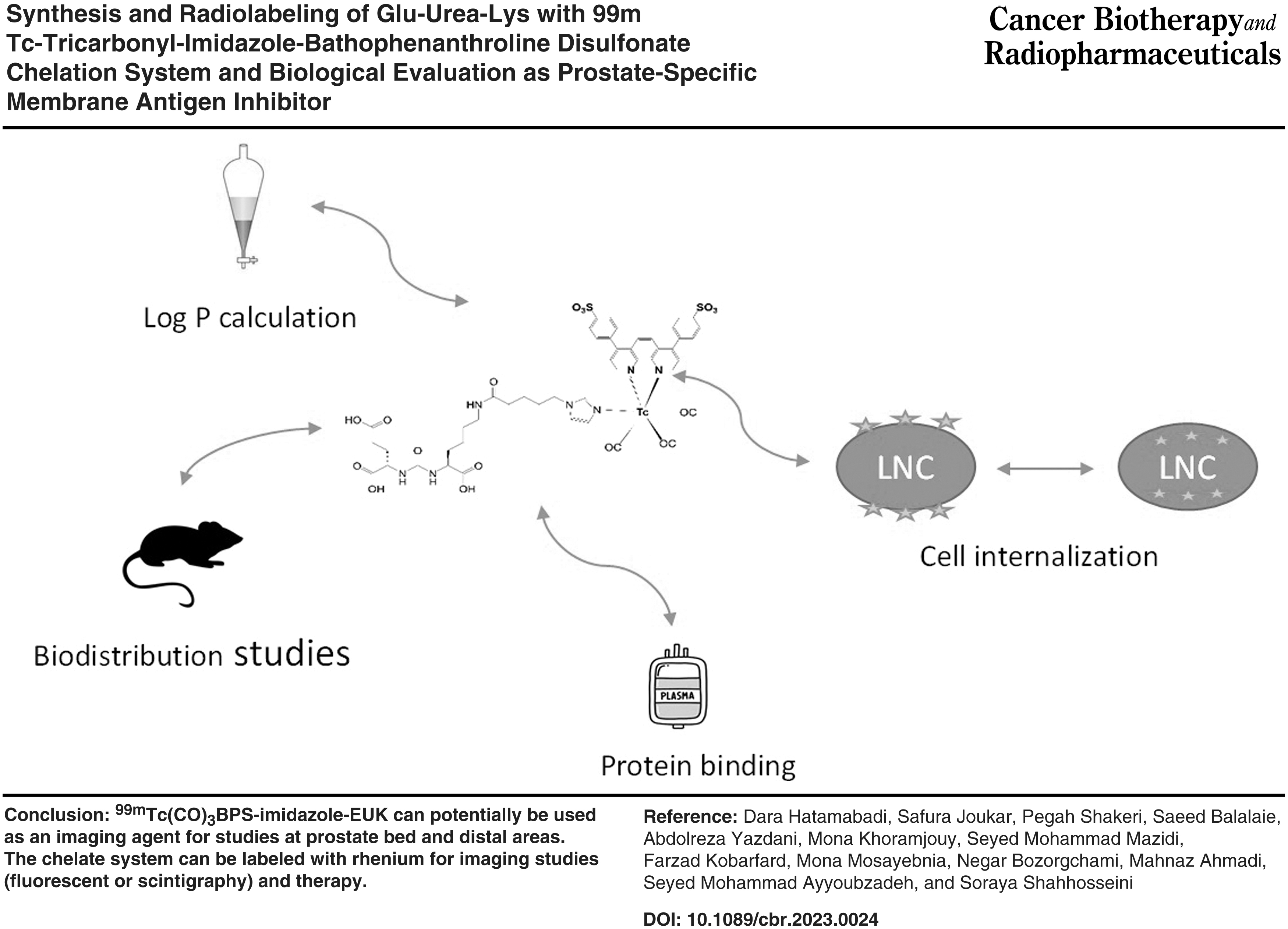
Abstract
Background:
The Glu-Urea-Lys (EUK) pharmacophore as prostate-specific membrane antigen (PSMA)–targeted ligand was synthesized, radiolabeled with 99mTc-tricarbonyl-imidazole-BPS chelation system, and biological activities were evaluated. The strategy [2 + 1] ligand is applied for tricarbonyl labeling. (5-imidazole-1-yl)pentanoic acid as a monodentate ligand and bathophenanthroline disulfonate (BPS) as a bidentate ligand formed a chelate system with 99mTc-tricarbonyl. EUK-pentanoic acid-imidazole and EUK were evaluated for PSMA active site using AutoDock 4 software.
Materials and Methods:
EUK-pentanoic acid-imidazole was synthesized in two steps. BPS was radiolabeled with 99mTc-tricarbonyl at 100°C for 30 min. The purified 99mTc(CO)3(H2O)BPS was used to radiolabel EUK-pentanoic acid-imidazole at 100°C, 30 min. Radiochemical purity, Log P, and stability studies were carried out within 24 h. Affinity of 99mTc(CO)3BPS-imidazole-EUK was performed in the saturation binding studies using LNCaP cells at 37°C for 1 h with a range of 0.001–1000 nM radiolabeled compound range. Internalization studies were performed in LNCaP cells with 1000 nM radiolabeled compound incubated for (0–2) h at 37°C. Biodistribution was studied in normal male Balb/c mice. The artificial intelligence predicts the uptake of radiolabeled compound in tumor.
Results:
The structures of synthesized compounds were confirmed by mass spectroscopy. Radiochemical purity, Log P, and protein binding were ≥95%, −0.2%, and 23%, respectively. The radiolabeled compound was stable in saline and human plasma within 24 h with radiochemical purity ≥90%. There was no release of 99mTc within 4 h in competition with histidine. The affinity was 82 ± 26.38 nM, and the activity increased inside the cells over time. Biodistribution studies showed radioactivity accumulation in kidneys less than 99mTc-HYNIC-PSMA. There was a moderate accumulation of radioactivity in the liver and intestine.
Conclusion:
Based on the results, 99mTc(CO)3BPS-imidazole-EUK can potentially be used as an imaging agent for studies at prostate bed and distal areas. The chelate system can be potentially labeled with rhenium for imaging studies (fluorescent or scintigraphy) and therapy.
Introduction
Molecular imaging studies are essential in timely diagnosis and successful treatment. Nuclear medicine imaging studies using targeted radiopharmaceuticals are one of the best techniques in molecular imaging and noninvasive with high sensitivity, which provides physiological information about the size and location of the lesion.
Prostate cancer is one of the causes of death in men. If primary tumors and metastasis are diagnosed in early stages, appropriate treatment increases the life span and improves the quality of life. 1 –3 Prostate-specific membrane antigen (PSMA) is a membrane glycoprotein that expresses on the surface of all types of prostate cancer cells (sensitive or not sensitive to androgen, from undifferentiated to advanced stages) and not expresses in normal cells or hyperplasia prostate. PSMA is a glutamate carboxypeptidase (GCPII) that also expresses in the other tissues such as the brain, small intestine, kidneys, and salivary glands 100–1000 times less than prostate cancer. After the attachment of PSMA to its ligands, the complex internalizes (endocytosis), and PSMA is restored following the separation of the ligand. Considering PSMA characteristics mentioned previously and not being secreted into the blood, makes PSMA a good in vivo cell surface biomarker (target) for imaging studies of prostate cancer. 4 –7
Different targeted ligands have been designed for PSMA in the past decade, such as monoclonal antibodies and their fragments, peptides, and small molecules. Among them, small molecules are more interesting because they have low molecular weight with simple chemical structures, resulting in more predictable pharmacokinetics and pharmacodynamics properties. In imaging studies, the interaction of small molecules (with peptide or nonpeptide structures) with the extracellular domain of PSMA is preferred. 3 –5,8,9 Small molecules with different chemical structures based on phosphor, thiol, hydroxamate, or urea bond have been prepared and evaluated as targeted ligands of PSMA. Small molecules with urea bond are of interest because of their high affinity, simple synthesis, resistance to hydrolyze in the presence of human plasma, and fast and efficient internalization in the LNCaP cell line. Small molecules have been mostly designed based on urea bond such as Glu-Urea-Glu (EUE), Glu-Urea-Lys (EUK), Glu-Urea-Tyr (EUY), and Glu-Urea-Cys (EUC). Among these four urea bonds, EUK has attracted more attention. 5
Pomper et al., Foss et al., and Chen et al. designed and synthesized small molecules based on urea bond (EUC and EUK), which were radiolabeled with 11 C for PET studies, 125I and 99mTc for single-photon emission computed tomography (SPECT) imaging studies. 10 –12 A few small molecules based on EUK were developed by Molecular Insight Pharmaceuticals as PSMA inhibitors, which were radiolabeled with 123I (MIP-1095 and MIP-1072) or 99mTc (MIP-1404 and MIP-1405). The biological evaluation of these compounds showed high affinity for PSMA. The 99mTc radiolabeled MIP entered clinical trials in 2013. 5,6,13,14 Chen et al. synthesized [ 18 F]DCFPyL and published preclinical data. 15 Subsequently, 18 F-labeled PSMA inhibitor, 18 F-DCFPyL( 18 F-PSMA or 18 F-piflufolastat) was also approved by the Food and Drug Administration (FDA) in May 2021.
The PSMA-11 with EUK pharmacophore is radiolabeled with 68Ga for diagnosis of prostate cancer. 68Ga, a positron emitter radioisotope with 68 min half-life, available through 68Ge/68Ga generator, is very expensive and unreachable in all nuclear medicine centers. 68Ga-PSMA-11 has been approved by the FDA and is applied in imaging studies of prostate cancer using PET cameras. 5,16 –19
Radioisotope 99mTc with appropriate physical radiation characteristics (6 h half-life, 140 keV [90%], IT) is cheap and available in all nuclear medicine centers through 99Mo/99mTc generators. The chemical properties of technetium as a transition metal are well studied, and different chemical structures can be radiolabeled with 99mTc in simple methods with high stability in human plasma. 99mTc-radiolabeled compounds are studied by SPECT cameras in imaging studies, which are cheaper and more reachable than PET (positron emission tomography) cameras. 20 The development of prostate cancer radiopharmaceuticals with high sensitivity and specificity, cheap, and available is of interest. Considering the availability of 99mTc and SPECT systems, the costs of SPECT compared with PET, 99mTc-radiopharmaceuticals are preferred. A few PSMA ligands have been radiolabeled with 99mTc and evaluated in prostate cancer patients. 1,7,14,21,22
The pharmacokinetics of radiolabeled compound affects the interpretation of images. If the study focuses on the prostate bed, the high accumulation of radiotracer in the kidneys is a problem. But suppose the diagnosis and location of metastasis at the lower spine, pelvis, and lymph nodes within the abdominal cavity is desired, then high accumulation in the liver and slow clearance from gastrointestinal (GIT) is a problem. 23 So, it is a challenge to design a radiotracer to avoid its high uptake in the kidneys, liver, and GIT. The hydrophilicity/lipophilicity of radiotracer affects its pharmacokinetic fate. The more hydrophilic the radiotracer, the more clearance from the kidneys.
Based on clinical results, 99mTc-HYNIC-PSMA, with hydrophilic nature, has high accumulation in kidneys making the interpretation of images difficult when the focus is on prostate bed rather than distal areas. 2,24,25 Cwikła et al. in 2021 reported the clinical trial of 99mTc-PSMA-T4 developed by POLATOM in small numbers of patients. PSMA-T4 was designed with a new linker to increase tumor uptake and decrease kidney accumulation. 22 Tricarbonyl, a lipophilic core of 99mTc, contributes to hepatic accumulation and hepatobiliary clearance. Maresca et al. in 2010 and Hillier et al. in 2013 introduced novel single amino acid chelates functionalized with polar lysine imidazole derivatives to decrease the lipophilicity and enhance the renal clearance. 23,26 MIP-1404 with an extra carboxylic acid group radiolabeled with 99mTc-tricarbonyl and in clinical studies showed better clearance from the kidneys and lower uptake in the liver and intestine. García-Pérez et al. in 2017 and 2018 evaluated 99mTc-EDDA/HYNIC-iPSMA in vitro and in vivo. Based on the results 99mTc-EDDA/HYNIC-iPSMA showed less liver uptake than 99mTc-MIP-1404. 2,7,13,14,23,27
This study aimed to radiolabel EUK with 99m Tc-tricarbonyl using a new chelate system to improve the pharmacokinetics and efficiency of detection at the prostatic bed level and distal areas. So, a polar derivative of lysine was synthesized by conjugating of EUK with (5-imidazole-1-yl)pentanoic acid as a linker and a monodentate ligand. BPS was selected as a bidentate ligand. 28 The [2 + 1] ligand strategy was chosen for radiolabeling of EUK with 99mTc-tricarbonyl using an imidazole-BPS chelation system.
Molecular modeling and docking studies were carried out on the EUK and EUK-pentanoic acid-imidazole based on fundamental interactions of amino acids at the PSMA active site and urea compound. EUK was synthesized and coupled to (5-imidazole-1-yl)pentanoic acid. The resulting compound was radiolabeled with 99mTc-tricarbonyl using BPS. Radiochemical purity and Log P of the radiolabeled compound were determined. Stability studies were carried out in saline, human plasma, and in competition with histidine. Biodistribution studies were performed in normal mice. The affinity to PSMA and internalization studies was determined in LNCaP cells. The uptake of radiolabeled compound in tumor model mice was predicted using artificial intelligence.
Materials and Methods
Organic reactions were performed in dry glassware under inert argon pressure. All reagents and solvent were purchased from Sigma-Aldrich, Merck and Bachem.
Docking studies
Docking studies were carried out based on our previous studies using AutoDock 4.
29
In brief, X-ray crystallographic structure of PSMA was taken from
Synthesis of EUK
In 50 mL of dry dichloromethane (DCM), triphosgene (10 mmol) was suspended and stirred for 10 min at 0°C.
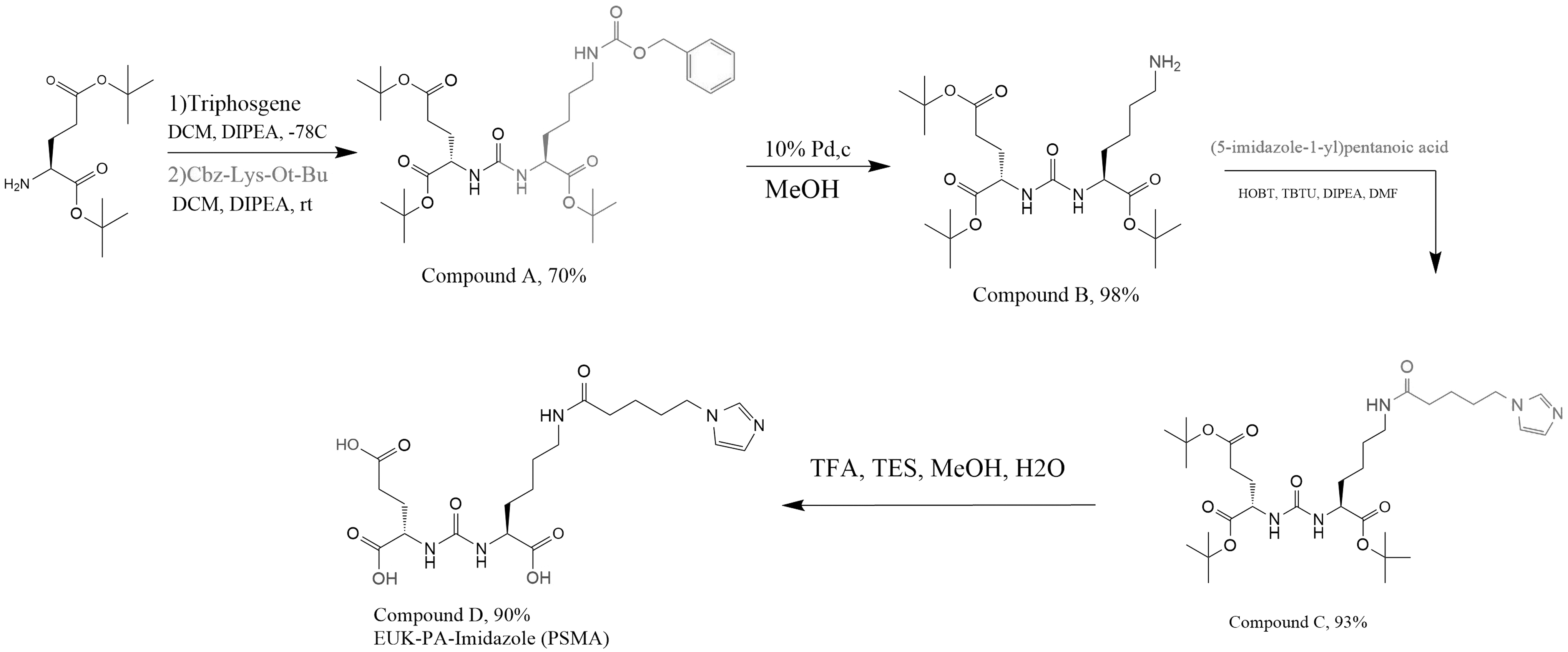
The synthesis steps of compound D.
A rotary evaporator removed the solvent, and the concentrate was dissolved in 100 mL ethyl acetate. Hydrophilic impurities were removed by washing the solution with 2 N NaHSO4 and saturated NaCl. After removing ethyl acetate, the resulting residue, which was a yellow oily substance, was dried using sodium sulfate. In the next step, column chromatography (SiO2 as the stationary phase and ethyl acetate:n-hexane 1:1 as the mobile phase) was applied for the purification of EUK.
One equivalent of EUK (compound A) and 0.1 equivalent of Pd/c were added to methanol (25 mL) and stirred under an argon atmosphere for 24 h. TLC was performed to confirm the completion of the reaction. The mixture was filtered, and the methanol was removed using a rotary evaporator to obtain Glu-Urea-Lys-NH2 (compound B) (Fig. 1).
Coupling of (5-imidazole-1-yl)pentanoic acid with EUK
To a premixed solution of DIPEA (3.5 mmol), hydroxybenzotriazole (HOBT; 1.33 mmol), and O-(benzotriazol-1-yl)-N,N,N',N'-tetramethyluronium tetrafluoroborate (TBTU; 1.12 mmol) in dimethylformamide (DMF; 20 mL), (5-imidazole-1-yl)pentanoic acid (1.24 mmol) was added and stirred at room temperature to form the coupling complex. After 30 min, 200 mg of compound B (0.41 mmol) was dissolved in DMF (5 mL) and added to the above mixture and the resulting solution was stirred for the next 24 h. The reaction was controlled using TLC to ensure no compound B amount was left unreacted. The product was extracted using ethyl acetate and purified by column chromatography yielding the EUK-imidazole as a yellow solid (compound C) (Fig. 1).
Compound C was dissolved in a premixed solution containing trifluoroacetic acid:triisopropylsilane:methanol:water (TFA:TIS:MeOH:H2O with a ratio of 25%:15%:25%:25%) and stirred for 6 h to remove the tert-butyl protecting groups from C-terminals and obtaining compound D. The solvent was removed by rotary evaporator that resulted in a clear oily compound. To remove TFA residues, DCM (10 mL) was added to the product and evaporated (three times) (Fig. 1).
Preparation of 99mTc-tricarbonyl
Based on published articles, technetium tricarbonyl was prepared in-house using boranocarbonates (provided by Dr. Abdolreza Yazdani). 30 –32
For the preparation of technetium tricarbonyl [99mTc(CO)3(H2O)3]+, a mixture of 22 mg sodium tartrate (Na2C4H4O6) (Merck), 15 mg sodium carbonate (Na2CO3) (Merck), 20 mg sodium borate (NaBH4) (Merck), and 10 mg sodium boranocarbonate (Na2H3BCO2, in house preparation) was prepared by adding 2 GBq sodium pertechnetate (Na99mTcO4, 99Mo/99mTc generator; Pars Isotope Co., Tehran, Iran) solution and incubated at 90°C for 10 min. The pH of the mixture was adjusted to 7 using 1 M HCl.
The radiochemical purity of the [99mTc(CO)3(H2O)3]+ was determined using SG-TLC (1% HCl in methanol as mobile phase) and HPLC (C18 column, gradient mobile phase system: solvent A; H2O (0.1% trifluoroacetic acid [TFA] and 2% acetonitrile), solvent B; acetonitrile [0.1% TFA] with flow rate of 1 mL/min). Technetium tricarbonyl (20 μL) was injected into HPLC, and 1 mL/min fractions were collected and counted.
Radiolabeling of compound D with 99mTc-tricarbonyl
In brief, 4 mg BPS was incubated with an aqueous solution of [99mTc(CO)3(H2O)3]+ (370 MBq, 1000 μL, pH 7.0) at 100°C for 30 min. After cooling, the reaction mixture was purified by C18 cartridge Sep-Pak (elution with acetonitrile 15%, 25%, and 80% in ammonium formate 1 M, respectively). 99mTc(CO)3(H2O)BPS was obtained after elution with 80% acetonitrile in ammonium formate (Fig. 2). The radiochemical purity of 99mTc(CO)3(H2O)BPS was determined by TLC. After evaporation of the solvent, 4 mg compound D in 1 mL saline was added to the 99mTc(CO)3(H2O)BPS and radiolabeling was carried out at 100°C for 30 min (Fig. 2). The radiochemical purity was determined using TLC.
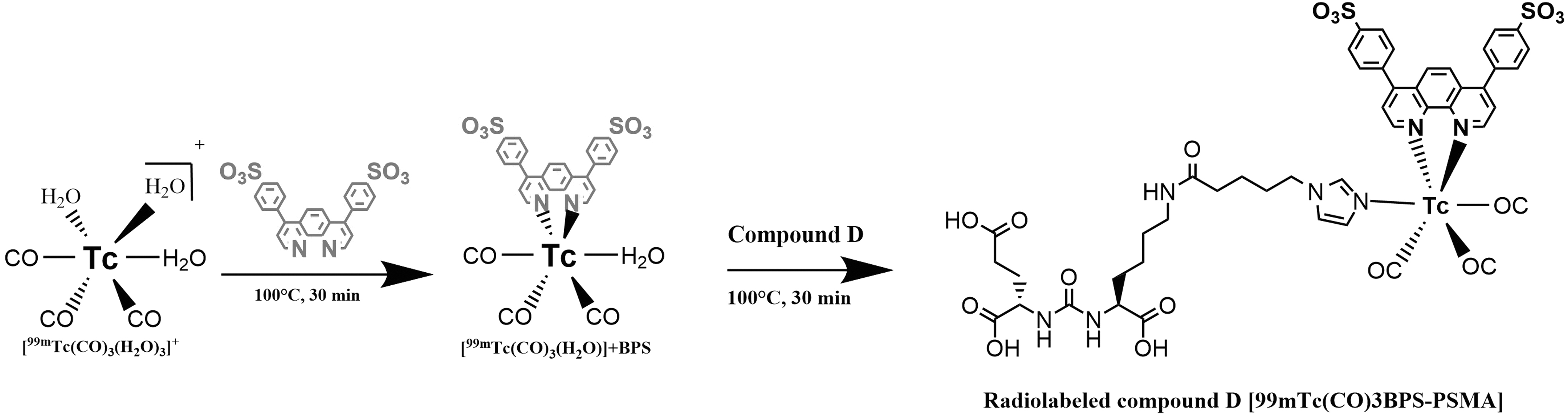
Radiolabeling of BPS with 99mTc-tricarbonyl followed by radiolabeling of compound D with 99mTc-tricarbonyl [2 + 1] strategy. BPS, bathophenanthroline disulfonate.
The lipophilicity of the radiolabeled compound D, 99mTc(CO)3BPS-imidazole-EUK was determined by calculating the partition coefficient (Log P). In brief, 100 μL 99mTc(CO)3BPS-imidazole-EUK was added to a vial containing n-octanol (1 mL) and phosphate-buffered saline (PBS; 1 mL, 0.1 mM, pH 7.4). The resulting mixture was vortexed and centrifuged at 4000 g for 5 min. Then, octanol and PBS were withdrawn and counted using a γ-counter. Log P was calculated by the ratio of n-octanol to PBS counts.
Saline
99mTc(CO)3BPS-imidazole-EUK was incubated in saline for 24 h at 37°C and radiochemical purity was calculated at different times up to 24 h by TLC analysis and a γ-counter.
Human serum
Stability studies in human serum were performed by addition of 100 μL 99mTc(CO)3BPS-imidazole-EUK to human serum (1 mL) and incubated at 37°C for 24 h. At 1 and 24 h, 100 μL of the reaction mixture was withdrawn and mixed with 100 μL absolute ethanol. The resulting residue was separated by centrifugation at 10,000 g for 10 min. The radiochemical purity of the supernatant was determined using TLC. Moreover, the protein binding of the 99mTc(CO)3BPS-imidazole-EUK was measured.
Histidine solution
Approximately 100 μL of 99mTc(CO)3BPS-imidazole-EUK was added to 0.5 mL buffered solution of 2 mM histidine (PBS, 0.1 mM, pH 7.4) and incubated for 1, 2, and 4 h at 37°C. TLC analyzed the radiochemical purity of the reaction mixture at 1, 2, and 4 h of incubation.
Biological properties of radiolabeled compound D
Specific binding assay
The affinity (Kd) of 99mTc(CO)3BPS-imidazole-EUK was determined using LNCaP cells (as a PSMA-positive cell line that was purchased from Pasteur Institute of Iran) and the saturation binding analysis. LNCaP cells were seeded in 12-well plates containing RPMI-1640 medium supplemented with 0.5% bovine serum albumin (∼4 × 10⁵ cells per well). After 24 h, the seeded cells were washed with PBS and incubated at 37°C for 1 h with a range of concentration 0.001–1000 nM radiolabeled compound. After this time, the cells were washed with PBS and then centrifuged at 700 g for 5 min and the amount of the bound radioactivity was measured using the γ-counter (total binding).
To determine nonspecific binding, the total binding protocol was performed in the presence of an excess of nonradiolabeled compound (10 μM). Specific binding was obtained as the difference between total binding and nonspecific binding. The values of Kd and the maximum number of binding sites (Bmax) were determined by nonlinear regression analysis using GraphPad Prism 8.0 software (GraphPad Software, Inc., La Jolla, CA). All tests were performed in triplicate.
Internalization
LNCaP cells were seeded in 12-well plates (∼4 × 10⁵ cells per well in duplicates). Cells were treated with 1000 nM 99mTc(CO)3BPS-imidazole-EUK and incubated for 0–2 h at 37°C. At the defined time, the medium was removed, and the cells were washed twice with PBS (pH = 7.4) and then incubated twice for 5 min with acidic PBS (pH = 2.3). Finally, the cells were lysed using 1 M NaOH solution and a γ-counter measured the amount of internalized radioactivity. The blocking study was carried out by HYNIC-PSMA provided by Pars Isotope Co.
Biodistribution studies
All animal studies were conducted by the guidelines established by the Ethical Committee of Shahid Beheshti University of Medical Sciences and the National Institute for Medical Research Development (NIMAD) with approval code IR.NIMAD.REC.1399.048. Biodistribution studies of the 99mTc(CO)3BPS-imidazole-EUK were performed in normal Balb/c mice (25–30 g, male). One hundred microliters of radiolabeled compound (8 MBq) was injected into the mice via the tail vein. The animals were killed at 30, 120, and 180 min postinjection. After the indicated times, the desired organs were removed, weighed, and the amount of the absorbed radioactivity was counted with a γ-counter. Data were expressed as a percentage of injected dose per gram of the organs (%ID/g, mean ± SEM, n = 3).
Owing to the limitations of experimental and animal studies, an artificial intelligence tool predicted the amount of tumor uptakes for radiolabeled compound. The tumor uptake of radiolabeled compound in tumor-bearing mice was predicted based on the data retrieved from similar articles. In this regard, support vector machine (SVM), as a type of machine learning method, was used to build a model. Then constructed SVM model was applied to predict tumor uptake based on Log P and Kd of the radiolabeled compound. Rapid Miner software (version 9.10.008) was used to build the SVM model and predict tumor uptake.
Results and Discussion
According to the literature review, PSMA is one of the best targets in prostate cancer for diagnosis and therapy. Small molecules based on urea bond (EUK pharmacophore) as targeted ligands for PSMA have attracted more attention. Since 99mTc is one of the best radioisotopes for imaging studies in the clinic worldwide, the aim of this research was radiolabeling of EUK with 99mTc-tricarbonyl without significantly altering the structure and function of EUK. Tricarbonyl with a sphere shape and small size, forms very stable octahedral complexes with ligands such as heterocyclic rings (imidazoles, pyridines, pyrazoles), which makes this core very important for the preparation of new radiopharmaceuticals. Although tridentate ligands containing amine, aromatic N-heterocycles, and carboxylate donors are the most effective ligands for tricarbonyl core, [2 + 1] ligands form stable complexes with tricarbonyl as well. 33 –36
In this study, a combination of bidentate (BPS) and monodentate (imidazole) ligands was used for radiolabeling of EUK with technetium tricarbonyl to provide stable covalent coordination bonds with 99mTc, which protects technetium from ligand attack in vitro and in vivo. 35,36 (5-imidazole-1-yl)pentanoic acid was conjugated with EUK as the monodentate part of the [2 + 1] ligand. The selection of (5-imidazole-1-yl)pentanoic acid was based on previous studies, which suggested that N-alkyl imidazole ligands construct robust [2 + 1] 99mTc complexes and exhibited favorable in vitro and in vivo results. 28,33 The imidazole ring chelates with 99mTc while pentanoic acid as a linker prevents spatial hindrance between pharmacophore (EUK) and tricarbonyl. The effect of (5-imidazole-1-yl)pentanoic acid on EUK pharmacophore was determined by docking studies.
The results of docking studies showed that imidazolyl pentanoic acid has no significant effect on the pharmacodynamics of EUK. The main interactions with necessary amino acids of the PSMA-active site were the same, and the affinity binding energy of EUK and EUK-pentanoic acid-imidazole were −11.6 and −11.2, respectively (Fig. 3).
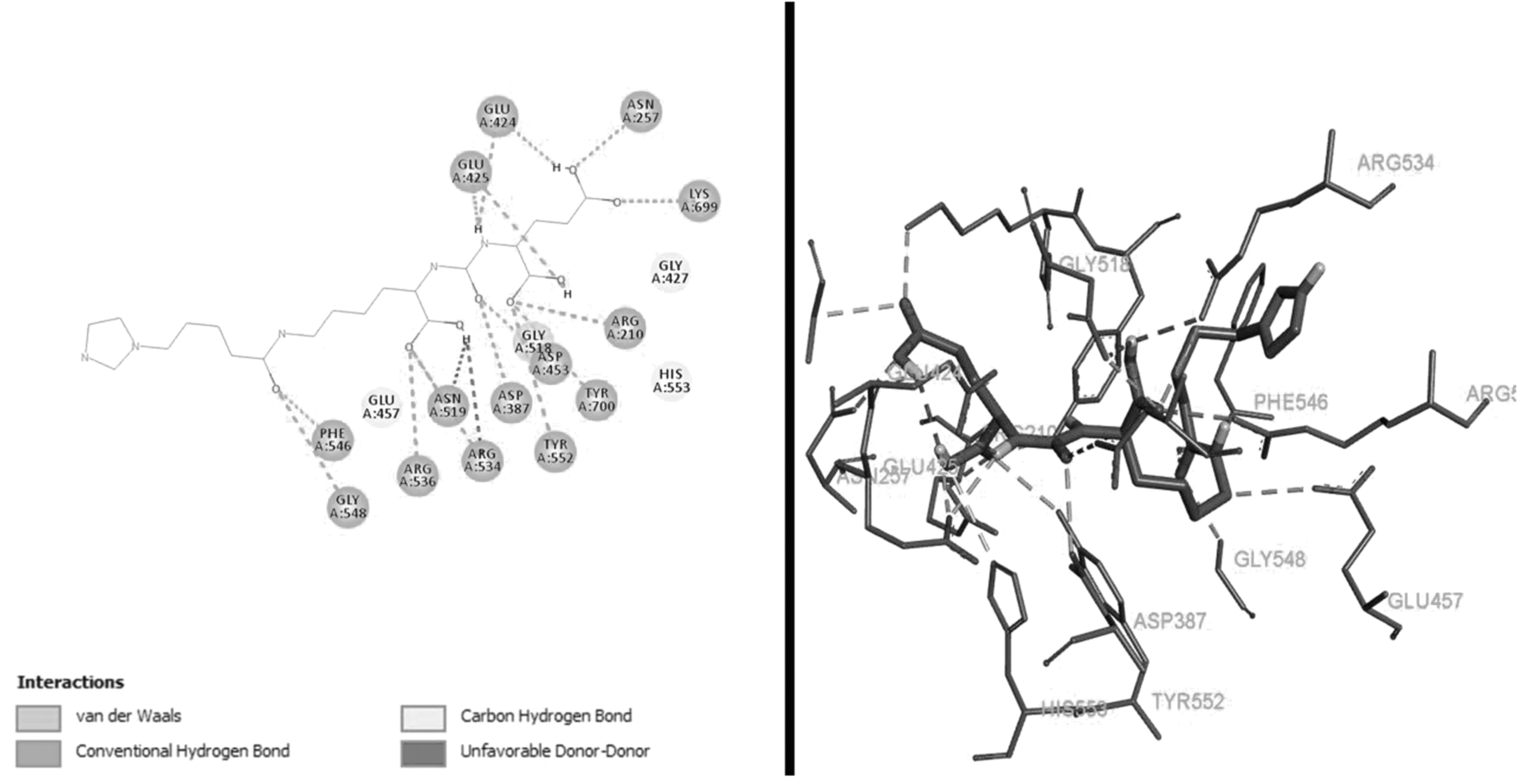
Interactions of EUK conjugated (5-imidazole-1-yl)pentanoic acid with amino acids of PSMA active site. EUK, Glu-Urea-Lys; PSMA, prostate-specific membrane antigen.
EUK was prepared in two steps. In the first step, isocyanate intermediate resulted from triphosgene's reaction with the accessible N-terminal of
Palladium on carbon Pd/C was used to remove the benzyl protection of the lysine side chain and make its N-terminal accessible for further reactions (Fig. 1). After performing the hydrogenolysis, the reaction mixture was filtered to separate the Pd/C particles, and the solvent was removed under reduced pressure to collect Glu-Urea-Lys-NH2 (compound B) as a clear oily compound (8.9 g, yield: 98%). The mass spectrum confirmed the preparation of compound B (Supplementary Fig. S2).
EUK was synthesized and (5-imidazole-1-yl)pentanoic acid was conjugated to the amine side chain of lysine in the EUK structure. General peptide coupling reagents [HOBT, and O-(benzotriazol-1-yl)-TBTU] were utilized to attach the (5-imidazole-1-yl)pentanoic acid to the N-terminal of lysine side chain (Fig. 1) following the completion of the coupling reaction, and the crude product was collected and purified using silica gel column chromatography to yield compound C (10.82 g, 93%) that was confirmed by mass spectrum (Supplementary Fig. S3).
The general procedure for removing tert-butyl groups was performed using trifluoroacetic acid (TFA) and radical scavengers in a solution of H2O and methanol. De-protection of C-terminals increases the compound's hydrophilicity, which is desirable for radiolabeling reactions and in vivo experiments. After tert-butyl de-protection and purification, compound D was isolated (7.17 g, 90%) (Fig. 1) and confirmed by mass spectrum (Supplementary Fig. S4).
Technetium tricarbonyl [99mTc(H2O)3(CO)3]+ was prepared from boranocarbonate based on our previous studies. 32 Radiochemical purity of 99mTc-tricarbonyl was determined (98.5% ± 0.03%) by TLC (1% HCl in methanol:Rf 99mTcO4 = 0.9–1, Rf 99mTc-tricarbonyl = 0.4) and HPLC (Rt 99mTc-tricarbonyl = 5 min).
Compound D was radiolabeled in two steps with 99mTc-tricarbonyl. In the first step, BPS as a bidentate ligand was radiolabeled with 99mTc-tricarbonyl at 100°C for 30 min (Fig. 2). Hyperconjugation of 99mTc(CO)3(H2O)BPS complex changed the color of the reaction mixture to light pink. Two water molecules of tricarbonyl were replaced with BPS. 99mTc(CO)3(H2O)BPS was purified by C18 Sep-Pak cartridge using 80% acetonitrile in ammonium formate 1 M with radiochemical purity of (99% ± 0.03%): TLC (1% HCl in methanol: Rf 99mTc-tricarbonyl = 0.4 and Rf 99mTc(CO)3(H2O)BPS = 0.8). The impurities were washed with 15% and 25% of acetonitrile in ammonium formate 1 M. In the second step, compound D was added to the 99mTc(CO)3(H2O)BPS solution. The imidazole of compound D reacted with 99mTc(CO)3(H2O)BPS (100°C, 30 min) and replaced the water molecule resulting in 99mTc(CO)3BPS-imidazole-EUK complex (Fig. 2). The radiochemical purity of the complex was 95% ± 1% using TLC with R f = 0.3 (Fig. 4). The molar activity was 544 Ci/mole.
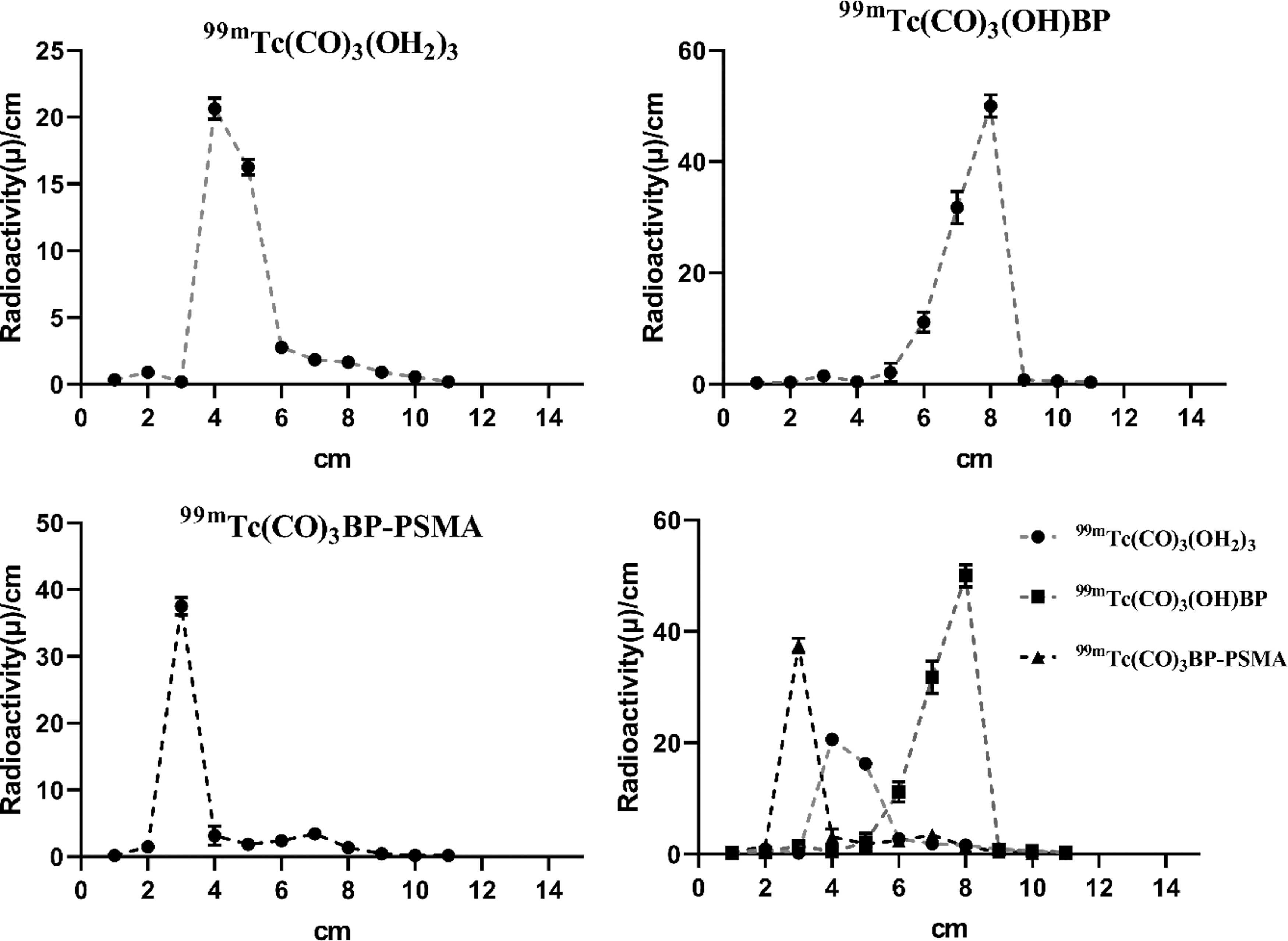
TLC Radioactivity profile (mean ± SEM, n = 3) of the 99mTc complexes; the peaks at 4, 8 and 3 cm are related to [99mTc(CO)3(H2O)3]+, 99mTc(CO)3(H2O)BPS and 99mTc(CO)3BPS-imidazole-EUK, respectively.
The Log P value of the complex was obtained at −0.2, which indicates the less hydrophilic nature of the compound compared with 68Ga-PSMA-11 and 99mTc-HYNIC-PSMA with Log P between −2 and −3. 21,29,37
The complex was stable in saline and human plasma for 24 h with radiochemical purity >90%. There was no release of technetium in competition with histidine within 4 h as well (Fig. 5). The protein binding of the radiolabeled compound was 23%.
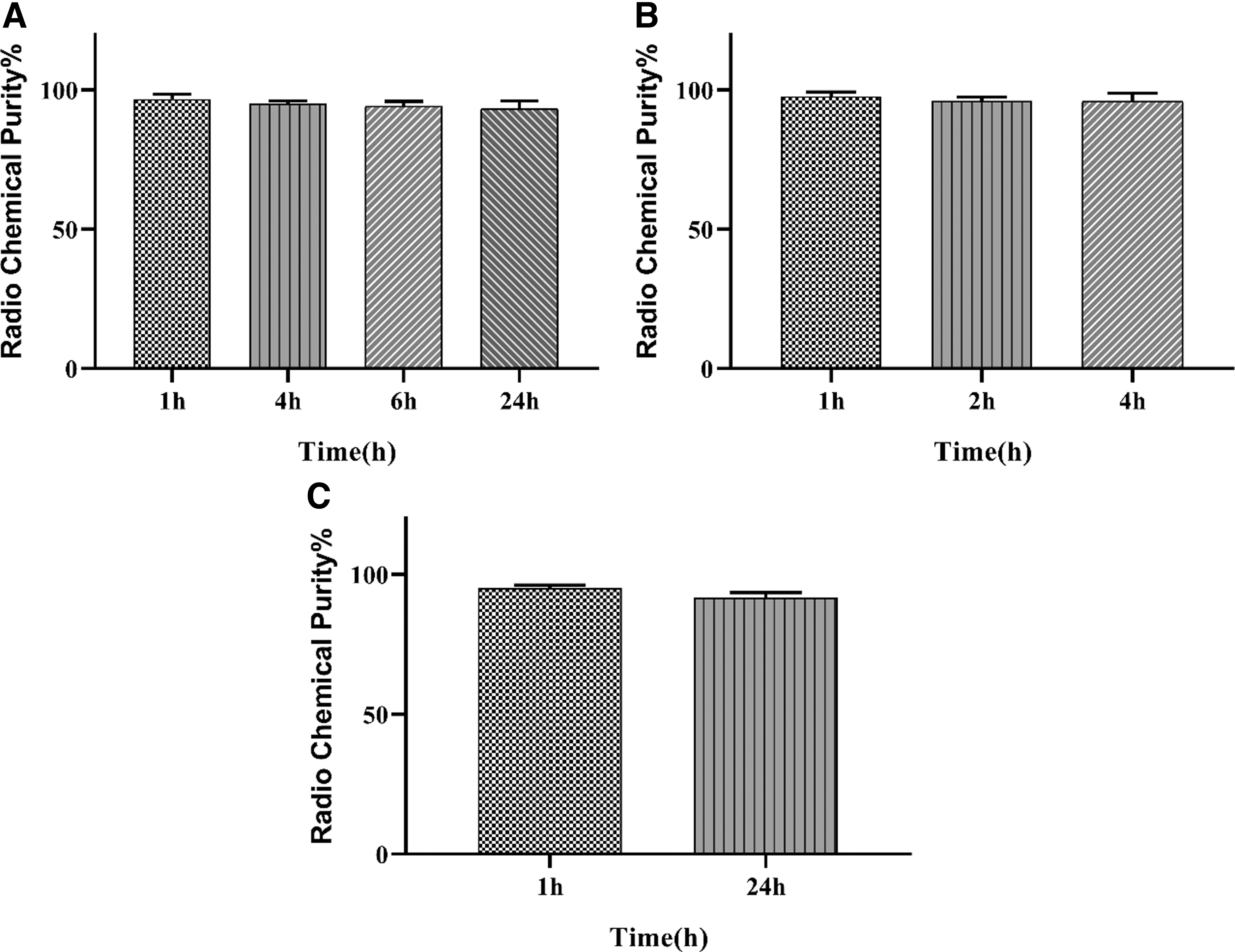
Stability studies of 99mTc(CO)3BPS- imidazole-EUK in
The affinity of the complex (Kd) for PSMA was determined in saturation binding studies using LNCaP cells over expressed PSMA. The data provided in Figure 6A were curve-fitted using nonlinear regression analysis, binding saturation, and one site-specific binding (GraphPad Prism software). The Kd value was calculated (82 ± 26.38) nM. Although the affinity obtained is less than [68Ga-PSMA-11] (K d = 12 ± 2.8 nM), it is still in the nanomolar range. The Bmax was 251896 ± 20445.717 cpm/105 cells (4.22 × 10¹² binding sites/cell).
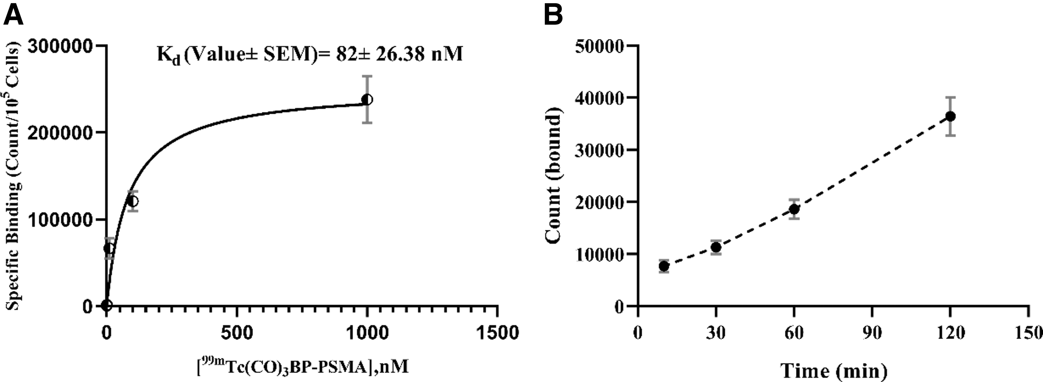
These two compounds 99mTc(CO)3BPS-imidazole-EUK and 68Ga-PSMA-11 have identical pharmacophore (EUK), but different linkers, chelates, and radioisotopes resulted in different affinities and perhaps in vivo different pharmacokinetic parameters.
99mTc(CO)3BPS-imidazole-EUK was shown to internalize at 37°C (Fig. 6B), which is important for imaging and therapy studies.
Biodistribution studies were carried out in normal Balb/c mice. According to Table 1, 99mTc(CO)3BPS-imidazole-EUK rapidly cleared from the blood circulation. The %ID/g of the radiolabeled compound in blood 30 min after injection was 2.49 reaching 0.26 at 180 min postinjection showing rapid clearance from blood circulation. The radiolabeled compound accumulates in the kidneys and liver, representing the kidneys and hepatobiliary system as the main routes of elimination. The %ID/g of the radiolabeled compound in kidneys was ∼(1–2)% higher than that of the liver at all times (Table 1), which is consistent with the Log P value (−0.2) and protein binding (23%).
Biodistribution Studies of 99mTc(CO)3BPS-Imidazole-EUK at 30, 120, and 180 Min After Injection in Normal Mice as %ID/g of Tissue, Mean ± SEM, n = 3
One of the effective factors on the biodistribution behavior of compounds is Log P. The more negative value of Log P, the more hydrophile the compound. The main drawback of hydrophilic compounds of 68Ga-PSMA-11 and 99mTc-HYNIC-PSMA is high kidney and bladder accumulation, which causes difficulties for imaging studies at the prostate bed. 24,25,38 The %ID/g of 99mTc(CO)3BPS-imidazole-EUK in the kidneys and liver in comparison with 99mTc-HYNIC-iPSMA, 99mTc-HYNIC-ALUG, and 68Ga-PSMA-11 (Table 2) showed that the accumulation of the radiolabeled compound in kidneys was less than the others, which might improve the quality of scans at prostate bed. 7,17,21 Although the accumulation of the radiolabeled compound in liver was higher than the others, it is a moderate radioactivity.
Percent of Injected Dose Per Gram Tissue (%ID/g) of Radiolabeled Prostate Specific Membrane Antigen Derivatives in Kidneys and Liver in Mice
In this molecule 99mTc(CO)3BPS-imidazole-EUK, the pentanoic acid acted as a linker between pharmacophore EUK and 99mTc-tricarbonyl, and the imidazole ring contributed to the formation of the new chelate system [2 + 1] along with BPS. The combination of the linker and chelating system provided a radiolabeled hydrophile compound that contrasts to its nature, showing less accumulation in kidneys. There was moderate radioactivity in the liver. This molecule likely improves imaging studies at the prostate bed and distal areas. More studies are needed to reveal the pharmacokinetics of new radiolabeled compound.
The biodistribution studies were carried out in normal mice, and the percent of uptake in tumor model mice 4-h postinjection was predicted using artificial intelligence. The predicted quantity of tumor uptake for the radiolabeled compound in tumor-bearing mice was determined as 8.456 (%ID/g) based on the respective Log P and Kd parameters. The uptake of 99mTc-labeled PSMA ligands in LNCaP tumor xenografts was (7–12) %ID/g at 4 h. 7,23,29,38,39 The assumed uptake in tumor implies the potential of radiolabeled compound for diagnosis and therapy of prostate cancer.
In addition, the new chelate system can be potentially radiolabeled with 188/186Re (rhenium tricarbonyl) for imaging and therapy studies because rhenium and technetium have similar chemical properties. Moreover, rhenium complexes have suitable properties for cellular imaging such as fluorescent microscopy. 40,41
Conclusion
PSMA is one of the best targets in prostate cancer for diagnosis and therapy. In this study, EUK was radiolabeled with 99mTc-tricarbonyl-imidazole-BPS as a new chelate system using the [2 + 1] ligand strategy, and some of the biological activities were evaluated. Based on the results, the radiolabeled compound internalized in the LNCaP cells and showed an affinity for PSMA. The accumulation in the kidneys of normal mice was less than 99mTc-labeled PSMA derivatives, which might improve the quality of scans at the prostate bed. The percent of uptake in tumor model mice was predicted using artificial intelligence. This was a preliminary study, and more studies are ongoing. The chelate system can potentially be labeled with rhenium for cellular imaging and therapy.
Footnotes
Authors' Contributions
D.H., S.J., and P.S.: Experimental studies and data gathering, S.S., F.K., S.B. and A.Y.: Conceptualization and design the study, M.K. and S.M.M.: Animal studies, M.M. and N.B.: Literature review, M.A. and S.M.A.: Artificial intelligence studies, S.S.: Writing, reviewing and editing.
All authors are primarily engaged in teaching or medical research and are not directly funded by the government.
Disclosure Statement
The authors declare no conflict of interest.
Funding Information
This study was supported by the Elite Research Grant Committee under award number [987609] grant IR.NIMAD.REC.1399.048, from the National Institute for Medical Research Development (NIMAD), Tehran, Iran.
Supplementary Material
Supplementary Figure S1
Supplementary Figure S2
Supplementary Figure S3
Supplementary Figure S4
References
Supplementary Material
Please find the following supplemental material available below.
For Open Access articles published under a Creative Commons License, all supplemental material carries the same license as the article it is associated with.
For non-Open Access articles published, all supplemental material carries a non-exclusive license, and permission requests for re-use of supplemental material or any part of supplemental material shall be sent directly to the copyright owner as specified in the copyright notice associated with the article.