
Abstract
Introduction:
Many studies have reported the role of P-glycoprotein (Pgp) in chemoresistance in various pathological conditions such as cancer and neurodegenerative diseases, such as Alzheimer’s. In this study, we are reporting the high-performance liquid chromatography (HPLC)-based purification of fluorine-18 [18F]AVT-011 and its preclinical evaluation.
Methods:
AVT-011 was labeled with 18F using the nucleophilic substitution method by heating the reaction mixture at 110°C for 10 min, followed by purification using preparative HPLC and C18ec cartridge. The in vitro cell uptake study was carried out in U87 cells with and without an inhibitor. The preclinical toxicity was carried out in CD1 mice in three groups, including control, AVT-011 treated, and [18F]AVT-011 treated. The biodistribution study was done in CD1 mice (n = 12) after intravenous injection of 4–6 MBq [18F]AVT-011, and mice were sacrificed at various time intervals. A dose of 3.7 ± 0.7 MBq of [18F]AVT-011 was injected intravenously in the healthy Swiss albino mice, and the whole-body micro-positron emission tomography was acquired at 0-, 30-, 60-, and 120-min postinjection.
Results:
The radiochemical purity of [18F]AVT-011 was 97 ± 1.5% as evaluated by radio-HPLC with a yield of 14 ± 2% and was stable up to 95% under in vitro conditions in blood and in vivo conditions up to 4 h. The in vitro cell uptake study showed a significant difference in control (27.4 ± 2.1%) and blocked U987 cells (73.2 ± 3.2%) after incubation of 120 min. The tissue distribution in mice showed the highest uptake in the liver (17.3 ± 2.4%), kidneys (16.6 ± 3.1%), lungs (10.4 ± 2.9%), and spleen (5.6 ± 0.8%) at 15 min, and the activity was washed out with time. The radioactivity cleared through the hepatorenal pathway. The animal imaging study also demonstrates a similar biodistribution pattern.
Conclusions:
[18F]AVT-011 showed higher specific activity than the cartridge-based method but showed similar biological activity.
Introduction
Drug resistance is one of the major causes of the failure of treatment in cancers. Drug resistance can be due to various reasons such as genetic factors, stem cells, and efflux proteins. The overexpression of efflux protein-like multiple drug resistance (MDR) is considered one of the major factors for drug resistance. 1 –3 P-glycoprotein (Pgp), or multidrug resistance protein-1, is a unidirectional efflux pump that can actively efflux out any substance against its concentration. It plays a key role in drug resistance. Its overexpression and underexpression may cause pathological conditions. The resistance may develop before the therapy or during the course of treatment. 4 –6 The diagnosis of drug resistance at an early stage is the prime target for many researchers as it will directly benefit the patient and clinician in deciding on the treatment regimen. Liu et al. have synthesized a library of fluorescent nanoparticles with various sizes, surface charges, and compositions (SiO2 nanoparticles, organic PS-co-PAA nanoparticles, and ZIF-8 nanoparticles) and studied their accumulation in drug-resistant and sensitive cancer cells. 7 These nanoparticles were used to diagnose the resistance in a single cell. Therefore, a noninvasive tool is required to detect the Pgp activity and monitor the treatment response.
The blood–brain barrier (BBB) is a membrane-based structure of the human brain. It controls the exchange of compounds between the blood and brain parenchyma to maintain homeostasis. BBB comprises many types of cells along with transporters, which include efflux proteins such as Pgp. 8,9 Pgp is an efflux protein that regulates the transport across the BBB and efflux out small hydrophobic xenobiotics. It prevents the brain from the toxicity of the substances and limits access to many types of drugs, such as anticancer, antidepressants, and antiepileptics. The regulation of the Pgp transporter is crucial as over- and underexpression/functioning of this pump can cause pathological conditions. The overexpression leads to conditions such as drug resistance, and underexpression can affect the clearance of β-amyloid inside the cells, which can lead to neurodegeneration and cause pathological conditions such as Alzheimer’s disease. 10,11 Therefore, the evaluation of the Pgp function can be a biomarker to evaluate the disease condition. Hence, imaging the Pgp expression may be a biomarker to assess the chemoresistance at an early stage.
Many radiopharmaceuticals have developed to image the Pgp expression and function, but out of that, carbon-11[11C]verapamil is the most successful, but a short half-life of 11C and other inherent disadvantages lead the quest to develop a better tracer. Fluorine-18[18F]MC-225 is another promising tracer studied at the preclinical levels, and few are under clinical trial. 12 –15 Kannan et al. 16 have developed and characterized [18F]AVT-011 using a precursor 6-o-desmethyl tariquidar. It was developed as a new radiotracer for tumor imaging of MDR. The study has shown the potential of [18F]AVT-011 to measure ABCB1 function in tumors. The major limitation of the study was low yield, which may be due to multistep (two-step) radiolabeling. 16 Our laboratory has used the modified precursor [toluene-4-sulfonic acid 2-{6-[2-(4-{4,5-dimethoxy-2-[(quinoline-2-carbonyl)amino]benzoylamino}-phenyl)–ethyl]-3-methoxy-5,6,7,8-tetrahydro-naphthalen-2-yloxy}-ethyl ester] to make a single-step labeling procedure. Our laboratory has also synthesized and labeled AVT-011 precursor with 18F using the cartridge-based purification method. 17
In this study, we have used an high-performance liquid chromatography (HPLC) purification-based method to synthesize [18F]AVT-011 to increase the specific activity of the radiotracer. The other goal was to compare the two methods of synthesis and their biological activity.
Materials and Methods
All the reagents and chemicals were procured from Sigma-Aldrich. The radionuclide 18F was produced by bombarding proton onto the liquid target O-18 (H2O18) water using [18O (p,n) 18F] reaction. The beam current was kept in the range of 30–65 µA for 10–30 min; the total bombardment time depended upon the requirement of the radioactivity. The synthesis of the AVT-011 precursor and cold standard of AVT-011 was described in our previous publication. 17 The radioactivity labeling reaction was carried out in the chemistry module (Tracerlab FX2N, GE Healthcare) equipped with HPLC preparative column (C18 OBD preparative column, SunFire, 100 Å, 10 µm, 10 × 250 mm, Waters India), and 18F was transferred to the module using the Helium (UHP-5.5) as a carrier gas. The preconditioned QMA cartridges (ABX) were used to trap 18F, and C18ec (chromatic, Macherey-Nagel) was used for the final purification of [18F]AVT-011 from the crude reaction mixture. The elution solvent was prepared by dissolving 15.0 mg of kryptofix (K222) in 900 µL of acetonitrile, followed by the addition of 100 µL of potassium carbonate (K2CO3) from a stock solution of 30 mg/mL in water. The radiochemical purity was tested by radio-HPLC equipped with a radioactive and ultraviolet-visible (UV-Vis) coupled detector (Dionex). The quantitative HPLC was performed on C18 column (5um, 4.6 × 250 mm, Shim-pack GWS, Shimadzu) using mobile phase composition of acetonitrile (0.1% trifluoroacetic acid) and water (0.1% trifluoroacetic acid), starting with 5% acetonitrile (0–5 min), 5%–100% acetonitrile (5–20 min), then 100% acetonitrile (20–25 min), and again at 5% acetonitrile (25–30 min). The residual solvents were measured using gas chromatography (GC) (Scion 436 GC) with a flame ionization detector. The column was operated initially at 40°C for the first 3 min and then rose 50°C/min up to 8 min, and the final temperature was set at 240°C and the column was BR-200ms, 0.32 mm ID. The makeup gas consists of helium (28 mL/min), zero air (300 mL/min), and nitrogen gas (30 mL/min) flow at the rate of 2 mL/min. The radioactive counts were measured on a γ-counter (Capnitec Inc.). All animal experiments were conducted after obtaining the ethical approval from the institute (Ref: AEC/74/486/NIIR).
Radiochemistry
The conditions for labeling AVT-011 precursor with 18F have been standardized and published elsewhere. 17 In this study, they have synthesized [18F]AVT-011 using the FX2N chemistry module using the preparative HPLC method to observe its effect on labeling efficiency and specific activity. The chemistry module was prepared before starting the synthesis. The preconditioned QMA was fixed on the QMA port. The main changes are shown in Figure 1; the VX2L tube was connected to VZ1 to push the crude mixture to the sample tube (for HPLC purification). The sample tube pushed the sample to the preparative HPLC. The output of preparative HPLC was connected to V18, the round bottom flask, and C18ec on the C18 (2) port for final purification.
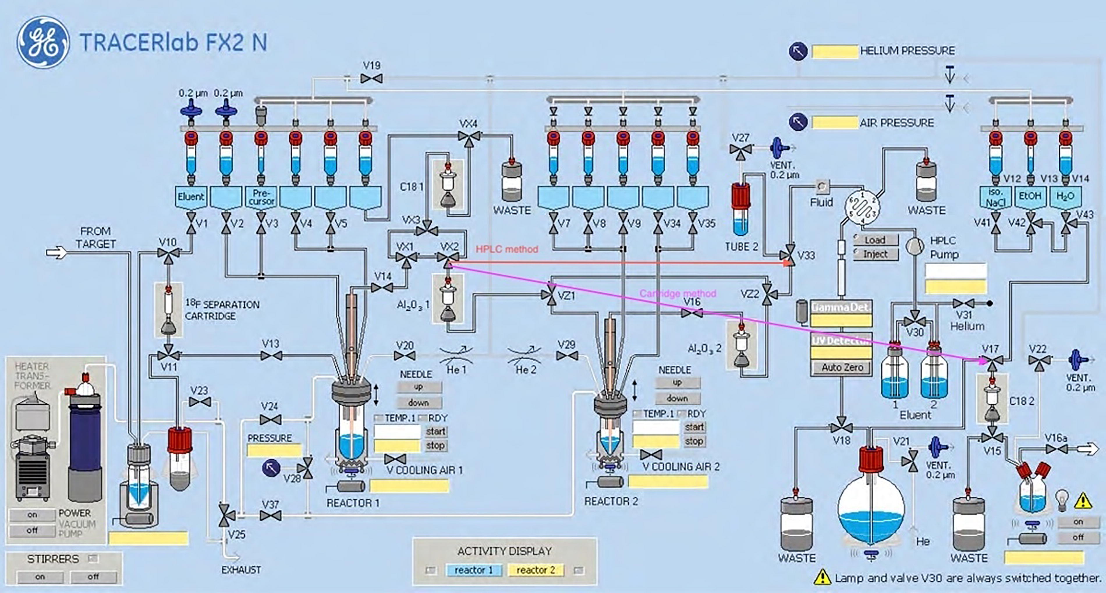
Schematic flow diagram of chemistry module (FX2N, Tracerlab). In HPLC method, VX2L was connected to V3 via VZ1 to VZ2 (orange color). In cartridge based, VX2L was directly connected to V17 (magenta line). HPLC, high-performance liquid chromatography.
Briefly, 800 ± 200 mCi of 18F was added into the V-vial, passed through V10 and V11 under vacuum, and enriched water was recovered in an 18O-water recovery vial. 18F was trapped in QMA and eluted with 1 mL of K222/K2CO3 (R1) solution in the reactor vial. The acetonitrile solvent was heated azeotropically at 95°C for 4 min under helium flow and vacuum with a total time of 16 min. The reactor vial was cooled to 65°C, and a precursor (vial 3) was added. The reaction was carried out at 110°C for 10 min under closed conditions. After 10 min, the reaction mixture was cooled to 45°C, 5 mL of acidic water (vial 5) was added to it, and the reaction mixture was transferred to the HPLC column (in-line module). The product was separated on an HPLC preparative column using 40% acetonitrile (mobile phase) solvent flow at 4 mL/min. The separation of the peak was performed to collect maximum activity in a round bottom flask. It was pushed onto the C18ec cartridge with the help of helium; 5 mL of water (vial 12) was passed through the C18ec cartridge, followed by elution with 1.5 mL of ethanol (vial 14) in the product collection vial. It was pushed to the dispensing vial in the dispenser. The vial was sent to quality control and used for experiments.
Quality control
The radiochemical purity of the preparation was estimated by radio-HPLC. The retention time of the cold AVT-011 standard was measured at wavelength (λmax) of 254 nm using a UV-VIS detector, whereas the [18F]AVT-011 was detected by using the radioactive detector. The quality control parameters such as physical appearance, pH, radiochemical purity, radionuclide purity, chemical purities, residual solvents, sterility, endotoxin test, and stability (in vitro) were done as per the standard protocol described elsewhere. 18 The specific activity was calculated from a standard curve derived by HPLC of variable concentration of cold standard AVT-011.
Stability studies
In vitro stability
[18F]AVT-011 was diluted with water, saline, and buffer and kept at room temperature up to 4 h, and radiochemical purity was measured by radio-HPLC for stability.
Plasma stability
Around 4 mL of blood was collected from the human volunteer (Institutional Ethics Ref no. SSCHRC/IEC10/55) in a heparinized tube. After 1 h, the blood was centrifuged, and the plasma was separated. Around 20 µL of [18F]AVT-011 was mixed with 180 µL of plasma and incubated at 37°C for 1 h. Around 200 µL of chloroform/methanol (4:1) was added to centrifuge proteins and centrifuged at 3000 rpm for 10 min. After centrifugation, the supernatant and pellet were separated. The supernatant was subjected to the HPLC.
In vivo stability
The mice (CD1, male) were injected intravenously (tail vein) with 150 ± 20 µCi of [18F]AVT-011 (n = 3). The cardiac blood was collected from the mice in the heparinized vials after sacrificing the mice at 1 h and 4 h. The plasma is separated and processed in the same way as described in the upper section. The supernatant was subjected to HPLC.
Cell-binding assay
The cell binding was studied in the U-87 MG cancer cell line. U-87 MG cells were purchased from the National Center for Cell Science, Pune. The cells were cultured in Dulbecco’s modified Eagle’s medium high glucose containing 10% fetal bovine serum and 1% antibiotic in a T75 flask. The cells (suspended in media) were cultured in a T75 flask and placed in a humidified incubator at 37°C under 5% carbon dioxide/95% air. The cells, when confluent, were detached using 0.25% trypsin–ethylenediamine tetraacetic acid, washed, and resuspended in 1× phosphate-buffered saline at a concentration of approximately 5 × 10⁶ cells/mL. Each test tube contained 1 mL of cell suspension into which 10 µL (∼0.74 MBq) of [18F]AVT-011 was added and incubated for 15 min, 30 min, 60 min, and 120 min at 37°C in a water bath.
The blocking studies were done by incubating the same cell suspension (1 mL) with a 500-fold excess of Pgp blocking agent, that is, tariquidar solution, for 30 min at 37°C in a water bath. After 30 min, 10 µL (∼0.74 MBq) of [18F]AVT-011 was added and incubated for 15 min, 30 min, 60 min, and 120 min at 37°C in a water bath. The incubation was terminated by adding 0.5 mL of cold normal saline. The mixture was centrifuged at 3000 rpm for 10 min, and the supernatant from each wash was collected in marked test tubes. The pellet was washed thrice with normal saline, and the supernatant was collected separately in the marked tubes. The radioactivity associated with cells and supernatant was counted in a γ-counter (Capintec Inc.), and the percentage of radioactivity bound to the cells was calculated.
MTT assay
Cytotoxicity (in vitro) was determined using the MTT [3-(4,5-dimethylthiazole-2-yl)-2,5-diphenyl-2H-tetrazolium bromide] assay. The cells (U87 MG) were grown plated in a 96-well microtiter plate at a uniform cell density of 10,000 cells/well, 24 h before treatment. Cells were treated with a maximal concentration of AVT-011 (12 mM), and MTT assays were performed. At 24 h, cells were treated with MTT (at a final concentration of 0.05 mg/mL) for 2 h at 37°C, and the medium was removed. The cells were lysed, and the formazan crystals were dissolved by adding 100 μL of DMSO. Optical density was measured on 150 μL of extracts at 570 nm (reference filter: 690 nm). The cytotoxicity was evaluated as the percentage of metabolically active in relation to untreated cells.
Preclinical toxicity
Acute toxicity was performed in a group of CD1 mice. A dose of 5 mg/kg body weight AVT-011 (diluted as 10% w/v in DMSO containing water for injection) was injected intraperitoneally into 6-week-old mice (weight 30–45 g, females, n = 5) along with a control group (weight 30–45 g, females, n = 5) injected intraperitoneally with water. Mice were monitored at initial hours 1, 2, and 4 h postinjection and then daily for 14 days for any clinical signs of toxicity. The weight of the mice was observed daily, and on the 14th day, the mice were euthanized by excess inhalation of isoflurane. A macroscopic autopsy was performed on both groups. A third batch of mice (n = 5) was injected intravenously (tail vein) with a decayed dose of [18F]AVT-011. After injections, the mice were observed for 14 days, and a macroscopic analysis was performed as described earlier.
Animal biodistribution
Animal biodistribution studies were carried out in normal Swiss albino mice (n = 12) after approval from the Institute Animal Ethics Committee. All experiments were performed in accordance with the Committee for the Purpose of Control and Supervision of Experiments on Animals and complied with the Animal Research: Reporting of In Vivo Experiments guidelines. A group of mice (15–20 weeks, 25–35 g) was injected intravenously (in tail vein) with 4–6 MBq of [18F]AVT-011 diluted with saline (in a total volume of 100 µL), and the mice were sacrificed (n = 3, each time point) at 15-, 30-, 60-, and 120 min postinjection. Mice were then euthanized by excess inhalation of isoflurane, and tissues were dissected, washed free of blood, dried, and weighed. Concomitant radioactivity was counted with an 18F standard solution, and the injected dose per gram (% ID/g) was then calculated.
Animal imaging
The animal imaging was acquired on beta eye (Bioemtech). Briefly, 3.7 ± 0.7 MBq of [18F]AVT-011 was injected intravenously (tail vein) of the healthy Swiss albino mice (n = 3) and a dynamic positron emission tomography (PET) scan was acquired for 0–30 min (30 s per frame) followed by a delayed scan at 60 and 120 min.
Results
Radiochemistry
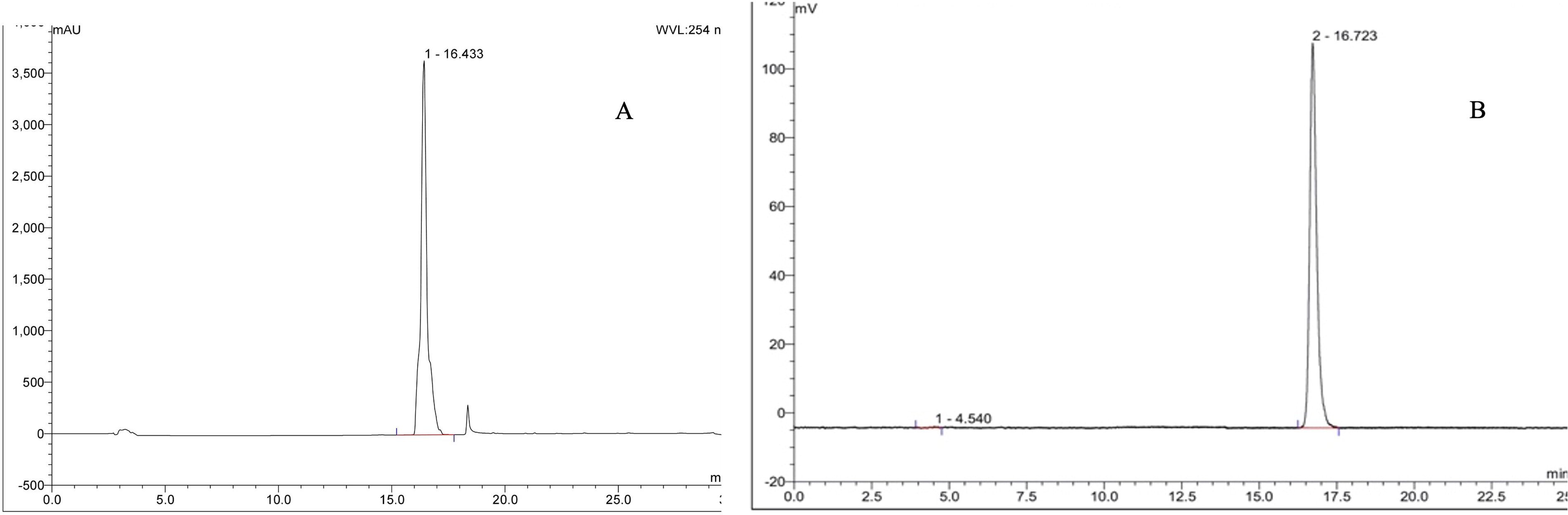
The [18F]AVT-011 was synthesized and purified using two methods: (a) cartridge only and (b) HPLC + cartridge-based methods. Method (a) was well defined in our previous publication. 17 The labeling reaction conditions were maintained in the same manner as published elsewhere. 17 The reagents were added to the module, as shown in Table 1. The elution efficiency of 18F from the QMA using K222/K2CO3 as eluent was 98 ± 1%. The [18F]cryptand was dried at 95°C for 4 min, and the reaction between the precursor solution and dried 18F was carried out at 110°C for 10 min. The product was passed through HPLC, and peak (retention time = 23–27 min, Fig 2) was collected in the round bottom flask (Fig 1). It was passed to the preconditioned C18ec cartridge, washed, and eluted with 1.5 mL of ethanol. The yield from the HPLC purification method was 14 ± 2% (n = 5) as compared with 42 ± 2% (n = 10) by the cartridge-based method with a radiochemical purity of 97 ± 1.5% (decay corrected). The specific activity was 935 ± 30 GBq/µmol as compared with 700 ± 60 GBq/µmol reported by the cartridge-based method. 17 The retention time of cold standard AVT-011 was 16.5 ± 1.2 min (Fig. 3a), whereas the radioactive [18F]AVT-011 peak was detected at 16.9 ± 0.8 min (Fig. 3b). All other quality parameters’ values were within the prescribed limit, and their values are given in Table 2. The [18F]AVT-011 was stable up to 95% under in-vitro conditions, in blood and under in vivo conditions up to 4 h.
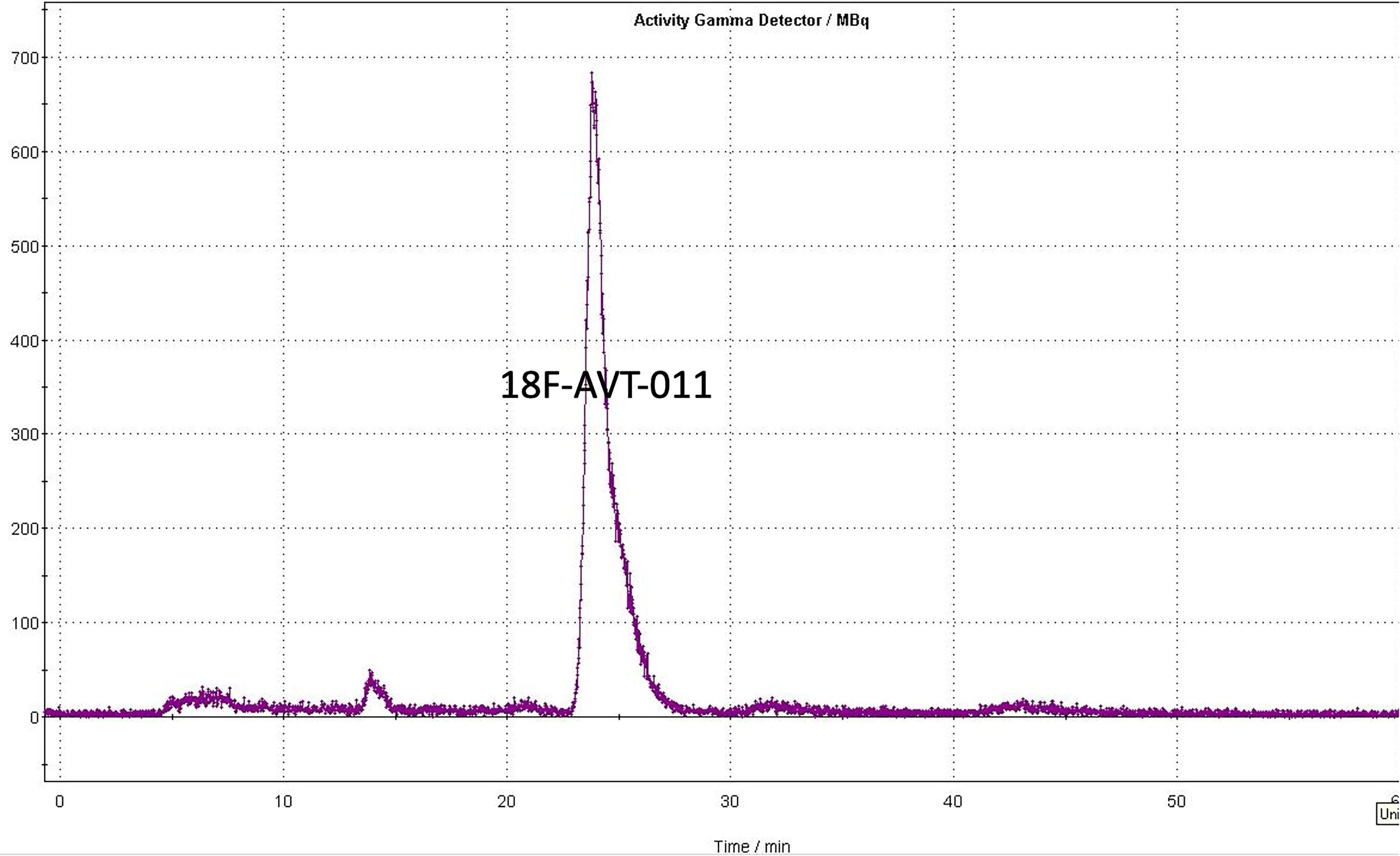
Preparative HPLC showing peaks of product eluting during the separation of crude mixture. Peak (Rt = 23–27 min) was collected and loaded onto the C18ec cartridge. HPLC, high-performance liquid chromatography.
Details of the Reagents in FX2N Module for Synthesis of [18F]AVT-011
HPLC, high-performance liquid chromatography.
Quality Control Parameters for [18F]AVT-011
RT-retetntion time, EU-endotoxin units.
In vitro studies
The in vitro cell uptake study showed efflux of radioactivity in the U87 cells, whereas blocked U87 cells showed retention of the radioactivity with time (Fig. 4). The activity decreased from 37.2 ± 1.5% (at 15 min) to 27.4 ± 2.1% (at 120 min) in U87 cells, whereas blocked U87 cells showed an increase in uptake from 48.2 ± 2.1% (at 15 min) to 73.2 ± 3.2% (at 120 min). The cell viability was calculated by MTT assay for the maximal concentration of AVT-011 (12 mM), which can be used as a precursor for synthesizing [18F]AVT-011. The viability was more than 94% as compared with the control experiment after 24 h incubation.
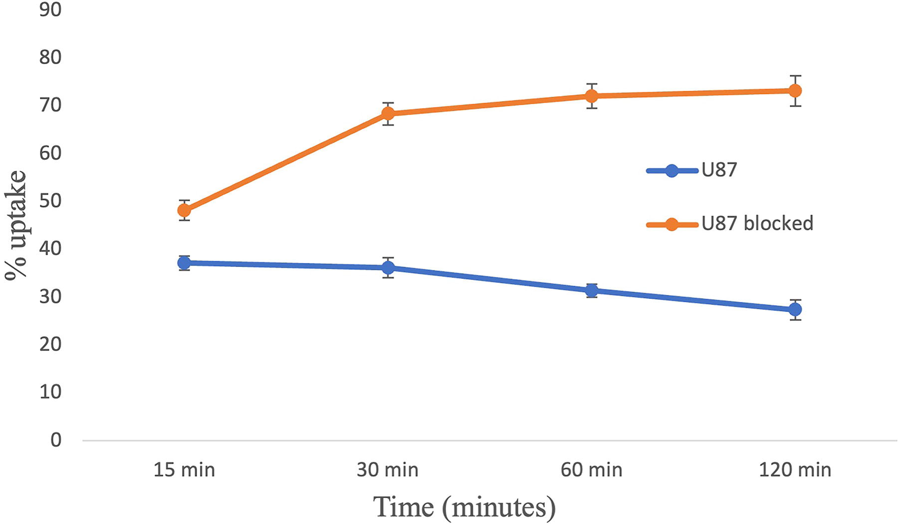
[18F]AVT-011 uptake in U87 cells (control and blocked cells).
Preclinical toxicity
There was neither a loss in weight nor any morbidity observed in any of the mice groups postintraperitoneal and intravenous injection until 14 days. All mice (control and treated group) showed normal gains in body weight. No clinical sign of any toxicity was observed over the observation period. No abnormality was observed on postmortem macroscopic examination.
Animal studies
The tissue distribution showed that the highest percentage of the injected dose was found in the liver (17.3 ± 2.4%), spleen (5.6 ± 0.8%), lungs (10.4 ± 2.9%), and kidneys (16.6 ± 3.1%) at 15 min (Fig. 5). With time, the activity was washed out from the liver, spleen, and lungs. The radioactivity was increased in the kidneys and intestine with time, indicating the hepatorenal excretory route of the radiotracer. The normal brain showed minimal uptake of the radiotracer.
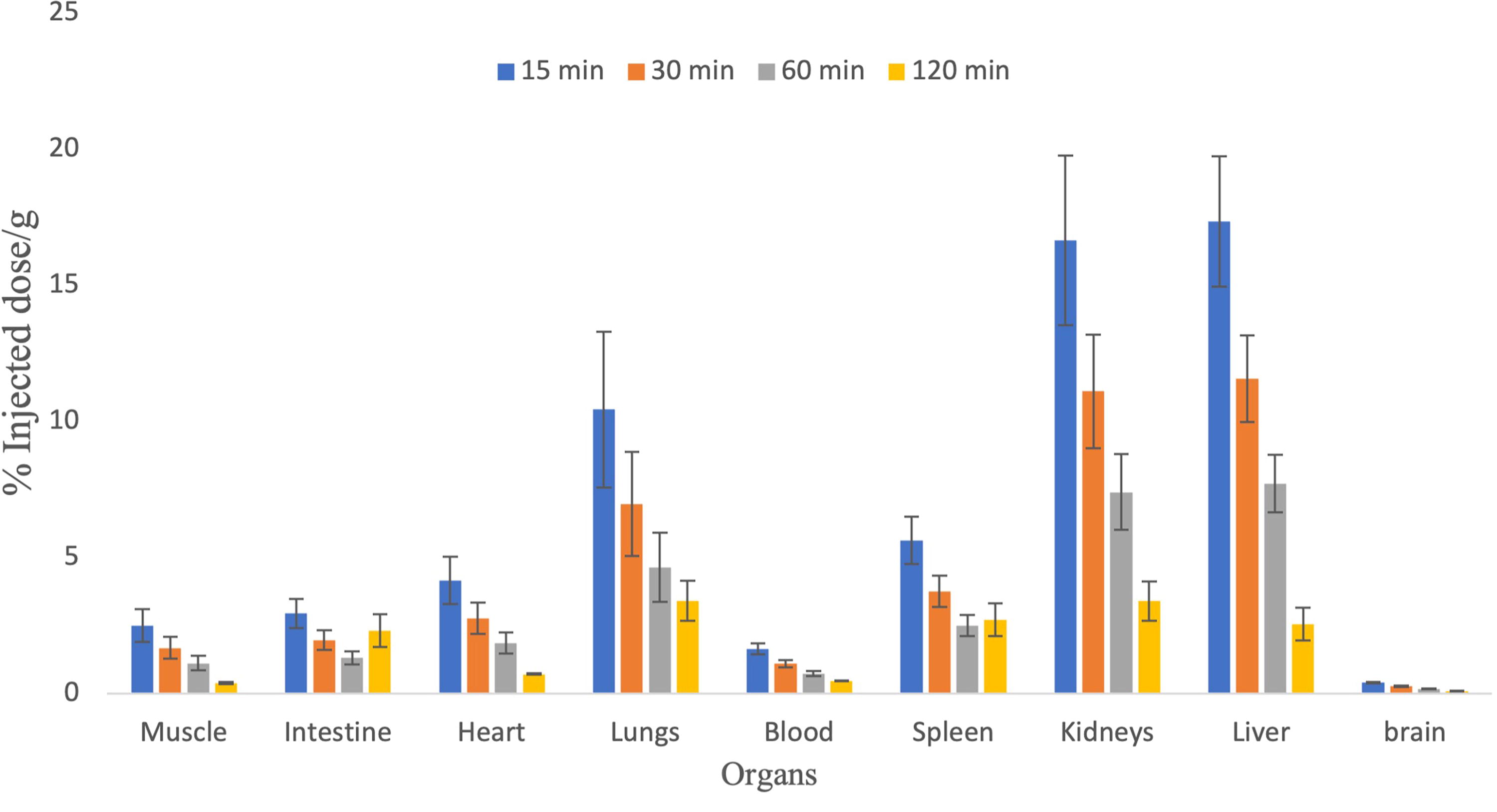
Biodistribution of [18F]AVT-011 in mice at various time intervals.
Animal PET/computed tomography imaging
The processed animal image was examined with naked eye and initial distribution was seen in lungs, which was washed with time and seen in liver at 30 min (Fig. 6a). The radioactivity was seen in intestine and bladder in the delayed scan at 60 and 120 min (Fig. 6b, c). It showed that the tracer was excreted through the hepatorenal pathway.
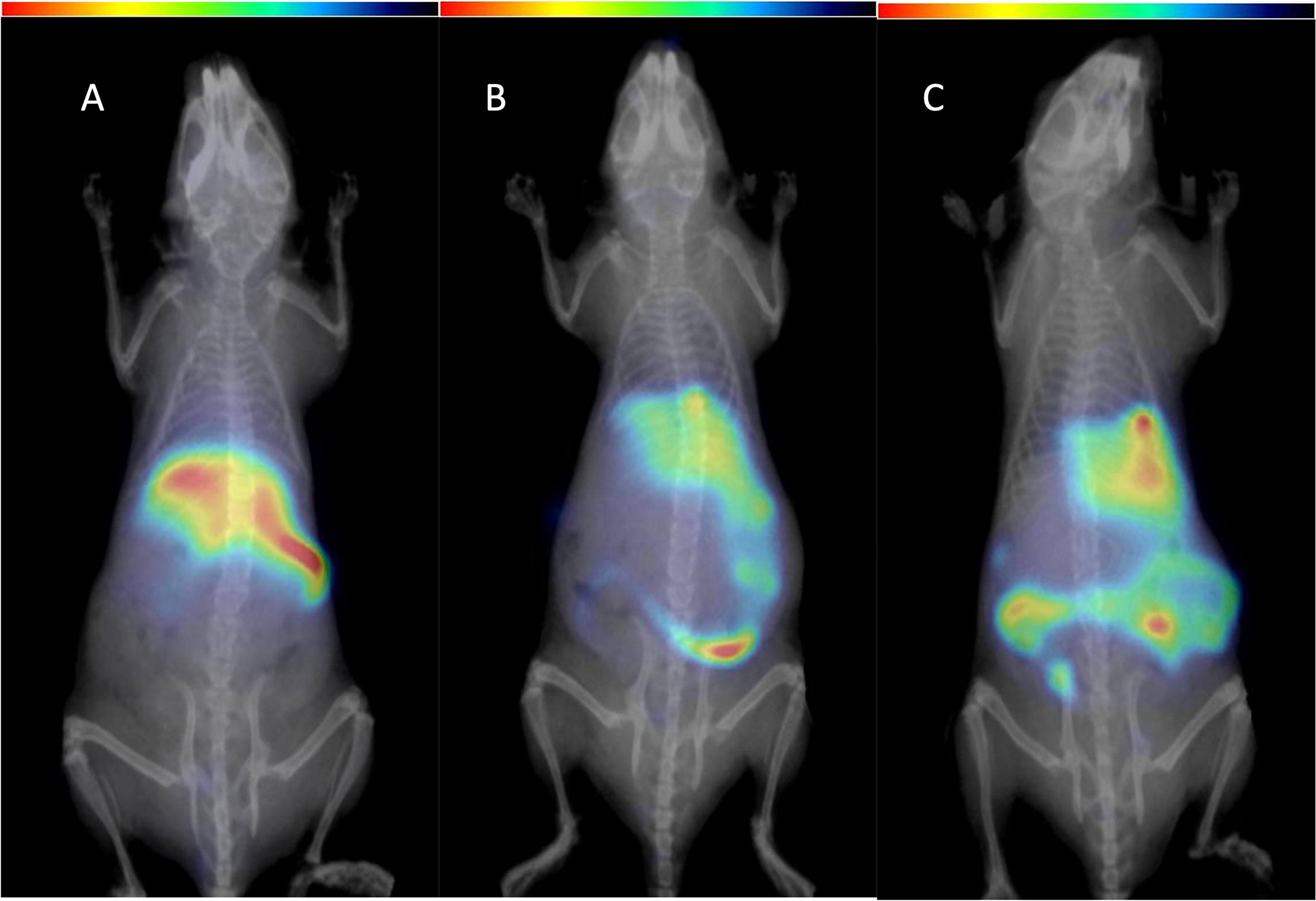
Animal PET imaging showing distribution in a healthy mouse:
Discussion
The two methods of purification, cartridge only and HPLC + cartridge, are common for the purification of the pure radiopharmaceutical from the crude mixture. In our previous study, we standardized the labeling of AVT-011 with 18F using a cartridge-based method. The preferred method is cartridge purification as it provides a much higher yield in less time, but it may have low specific activity. The specific activity matters mostly in receptor-binding radiotracers, especially in the case of neuroreceptor imaging. Therefore, in this study, we have performed the HPLC-based purification and synthesis of [18F]AVT-011 for Pgp receptor imaging. Pgp receptor imaging can provide an insight into the Pgp activity and can be observed in various disease conditions such as drug resistance and Alzheimer’s. In a cartridge-based method, we have standardized the concentration of Kryptofix, precursor, labeling conditions, and choice of cartridge. The synthesis of the AVT-011 precursor and cold standard AVT-011 was published elsewhere. 17 The same labeling reaction conditions were kept in the HPLC method. The crude mixture was separated using preparative HPLC, which showed a differential peak for [18F]AVT-011 and 18F or any other by-product. The radiochemical purity was greater than 95%, and stability under in vitro and in vivo conditions was greater than 95%. The radiochemical and stability pattern was similar in the cartridge-based method as well. The formulation was tested for all other required quality control parameters and found to be within prescribed limits. The time of synthesis for the HPLC method was 80 ± 5 min as compared with 45 ± 5 for the cartridge-based method due to extra purification steps. The challenges with the HPLC method were longer synthesis time, proper maintenance of the HPLC column, shift in flow rate, change in retention time, and many other factors. Therefore, an extra effort is required for the preparation of the chemistry module. However, the yield of our method (HPLC) was higher than in earlier reported studies. 16 The ethanol content in the final preparation was <10% (prescribed limit <10%) after diluting the formulation with water for injection. However, the European Medicines Agency and the U.S. Food and Drug Administration may accept high amounts of ethanol (class III residual solvent), provided they are a part of the procedure and good manufacturing practices are followed during the synthesis. As per guidelines for radiopharmaceutical use in humans, the content of ethanol shall be below 10%, and in most of the preparations, that volume was always below 10%. 19,20 The endotoxin level was <1.0 endotoxin units (EU)/dose compared with the prescribed limit of 175 EU/dose. The sterility test was done in soy broth, which showed any turbidity over 14 days. These tests confirmed the sterility of the final dose released for patient use.
The in vitro studies were carried out in U87 cells cultured in our laboratory and expressed Pgp receptors on the membrane. 21 The in vitro cell uptake study showed similar trends as our previous research article reported. 17 The [18F]AVT-011 uptake was observed in U87 cells and U87 blocked cells. The cells were blocked by incubating the cells with tariquidar solution followed by the addition of [18F]AVT-011. The tariquidar is a strong inhibitor for Pgp; hence, [18F]AVT-011 entered the cells via membrane but was unable to efflux out through Pgp as it was blocked by the tariquidar. The initial uptake of [18F]AVT-011 in U87 cells was 37.2 ± 1.5% at 15 min and decreased to 27.4 ± 2.1% at 120 min, and there was an efflux of around 27% in 2 h. The blocked U87 cells showed uptake of 48.2 ± 2.1% at 15 min and increased to 73.2 ± 3.2% at 120 min due to blockage of Pgp. This showed the specificity of the [18F]AVT-011 toward Pgp, and it did not efflux out through other receptors such as breast cancer resistance protein (BCRP). The [18F]AVT-011 showed differential uptake in control and blocked cells.
The MTT assay showed no toxicity effect of the cold and radioactive formulation on the growth of the cells. The mouse toxicity studies showed no acute effect on the mouse, and their weight remained consistent throughout the observation period. The macroscopic analysis showed that it is safe on intravenous administration as well. The dose injected was maximum, which can be injected into a human. All organs on macroscopic analysis were found to be normal. The tissue distribution was similar with both methods. The radioactivity was metabolized in the liver; hence, it showed major uptake, leading to excretion through the intestine. The initial activity was seen in the lungs and kidneys but was washed with time. Although, along with the intestine, kidneys were the site for excretion. Hence, it has a dual excretion (hepatorenal) pathway.
The biodistribution studies in mice and imaging data were in concordance with each other. The uptake was seen in the liver, lungs, and kidneys. The radioactivity was washed out from lungs in 30 min. The normal brain does not show any uptake as the activity may have effluxes out of it.
Although the affinity of [18F]AVT-011 for Pgp was already shown in an earlier published study, 16 their study had shown the potential of [18F]AVT-011 to measure ABCB1 function in tumors. The major limitation of their study was low yield (1%–2%) of [18F]AVT-011. We have developed a one-step labeling method (Supplementary Fig. S2) as compared with a two-step method (Supplementary Fig. S1) reported by Kannan et al. We have modified the precursor (Supplementary Fig. S2) and made it suitable for single-step radiolabeling with 18F. Our synthesized dose was enough to carry out the human trial study. However, we will carry out validation studies in mice model (transgenic or xenograft models) using micro-PET imaging.
This study demonstrates that [18F]AVT-011 produced from both methods showed similar biological behavior in the cells (in vitro uptake) and in animal studies.
Conclusions
The [18F]AVT-011 was synthesized using cartridge- and HPLC + cartridge–based methods. Both showed almost similar biological activity though differing in specific activity. Preclinical toxicity showed that [18F]AVT-011 is safe at the dose required for PET imaging. The residual solvents were within permissible limits; hence, it will be tolerable in preclinical and clinical studies. A cartridge method will be suitable for long-distance transport to other centers as it provided higher yield and similar biological uptake. HPLC-based purification shall be choice of use for in-house human imaging as it has higher specific activity.
Footnotes
Acknowledgments
The authors appreciate the support from the Central Animal Research Facility, NIMHANS.
Authors’ Contributions
Study conception and design: H.K.M., D.M., P.K., and A.M.K.M. Investigation and methodology: R.T., M.K., A.K., R.K.J., and P.K. In vitro experiments’ methodology: R.T., M.V., and P.K. Animal experiments’ imaging and biodistribution: R.T., P.K., A.K., B.M., and P.C. Article writing and reviewing: R.T., P.K., and M.K.
Disclosure Statement
The authors declare no conflict of interest.
Funding Information
The Indian Council of Medical Research has provided funding for the research fellow (R.T.) and the Board of Research in Nuclear Sciences has provided funding for the research project (sanction number 54/14/05/2022).
Supplementary Material
Supplementary Figure S1
Supplementary Figure S2
References
Supplementary Material
Please find the following supplemental material available below.
For Open Access articles published under a Creative Commons License, all supplemental material carries the same license as the article it is associated with.
For non-Open Access articles published, all supplemental material carries a non-exclusive license, and permission requests for re-use of supplemental material or any part of supplemental material shall be sent directly to the copyright owner as specified in the copyright notice associated with the article.