
Abstract
Abstract
Cartilage tissue engineering requires a porous biodegradable scaffold and nonimmunogenic cells with chondrogenic potential. In this study, the ability of the placenta-derived mesenchymal stem cells (PMSCs) to grow on silk fibroin (SF) biomaterial was determined, and the potential of a SF biomaterial serving as a delivery vehicle for human PMSCs in a rabbit articular cartilage defects model was evaluated. Human PMSCs were maintained in vitro in an allogeneic mixed lymphocyte reactions (MLR) system to investigate the suppressive effects on T cell proliferation. A total of 12 healthy adult New Zealand rabbits were implanted with a PMSC/SF biomaterial complex after articular cartilage defects of the femoral condyle in the knee were established. The repair of the articular cartilage defects was observed after 4 weeks, 8 weeks, and 12 weeks. Results from the MLR indicated that human PMSCs inhibited rabbit T cell responses. Knee damage was repaired by the newly formed hyaline cartilage, and within 12 weeks there was neither degeneration nor infiltration with lymphocytes or leukocytes, and no silk fibroin biomaterial residue was detected. In conclusion, the silk fibroin biomaterial can be applied as a new scaffold for cartilage tissue engineering, and implantation of human PMSCs on the cartilage can enhance repair of articular cartilage defects in a rabbit model.
Introduction
The biomaterial scaffolds used in cartilage tissue engineering should be biocompatible. Natural silk fibroin (SF) is a water-insoluble protein that contains up to 90% of the amino acids glycine, alanine, and serine, leading to significant antiparallel β-pleated sheet formation in fibers. SF is one of the most abundant natural proteins and can be obtained easily and inexpensively (Altman et al., 2003).1 In recent years, SF has been considered as biomedical material used in the field of biotechnology because of the unique mechanical properties of fibroin (Etienne et al, 2009; Kulig and Vacanti, 2004).4;8 Previous studies have indicated that SF matrices could promote the attachment and proliferation of human bone marrow stromal cells (BMSCs), the calcium deposition on them, and the development of bone-like trabeculae with cuboid cells (Kim et al., 2005).7
Tissue engineering means to combine the seed cells with biocompatible and biodegradable polymers to produce new tissue. Bone marrow–derived mesenchymal stem cells (BMSCs) have the ability to differentiate into chondrocytes, osteoblasts, adipocytes, and myoblasts, representing a promising option for future tissue engineering strategies (Le Blanc and Ringden, 2007; Mauck et al., 2003; Toma et al., 2002; Woodbury et al., 2000).10;13;25;26 However the amount of MSCs in bone marrow is extremely low and accounts for about 0.01–0.001% of the bone marrow–derived cells. More recently, MSCs with osteogenic potential have been reported to be isolated from different types of tissue, including adipose tissue (Zuk et al., 2001)28 and perinatal tissues such as umbilical cord (Sarugaser et al., 2005),22 placenta (Parolini et al., 2008),16 umbilical cord blood (Schuh et al., 2009),23 and amniotic fluid (de Coppi et al., 2007).2
Placenta, a temporary organ, is important for maintaining maternal and fetal oxygen and nutrients during the embryonic development. Placenta contains relatively immature embryonic stem cells (ESCs) and mature adult stem cells that have unique immunological characteristics. Previous findings indicate that placenta-derived mesenchymal stem cells (PMSCs) having the similar biological characteristics as BMSCs can be obtained and expanded in vitro, and the biological characteristics are still well maintained, similar to those of BMSCs. In addition, the cell bank of PMSCs can be set up in advance for clinical trials, suggesting PMSCs may have wide application (Miao et al., 2006).14 In this study, the SF biomaterial was prepared, and the combined potential of PMSCs and the SF biomaterial in construction of tissue engineering cartilage was evaluated.
Materials and Methods
Preparation of SF biomaterial
Domestic (Bombyx mori) silk fibers were degummed four times with 0.5% (wt/wt) Na2CO3 solution at 90–100°C for 30 min, and then rinsed thoroughly and air dried. The extracted SF was then dissolved in a ternary solvent system containing CaCl2/CH3CH2OH/H2O (1:2:8 in molar ration) at 70±2°C for 1 h. Regenerated SF solution with the concentration of about 3 wt% was obtained after dialysis and filtration.
Isolation and culture of PMSCs
PMSCs were isolated and expanded as our previously reported (Miao et al., 2006).14 Briefly, placentas were obtained from healthy mothers at 38–40 weeks of gestation after cesarean delivery from The First Affiliated Hospital of Soochow University with informed consent and according to the procedures approved by the institutional review board. The tissue was mechanically minced and digested enzymatically with 0.25% trypsin-EDTA (Gibco-Invitrogen Corp., Grand Island, NY, USA) for approximately 10 min at 37°C. The digested tissues were filtered twice through a 100-μm nylon membrane to eliminate undigested tissues. The homogenate was subsequently centrifuged and suspended in complete medium, which consisted of Dulbecco's modified Eagle's medium (DMEM; Gibco-Invitrogen) supplemented with 10% fetal bovine serum (FBS; HyClone, Logan, UT, USA), 100 U/mL penicillin-streptomycin (Gibco-Invitrogen Corp.), and 5 ng/mL basic fibroblast growth factor (bFGF; CYTOLAB). Cells were maintained at 37°C in a humidified atmosphere with 5% CO2, and medium was replaced every other day.
Flow cytometric analysis
PMSCs were stained with various monoclonal antibodies conjugated to fluorescein isothiocyanate (FITC) or phycoerythrin (PE): CD105-PE, CD45-PE, CD14-PE (1:500, Biolegend); CD73-PE, CD90-PE, CD34-FITC (1:500, BD Biosciences); and HLA-DR-PE (1:500, eBioscience). At least 15,000 events were analyzed by flow cytometry (FACScan, BD Biosciences) with the Cellquest software.
Mesodermal lineage differentiation
PMSCs were plated in a six-well plate at a density of 2×10⁴ cells/cm2 in complete medium. The differentiation medium was prepared as follows. Osteogenic differentiation was achieved by complete medium containing 0.1 mM dexamethasone, 0.2 mM ascorbic acid, and 10 mM β-glycerophosphate (Sigma-Aldrich). Adipogenic differentiation was induced by complete medium supplemented with 0.5 mM dexamethasone, 0.5 mM 3-isobutyl-1-methylxanthine, and 0.1 mM indomethacine (Sigma-Aldrich). Chondrogenic differentiation was induced by complete medium containing 50 μM ascorbic acid, 0.1 mM dexamethasone, 10 ng/mL transforming growth factor-β (TGF-β; R&D Systems), 40 ng/mL
Mixed lymphocyte reactions
Human peripheral blood mononuclear cells (PBMCs) were obtained from heparinized whole blood samples or buffy coats donated by healthy subjects, after informed consent was obtained, using density gradient centrifugation (density 1.077). The same method was used to obtain PBMCs from rabbit peripheral blood, but the separation medium was specific for rabbit (density 1.0965).
For the mixed lymphocyte reaction (MLR), 1×10⁵ fresh or cultured PMSCs were maintained in DMEM complete medium in a 96-well plate overnight. On the next day, PMSCs were γ-irradiated (3000 cGy), and an equal number of “responder” PBMCs or T cells was added, together with an equal number of γ-irradiated (3000 cGy) allogeneic “stimulator” PBMCs. MLRs without PMSCs were used as controls. Experiments were performed with ratios of PMSCs to PBMCs of 1:5, 1:10, 1:20, 1:50, 1:100, and 1:500. All cultures were carried out in triplicate. Cell proliferation was assessed after 5 days of culture using incubation with [3H]thymidine (INC Biomedicals) (1 μCi/well) for 16–18 h. Cells were then harvested with a Filtermate Harvester (PerkinElmer), and thymidine incorporation was measured using a microplate scintillation and luminescence counter (Top Count NXT; PerkinElmer).
Cultivation of bromodeoxyuridine–labeled PMSCs on the SF biomaterial
The PMSCs of passage 3 were plated in a culture flask for 48 h at a density of 1×10⁵cell/mL with bromodeoxyuridine (BrdU) (10 μg/mL) in complete medium. The SF biomaterial was sterilized by 60Co and placed into 24-well plates and seeded with cells (100 μL of cell suspension with BrdU-labeled PMSCs with a density of 1×10⁸ cells/mL). The cells were cultured in an incubator for 1 h, then the SF biomaterial was inverted and another 100 μL of cell suspension was added on the upper side and incubated for another 1 h. Next, 2 mL of complete medium was added to each well, covering the biomaterial. The cells were cultured in vitro for 8 days before transplantation, and the complete medium was refreshed every 2 days.
Observation using scanning electron microscopy
After 1, 4, and 8 days of culture, the PMSC/SF biomaterial complex samples were collected and then fixed in 2% glutaraldehyde, stained with osmium tetroxide, dehydrated in a progressive ethanol solution, and sputter-coated with gold. The samples were observed with scanning electron microscopy (School of Biology and Basic Medical Sciences, Soochow University, PhilipsXL220) and photographed.
Animal model construction and grouping
Twelve skeletally mature New Zealand rabbits were used in this study. After anesthesia, the right knee joint of the rabbits was revealed through medial parapatellar incision, and the patella was dislocated laterally. Full-thickness osteochondral defects (5×5 mm wide, 3 mm deep) were created in the trochlear groove of the femur, and the PMSC/SF biomaterial complex was implanted. After the operation, rabbits were allowed to move freely in their single cages and fed with standard food. Four of the rabbits were killed on each of three time points (4, 8, and 12 weeks postoperation), and samples of articular cartilage were taken for gross anatomy and histology observation. The Animal and Ethics Review Committee at the Soochow University evaluated and approved the protocol used in this study.
Immunofluorescent histology
Immunohistochemistry was carried out to identify the engrafted PMSCs on the basis of BrdU signal. Slides were washed with phosphate-buffered saline (PBS) and fixed with 4% paraformaldehyde for 10 min at room temperature. The slides were then washed with PBS containing 0.3% Triton X-100 (Sigma-Aldrich) and subjected to blocking solution of 10% donkey serum for 30 min at room temperature. They were incubated with the BrdU primary antibody for 1 h at 37°C, followed by a PBS wash and subsequent incubation with FITC-conjugated donkey anti-goat immunoglobulin G (IgG; 1:1000, Invitrogen) for 1 h at room temperature. Negative control sections were incubated with same isotype control antibodies as the primary antibody. Slides were analyzed, and photomicrographs were taken using a fluorescent microscope (Olympus BX-51).
Statistical analysis
Data are presented as mean±standard error, and statistical analysis was performed in Microsoft Excel for Mac using an unpaired t-test. Comparisons between groups were carried out by analysis of variance (ANOVA). p<0.05 was considered statistically significant.
Results
The culture and differentiation of PMSCs in vitro
The PMSCs showed the same fibroblast spindle-shaped morphology as BMSCs. Flow cytometry showed the undifferentiated PMSCs at passage 3 were CD73, CD90, and CD105 positive, and CD14, CD34, CD45, and HLA-DR negative (Fig. 1A).
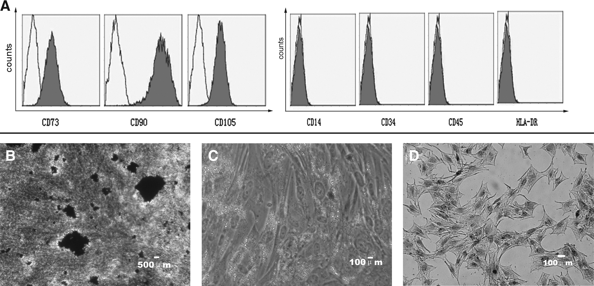
Cell-surface markers of hPMSCs and its differentiation ability. (
In osteogenic differentiation medium, the secretion of extracellular calcium crystals was identified by von Kossa staining, indicating osteogenic differentiation (Fig. 1B). In adipogenic differentiation medium, intracytoplasmic lipid vacuoles were observed from day 14 and confirmed by Oil Red O staining (Fig. 1C). After 30 days of cell pellet culture in chondrogenic inductive medium, chondrogenic differentiation was demonstrated by positive type II collagen staining (Fig. 1D).
Immunological regulation of PMSCs
In this study, the proliferation of T lymphocytes was inhibited to different extents by adding various ratios of PMSCs to human or rabbit PBMCs, with the inhibition rate increasing as the ratio of PMSC to PBMC increases that was consistent with previous reports (Di Nicola et al., 2002; Xue et al., 2010).3;27 The inhibitory effect was extremely high at a 1:5 and 1:10 ratios of PMSCs to human PBMCs (p<0.05) (Fig. 2). The inhibitory effect was extremely high, which was at a 1:20 ratio of PMSCs to rabbit PBMCs (p<0.05) (Fig. 3). Furthermore, all the counts per minute (cpm) values from human cells were higher than those from animal cells.
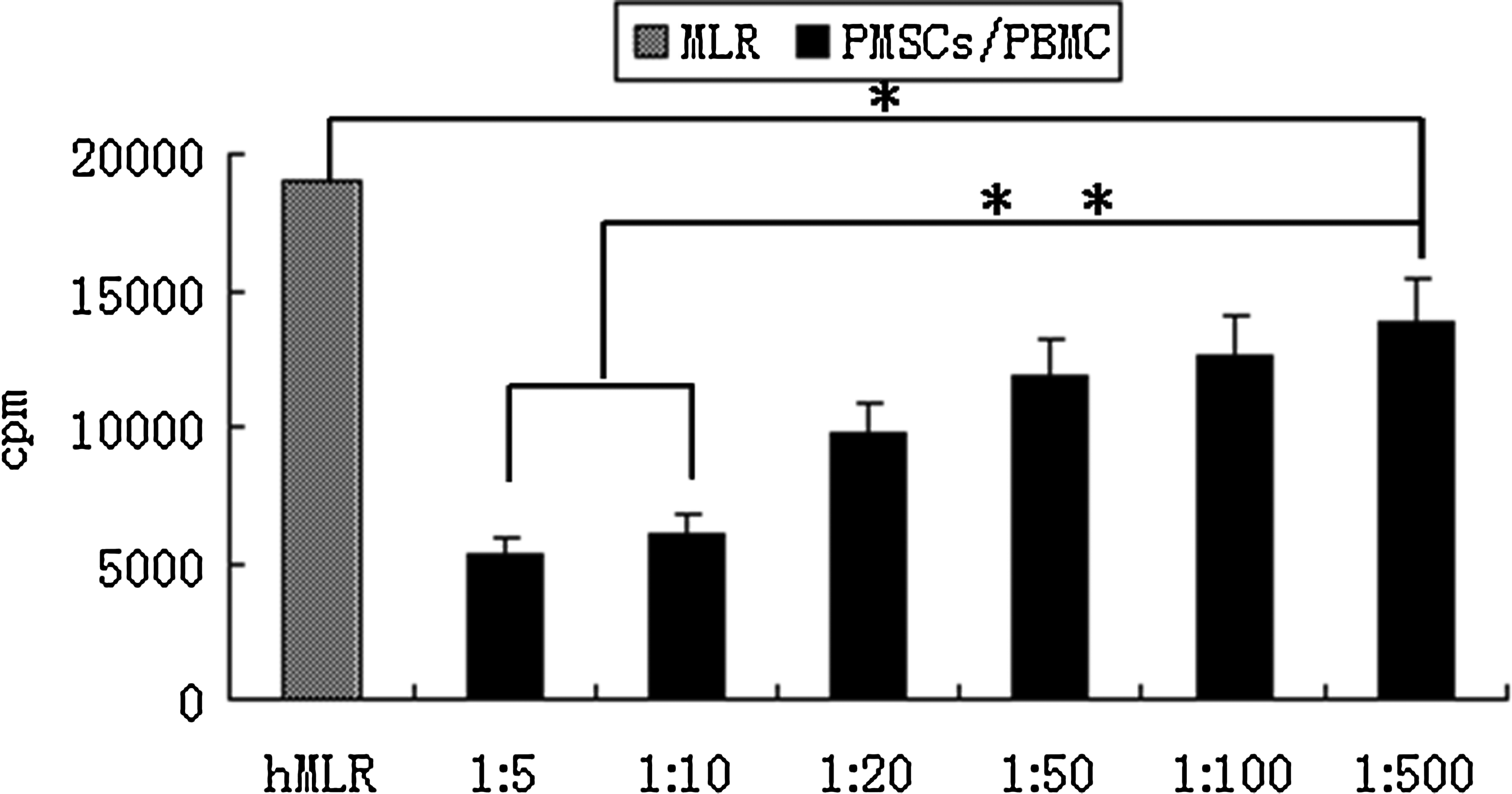
The effects of human PMSCs on hMLR (human PBMC), the proliferation of T lymphocytes was decreased after adding PMSCs. (*) p<0.05; (**) p<0.01.
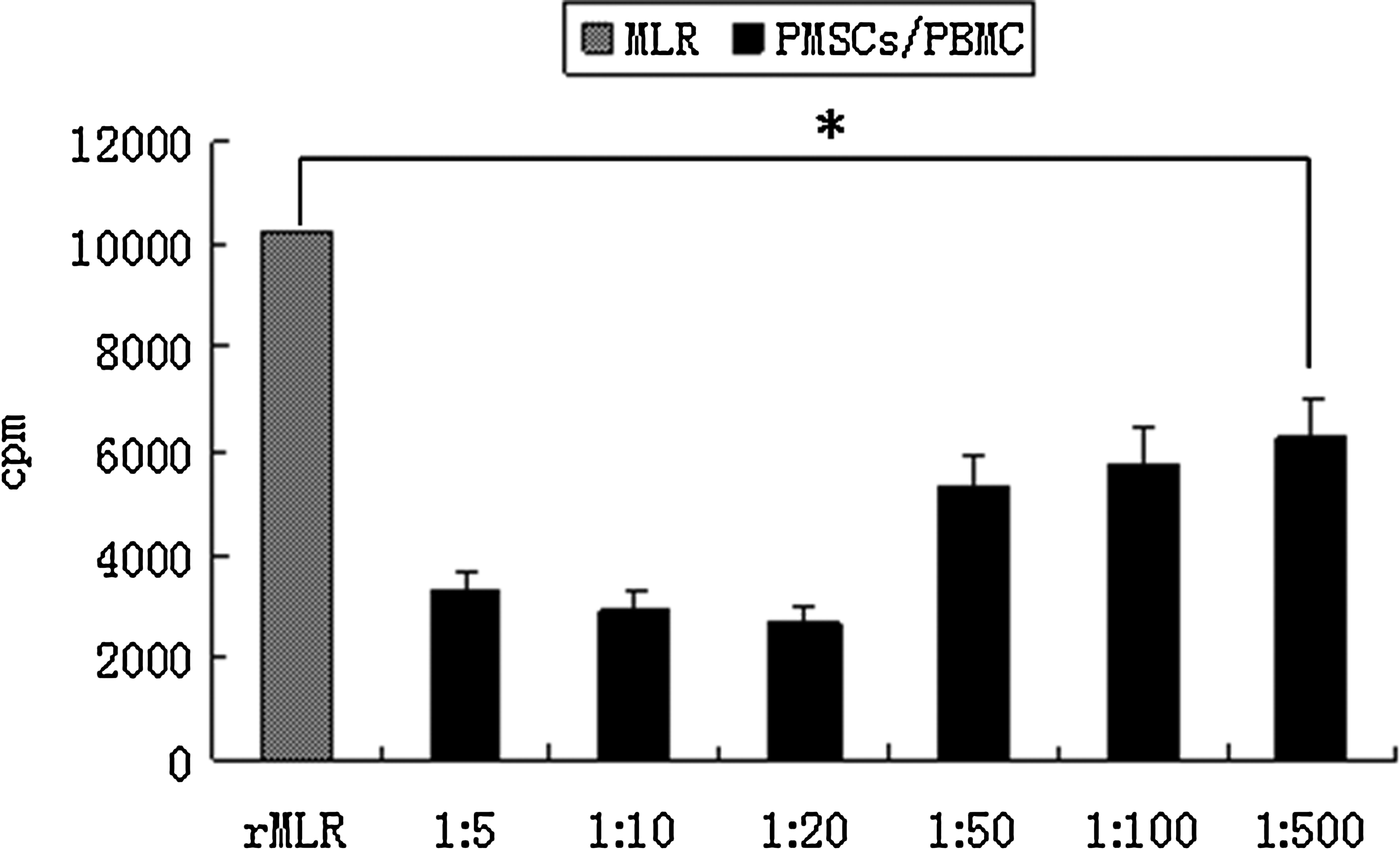
The effects of human PMSCs on rMLR (rabbit PBMCs), the strongest inhibition of proliferation was found in the group in which the ratio of PMSCs to PBMC was 1:20. (*) p<0.05.
Favorable biocompatibility of SF for human PMSCs in vitro
The surface of SF biomaterial is irregular, and the pore is large by simple scanning electron microscope. Some cells aggregated both on the material surface and in the pores after cell cultures were combined. On the first day following combination, the material surface showed relatively few adhered cells and little microvilli with microfilaments linking to the material. By the fourth day, the cells expanded, and granular and filamentous materials were detected both on the surface of the cells and their surroundings. By the eighth day, the shape and number of the cells changed slightly, whereas the secretion of the granular and reticular matrix increased and the material gap was filled with the secreted matrix (Fig. 4).
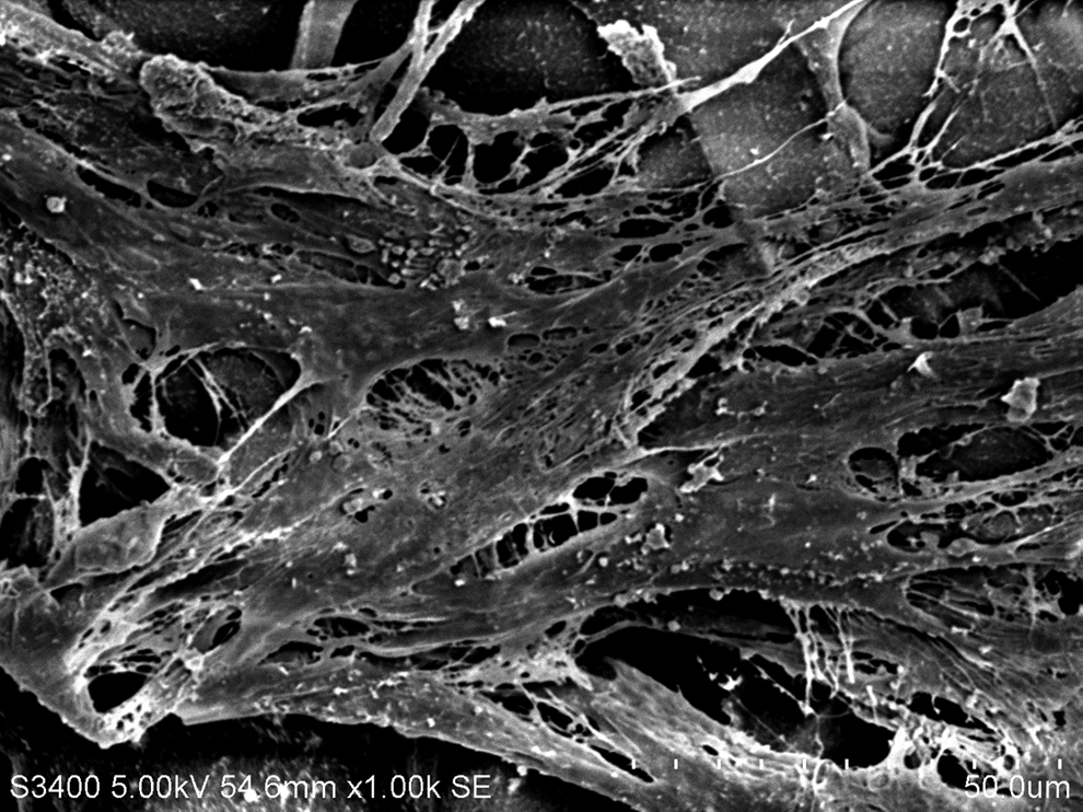
Scanning electron microscope result of the combination culture of human PMSCs and silk fibroin biomaterial on day 7. Magnification, 1000×,
Gross examination of the defect after transplantation
The grafts were well incorporated with the surrounding native cartilage. After 4 weeks, the defect was filled with semitransparent tissues. In the eighth week, recovery tissues appeared the same as the normal cartilage tissues, the boundary was obscured, and the surface was smooth (Fig. 5A). In the twelfth week, the color and luster of the recovered materials were similar to the surrounding normal cartilage, and the surface was smooth (Fig. 5B).
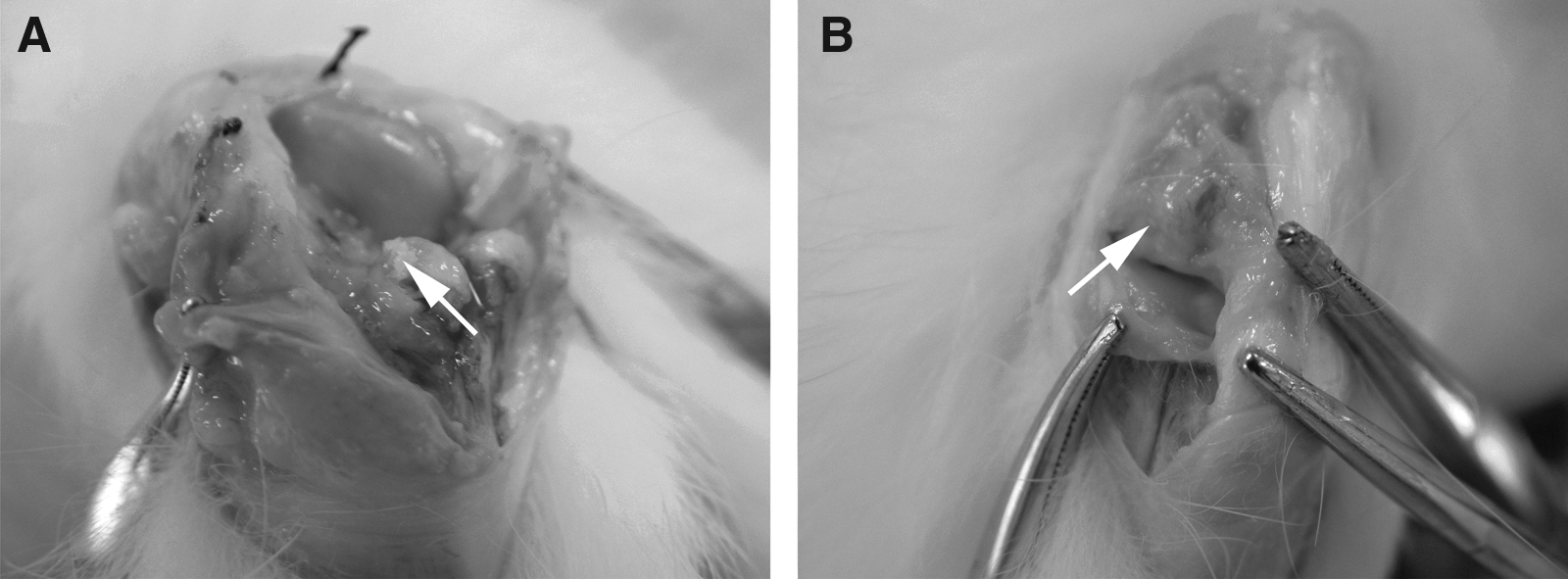
Macroscopic observation of cartilage defect after PMSCs/SF biomaterial transplantation. (
Histology and BrdU labeling of PMSCs in repaired tissues
Histological examination of the tissue-engineered cartilage graft grown as part of the subcutaneous tissue showed lobular cartilage. Hematoxylin & Eosin staining showed the cartilage to be highly cellular, with round-to-oval lacunae containing binucleate and single forms. Inflammation and foreign-body reaction were not noted. By the eighth week, a large part of recovery tissues had combined with surrounding tissues, cells increased in number and appeared as round hyaline cartilage-like cells, and cartilage lacuna and fusiform fibroblast-like cells were detected in the surface layer of the newly formed cartilage (Fig. 6A). By the twelfth week, recovery tissue appeared as hyaline cartilage with a thickness similar to the healthy endogenous tissue and integrated with the surrounding cartilage harmoniously. Fusiform fibroblast-like cells and cartilage lacuna appeared in the surface of the recovered tissue (Fig. 6B). A large amount of BrdU-labeled PMSCs were detected in the cartilage defects in the experimental group up until the eighth week. However, from the week 12 on, the number of BrdU-labeled PMSCs decreased profoundly. The results of immunofluorescent staining indicated favorable survival condition for PMSCs transplanted with scaffold (Fig.7A,B).
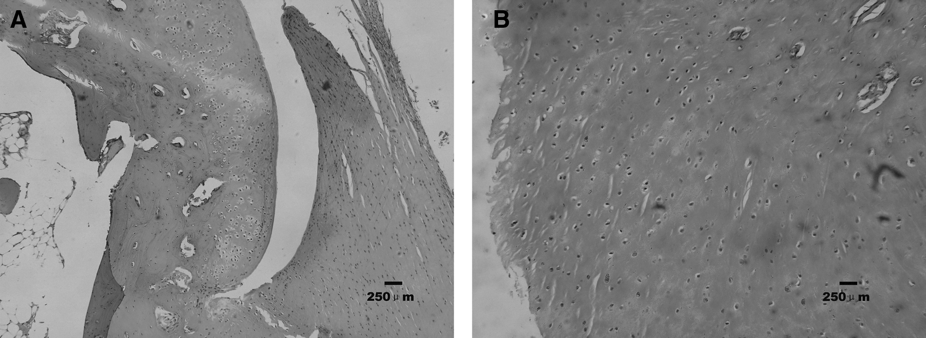
Histological examination of cartilage defect after PMSCs/SF biomaterial transplantation 8 weeks (
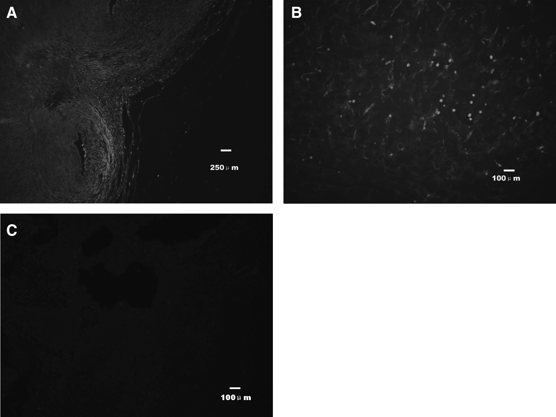
Results of immunofluorescence staining of BrdU-labeled human PMSCs. (
Discussion
In this study, we described the successful use of SF biomaterial and human PMSCs for repair of cartilage defects in rabbits. Our results demonstrate that human PMSCs can be used as favorable seed cells in tissue engineering because they exhibit potent immunosuppressive effects. Histological observations suggest that the transplanted cells survived in their new microenvironment and contributed to the repair of cartilage defects in spite of xenogeneic cells. In addition, SF exhibited favorable cellular affinity, and SF together with PMSCs promoted cartilage repair. Thus, SF in combination with PMSCs may provide a novel strategy for cartilage tissue engineering.
Articular cartilage is an avascular connective tissue that primarily functions to bear forces within joints. The unique material properties of the tissue permit it to resist compressive loads of relatively large magnitude (Pittenger et al., 1999).17 However, the avascular nature of cartilage results in a limited capacity for repair once damaged. Improved methods of cartilage repair may reduce the need for joint replacement. Tissue engineering has been investigated as a potentially promising method for reconstructing cartilage tissue in vitro for implantation (Temenoff et al., 2000).24 With recent advances in tissue-engineering techniques, the use of stem cells to produce cartilage and bone tissue may soon be a viable therapeutic option for the treatment of osteochondral defects (Pountos et al., 2007).18
The PMSCs have cell biological characteristics similar to BMSCs (Miao et al., 2006).14 In addition, using two-way MLR, the immunological regulation of PMSCs on allergenic T cells could be observed through inhibition of T cell proliferation. Taken together, these results indicate that PMSCs can confer immune regulatory effects and that they are superior to allografts as a favorable source for tissue engineering seed cells. Along with the ease of accessibility, lack of ethical concerns, and richness in cell source (O'Donoghue et al., 2003; Ringdén et al., 2006)15;21, PMSCs may be attractive alternative seed cells for regenerative medicine research. Further studies are required to better understand the precise nature of placenta-derived cells and to explore their potential clinical applications.
During our experiment, we observed that as the cartilage defects were repaired in rabbits, the BrdU-labeled transplanted cells were reduced in number. On the one hand, this might be the degradation of BrdU itself. On the other hand, this phenomenon probably indicates that it is not the transplanted stem cells, but rather something secreted by the stem cells, such as soluble factors, or even the microvesicles, which are small, spherical membrane fragments shed from the cell surface or secreted from the endosomal compartment, that play an important and underappreciated role in improving the function of damaged organs (Herrera et al., 2010; Kelly et al., 2011; Maria et al., 2010; Ratajczak et al., 2006; Ratajczak et al., 2011).6;9;12;19;20
Conclusions
In this study, we transplanted PMSCs combined with SF biomaterial into injured rabbit knees and demonstrated formation of cartilage cells and an abundance of extracellular matrix 8 weeks later. This suggests that SF biomaterials facilitate the propagation and differentiation of PMSCs. The observation that experimental animals exhibited good physical recovery indicates that the degeneration rate of SF biomaterial and the newly born rate of cartilage maintain balance and do not exhibit untimely disintegration. Our results show that the combination of PMSCs and SF biomaterial in vivo can form hyaline cartilage, which can repair full-thickness defects of knees, thus providing a potential approach for clinical treatment for reparation of cartilage defect.
Footnotes
Acknowledgments
We thank Professor Tianyi Zhang for electron microscopy and Professor Jianzhong Zhu for his constructive advice and technical support. We especially thank Dr. Lei Chen for valuable suggestions and critical review of this manuscript.