Abstract

A major area of focus for cancer research is the search for informative biomarkers to allow stratification of patients in clinical research and the eventual development of companion diagnostics for personalized therapies. But that search is limited by the lack of sensitivity and consistency in conventional immunohistochemistry (IHC) assays used by pathologists to evaluate solid tumor tissue samples—not to mention the scarcity of good quality biopsy samples in early-stage clinical studies. Until now, IHC has worked well to diagnose cancer, determine the stage and grade of a tumor, and identify cell type and origin. It has also been useful in monitoring treatment efficacy. Personalized medicine, however, requires IHC to act as a robust assay.
That's where high-performance computing comes in. Researchers at Definiens have developed a method of applying advanced computing tools to solid tumor image analysis, pioneering a new field called tissue phenomics. Tissue phenomics is the discipline of mining tissue images to identify patterns that are related to clinical outcome, providing potential prognostic and predictive value. It utilizes artificial intelligence (AI)—such as machine learning (ML) and deep learning (DL)—to quantify tumor pathology, setting the stage for tissue-based companion diagnostics.
“Tissue phenomics goes beyond conventional IHC by allowing the inclusion of spatial information, which is critical to understanding the landscape of the cancer,” says Ralf Huss, M.D., Definiens' chief medical officer. Automated tumor image analysis has the ability to find unique features that, after further investigation, might turn out to be clinically relevant. “These are features we could not detect otherwise, that the pathologist just has not been trained to read from images,” says Huss.
In addition to discovering novel features, tissue phenomics provides useful information about whether immune cells are in close relationship with cancer cells. Proximity determines whether an immune cell recognizes a cancer cell and kills it or signals to other cells. “This relational information is what tissue phenomics technology is able to provide,” Huss says.
Definiens recognizes that the true power of tissue phenomics will only be realized when its data are combined with other data. Ongoing discussions in the field about which types of data are better than others are counterproductive; the key to making sense of the available data is integration.
“Tissue phenomics is part of the 'omics field,” says Ingrid Braenne, Ph.D., who leads Definiens' exploratory data science group. “The whole 'omics field is moving towards the multi-omics approach, where we combine all the different 'omics [technologies] to get a detailed profile of the tumor microenvironment.”
Based on latest literature, the six IO-Panel categories (left) are defined by distinct immune profiles (right). It is also possible to assess the heterogeneity of the tumor interactions with a regional profiling (middle). In the future, the goal is to support treatment decisions based on this categorization.
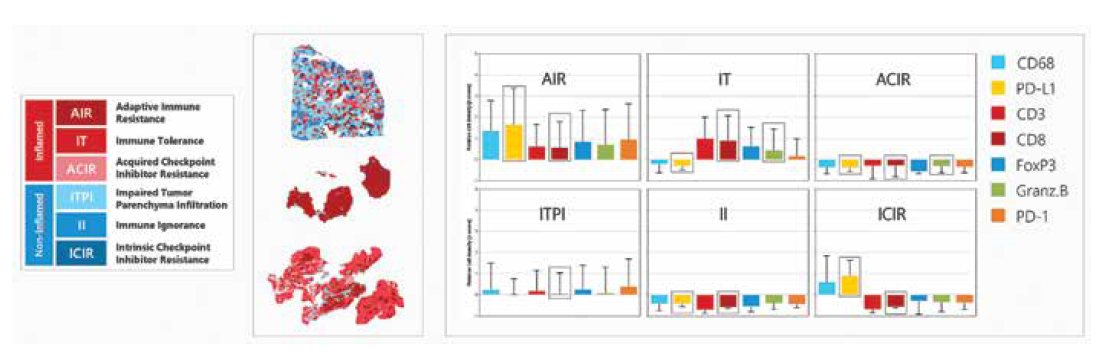
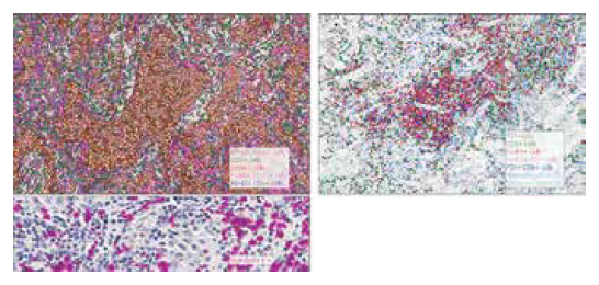
Artificial intelligence is used to mine the spatial relationships among seven different biomarkers in the tumor microenvironment. Using two Triplex and one Monostain, the different cell populations are identified and spatially placed in four different regions around and inside the tumor. This allows for a detailed profile mirroring the complex TME interactions and subsequent classification in one of six immune profile categories.
Slide Images: Courtesy Mosaic Laboratories, Lake Forest Image Analysis: Definiens
Definiens researchers have developed algorithms and other tools to understand in rich detail what happens in a tumor. These advanced tools are needed because the understanding of the tumor microenvironment increases with the different kinds of data included in an analysis. In addition to tissue phenomics, the use of 'omics data and multi-plex markers is what is going to enable and improve patient stratification. “We really believe that combining all the 'omics data is the only way to understand the phenotype of the cancer and how that affects the outcome of the patient,” Braenne says.
From Satellite Images to Tumor Sections
Definiens was founded 25 years ago by German scientist Gerd Binnig, Ph.D., who shared the 1986 Nobel Prize in Physics for designing the scanning tunneling microscope. Binnig started the company to develop Cognition Network Technology (CNT), an intelligent pattern recognition approach to image analysis that can describe objects in their contextual relationship—a technology first developed to analyze satellite images.
Early on, Definiens applied CNT software to object-based image analysis, including radiological images. In 2007, it introduced Tissue Map, a user-friendly tissue image analysis tool for oncology research. In 2009, the company introduced an ML platform for biomarker research called Tissue Studio. Definiens' researchers began to see the possibilities for improving IHC as the tools of AI via HPC and increased data storage became available.
Five years ago, Definiens began focusing on immunooncology. “Due to the increasing complexity of the data, we felt it best that we serve our customers by bringing expertise in-house,” Huss says. Definiens scientists also saw a need to ensure quality control of tissue samples and to standardize all data. In order to provide greater guidance to its customers, Definiens shifted its focus to consulting, offering Insight Services from its experts in computational science, bioinformatics, biostatistics, and pathology. Insights Services includes consultation, image analysis, data mining, and big data analytics. In 2017, the company introduced its Insights Portal, a web-based delivery platform for Insight Service projects. It allows customers to track the progress of the project and, when the project is completed, customers can view results, interrogate data, and share.
Multi-plex IO-Panel
Also in 2017, Definiens introduced its IO-Panel to overcome the current limitations of IHC assays. The IO-Panel offers partners and customers a standardized assay to profile the immune microenvironment of the tumor. It accelerates imaging studies across tissue samples by eliminating the variables associated with multiple testing kits. The panel allows the identification of the interactions between the immune system and the tumor on a single-cell resolution level in the context of the entire tumor microenvironment.
Using its tissue phenomics-based approach, the Definiens IO-Panel currently measures expression of seven well-characterized multi-plex biomarkers to efficiently categorize immune status of tumor samples. High-level results provide a quick and conclusive understanding of the tumor immune status, supported by multiple layers of comprehensive analysis. The Definiens IO-Panel allows researchers to compare current and future clinical studies across drug development portfolios.
According to Huss, the future of precision cancer therapy will only become more complex. Eventually, dozens of markers could be included in panels. Definiens offers its partners and customers customized solutions for these challenges.
For Definiens, the next stage in its evolution will be the creation of tissue-based companion diagnostics. “The ultimate goal will be for each cancer patient to undergo these detailed analyses,” Huss says. “This will not be trial-and-error with respect to cancer therapeutics, but instead, treatment selection based on standardized patient profiling.”