
Abstract
The Editor-in-Chief of DNA and Cell Biology officially retracts the article entitled, “H19 Functions as a ceRNA in Promoting Metastasis Through Decreasing miR-200s Activity in Osteosarcoma,” by Min Li, Hanwen Chen, Yuejiang Zhao, Shuming Gao, and Cai Cheng (DNA Cell Biol 35(5):235–240; doi: 10.1089/dna.2015.3171) after receiving alerts from the PubPeer platform1 and comments on Twitter2 regarding multiple image duplications between Figure 1A-E and Figure 4A-B, and also with a separately published manuscript from a different author group (Figure 4B).3
The journal publisher independently confirmed the suspected duplications.
The corresponding author of the paper, Dr. Min Li, along with all co-authors, were contacted for an explanation but no response was received.
DNA and Cell Biology
is committed to upholding the rigors of scientific publishing and the veracity of the literature.
1. PubPeer. Actinopolyspora biskrensis.
2. Twitter. @Thatsregrettab1.
3. Zhao D, Jiang Z, Wang Z, Gao J. Retinoid interferon-induced mortality19 (GRIM19) inhibits proliferation and invasion in rheumatoid arthritis fibroblast-like synoviocytes. Biomed Pharmacother 2018;98:719-725; doi: 10.1016/j.biopha.2017.12.114
Introduction
Osteosarcoma is the most common primary bone sarcoma around the world (Mirabello et al., 2009). Despite the improvement of several treatments, the total survival rate of osteosarcoma patients is still poor (Yan et al., 2012). The poor prognosis and high recurrence rate of osteosarcoma are largely due to the high rate of pulmonary metastasis (Rainusso et al., 2013). However, the exact molecular mechanism of metastasis remains largely unclear. Understanding the underlying molecular mechanisms may promote the development of effective metastasis-targeted therapy and improve the prognosis of patients with osteosarcoma.
Long noncoding RNAs (lncRNAs) are a class of transcripts longer than 200 nucleotides without protein-coding potential. Recently, increasing studies have shown that lncRNAs are frequently dysregulated in various cancers and have multiple biological functions on proliferation, apoptosis, or cell invasion (Yang et al., 2013; Yuan et al., 2014). In this regard, lncRNAs may function as decoys to titrate transcription factors or guides so that chromatin modifying enzymes can be recruited to target genes or serve as a “sponge” to titrate microRNAs (Wang and Chang, 2011; Ren et al., 2015). Among these lncRNAs, H19 was found to play a potential role in osteosarcoma progression (Chan et al., 2014). Previous studies have shown a close association between H19 and metastasis in several types of human cancers (Ma et al., 2014; Huang et al., 2015; Vennin et al., 2015). However, the underlying mechanisms by which H19 may contribute to the metastasis of osteosarcoma remain unclear.
In the present study, we found that H19 upregulated ZEB1 and ZEB2 by competitively binding the miR-200 family and then promoted migration and invasion. Thus, our findings suggest important roles of H19 in osteosarcoma metastasis and indicate its potential application in cancer therapy.
Materials and Methods
Cell lines and cell culture
The human osteosarcoma lines MG63 were purchased from the Cell Resource Center of the Institute of Basic Medical Sciences at the Chinese Academy of Medical Sciences and cultured in Dulbecco's modified Eagle's medium (Hyclone) that was supplemented with 10% fetal bovine serum (GIBCO).
RNA extraction and real-time quantitative polymerase chain reaction
The total RNA was isolated using TRIzol reagent (Invitrogen). First-strand cDNA was generated using the M-MLV Reverse Transcriptase (Invitrogen) and either gene-specific primers or random primers. Real-time quantitative polymerase chain reaction (qRT-PCR) was performed in the StepOne™ Real-Time PCR System using SYBR® Green (Takara). GAPDH was used as an endogenous control. The relative expression of RNAs was calculated using the comparative Ct method. The gene-specific primers are shown as follows: H19-F: ACCACCTCCCTCTTCTTCTT, -R: GTCGTGGAGGCTTTGAATCT, ZEB1-F: GGCTCCT ATAGCTCACACATAAG, -R: TGCTGAAAGAGACGG TGAAG, ZEB2-F: CGCCACGAGAAGAATGAAGA, -R: GATTACCTGCTCCTTGGGTTAG, and GAPDH-F: GAT TCCACCCATGGCAAATTC, -R: CTGGAAGATGGTGA TGGGATT.
Generation of a stable cell line
Recombinant lentiviruses containing control (Con), H19, shRNA-H19 (shH19), and miR-200a were purchased from the GeneChem Company. To generate the stable cell line, cells were transfected with lentiviruses and selected with 2 μg/mL puromycin for 1 week.
RNA immunoprecipitation
We performed RNA immunoprecipitation (RIP) assays using the Magna RIP™ RNA-Binding Protein Immunoprecipitation Kit (Millipore), according to the manufacturer's instructions. The AGO2 (Millipore) and Flag (Sigma) antibodies were used for RIP. The coprecipitated RNAs were detected by qRT-PCR.
Transwell assay
The transwell assays (Corning) were performed as previously described (Zhang et al., 2013). To assay the invasive capacity of tumor cells, the upper chamber of each transwell was coated with Matrigel (BD Biosciences) that was wiped out after incubating for 24 h using a cotton swab. Also, the cells migrating to the lower chamber were stained.
Luciferase reporter assay
pmirGLO, pmirGLO-H19, or pmirGLO-H19-mut was cotransfected with miR-200a or miR NC plasmid into MG63 cells by Lipofectamine-mediated gene transfer. pmirGLO or pmirGLO-ZEB1 or pmirGLO-ZEB2 was transfected into MG63 control or H19-overexpressed cells. The relative luciferase activity was normalized to the Renilla luciferase activity 48 h after transfection.
Statistical analyses
All statistical analyses were performed using the GraphPad Prism Software. For comparisons, Student's t-test (two tailed) was performed as indicated. p-Value less than 0.05 was considered significant.
Results
H19 promotes the migration and invasion of osteosarcoma cells in vitro
To investigate the role of H19 in metastasis of osteosarcoma cells, migration and invasion assays were performed. Figure 1A shows that H19 is overexpressed after transfection with H19 lentivirus in MG63 cells, while silenced after transfection with shH19 lentivirus. Functionally, overexpression of H19 promoted cell migration (Fig. 1B) and invasion (Fig. 1C) of MG63 cells compared with the control. In contrast, knockdown of H19 decreased cell migration (Fig. 1D) and invasion (Fig. 1E) of MG63 cells. These data suggest that H19 contributes to osteosarcoma cell migration and invasion in vitro.
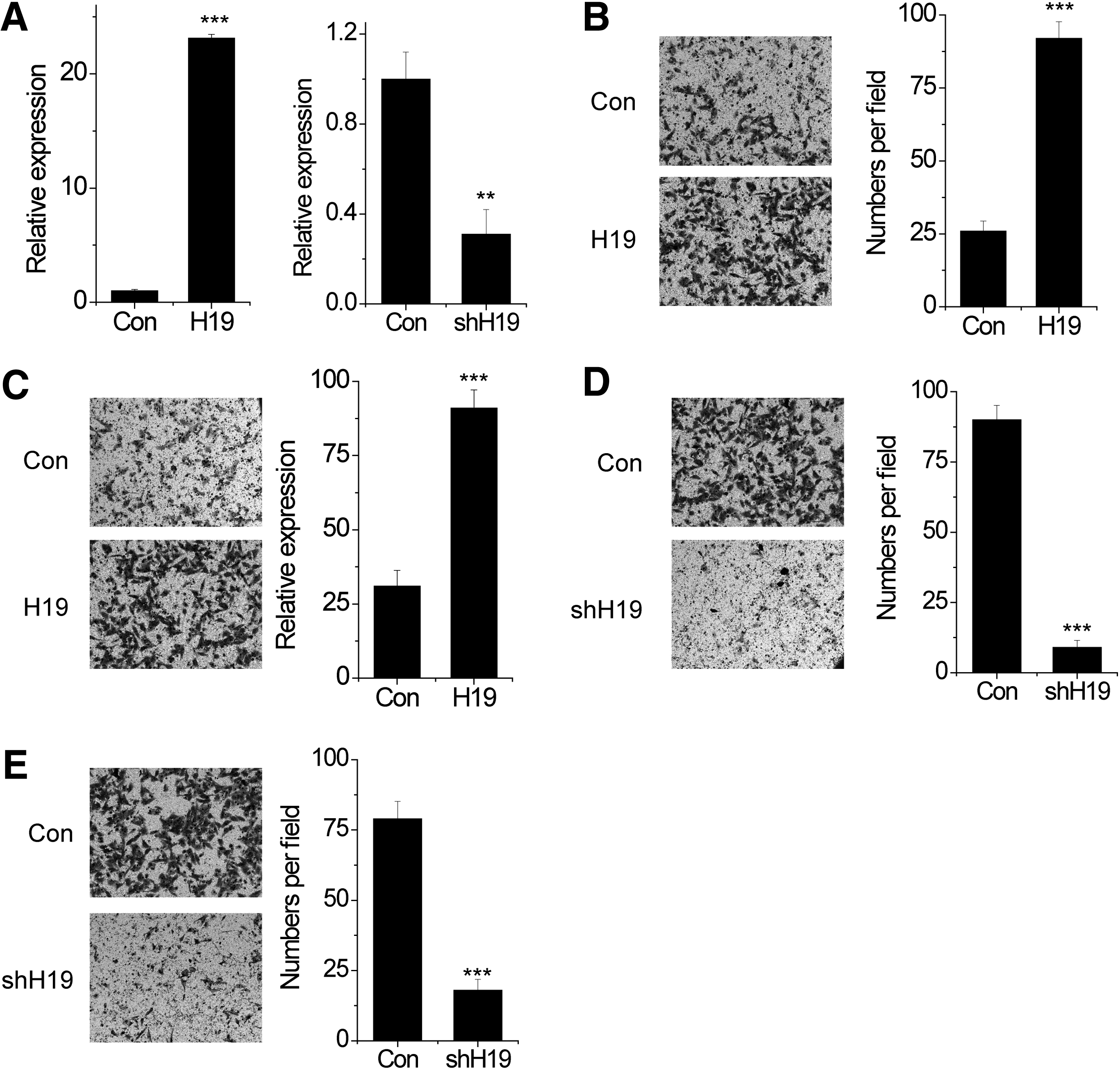
H19 promotes the migration and invasion of osteosarcoma cells in vitro.
H19 interacts with the miR-200 family
Recently, lncRNAs have been reported to function as competing endogenous RNAs (ceRNA) by competing with mRNA for common microRNAs. The miR-200 family, such as miR-200a, miR-200b, and miR-200c, has been reported to inhibit metastasis by targeting the 3′-untranslational regions (3′UTR) of ZEB1 and ZEB2 (Hill et al., 2013; Diaz-Lopez et al., 2015). Interestingly, H19 has these miR-200s-targeting sites (Fig. 2A), indicating a strong possibility as a ceRNA. To validate the direct binding between miR-200s and H19, we performed anti-MS2 RIP assay (Yuan et al., 2014) to pull down endogenous microRNAs interacted with H19. As shown in Figure 2B, the H19 RIP in MG63 cells was significantly enriched for miR-200s compared to the empty vector (MS2), IgG, and H19 with mutations in miR-200s-targeting sites (named H19-mut). In addition, we constructed luciferase reporters containing wild-type H19 (WT) or H19-mut. We found that transfection of miR-200s reduced the luciferase activities of the WT reporter vector, but not empty vector or mutant reporter vector (Fig. 2C).
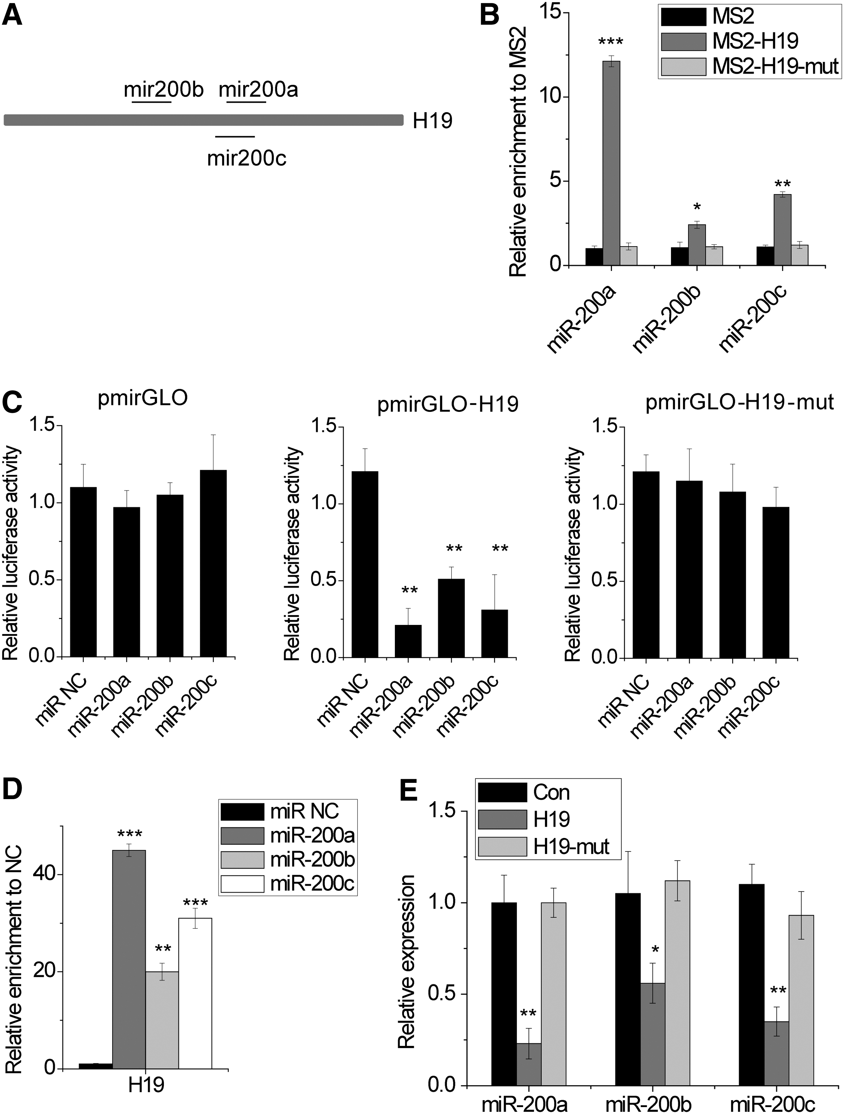
H19 interacts with the miR-200 family.
The microRNAs degrade mRNA in an AGO2-dependent manner (Shang et al., 2015). To investigate whether H19 was also regulated by miR-200s in this manner, we performed anti-AGO2 RIP in MG63 cells with overexpression of miR-200s. Endogenous H19 pull down by AGO2 was specifically enriched in miR-200s-overexpressed cells (Fig. 2D), which suggests that miR-200s are H19-targeting microRNAs. Finally, ectopically expressed H19 WT, but not the mutant, reduced the miR-200s expression (Fig. 2E). Our results suggested that H19 is associated with the miR-200 family and serves as a ceRNA.
H19 increases ZEB1 and ZEB2 expression through suppression of miR-200s
To investigate whether H19 can regulate ZEB1 and ZEB2 targeted by miR-200s, we detected the expression of ZEB1 and ZEB2 in H19 WT and H19-mut-overexpressed MG63 cells. Overexpression of H19 WT, but not the mutant, increased ZEB1 and ZEB2 mRNA and protein level (Fig. 3A). For the rescue experiment, we transfected miR-200a in H19-overexpressed cells. Overexpression of miR-200a abrogated the increase of ZEB1 and ZEB2 by H19 overexpression (Fig. 3B). In contrast, knockdown of H19 decreased ZEB1 and ZEB2 expression (Fig. 3C). Furthermore, inhibition of miR-200a overcame the suppression of ZEB1 and ZEB2 by H19 knockdown (Fig. 3D).
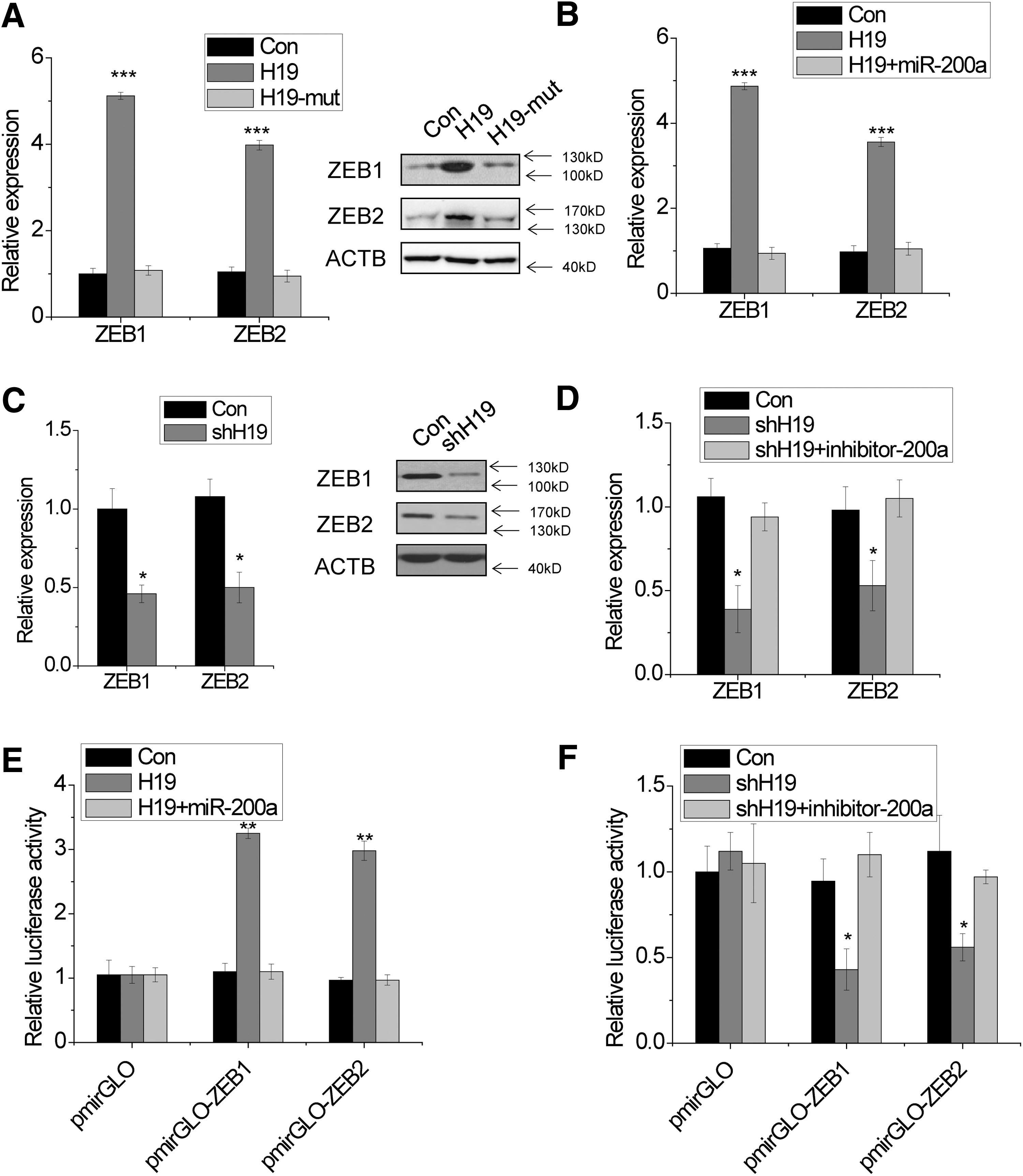
H19 increases ZEB1 and ZEB2 expression through suppression of miR-200s.
To confirm ZEB1 and ZEB2 3′UTR are involved in this regulation, we constructed luciferase reporters containing ZEB1 or ZEB2 3′UTR (pmirGLO-ZEB1 or pmirGLO-ZEB2). The luciferase activity of pmirGLO-ZEB1 and pmirGLO-ZEB2 is increased by H19, while abolished by miR-200a overexpression (Fig. 3E). In contrast, the knockdown of H19 decreased the luciferase activity of pmirGLO-ZEB1 and pmirGLO-ZEB2, which were rescued by miR-200a silence (Fig. 3F). All these results suggest an important role of H19 in regulating ZEB1 and ZEB2 by competitively binding miR-200s.
H19 promotes migration and invasion through upregulation of ZEB1 and ZEB2
Finally, we suspected that H19 may promote migration and invasion through upregulation of ZEB1 and ZEB2. To test this hypothesis, rescue experiments were performed. We found that knockdown of ZEB1 or ZEB2 expression significantly rescued the migration and invasion phenotypes that were induced by H19 overexpression (Fig. 4A, B). Together, these results demonstrated that H19 exerts its function at least, in part, through regulating ZEB1 and ZEB2 expression.
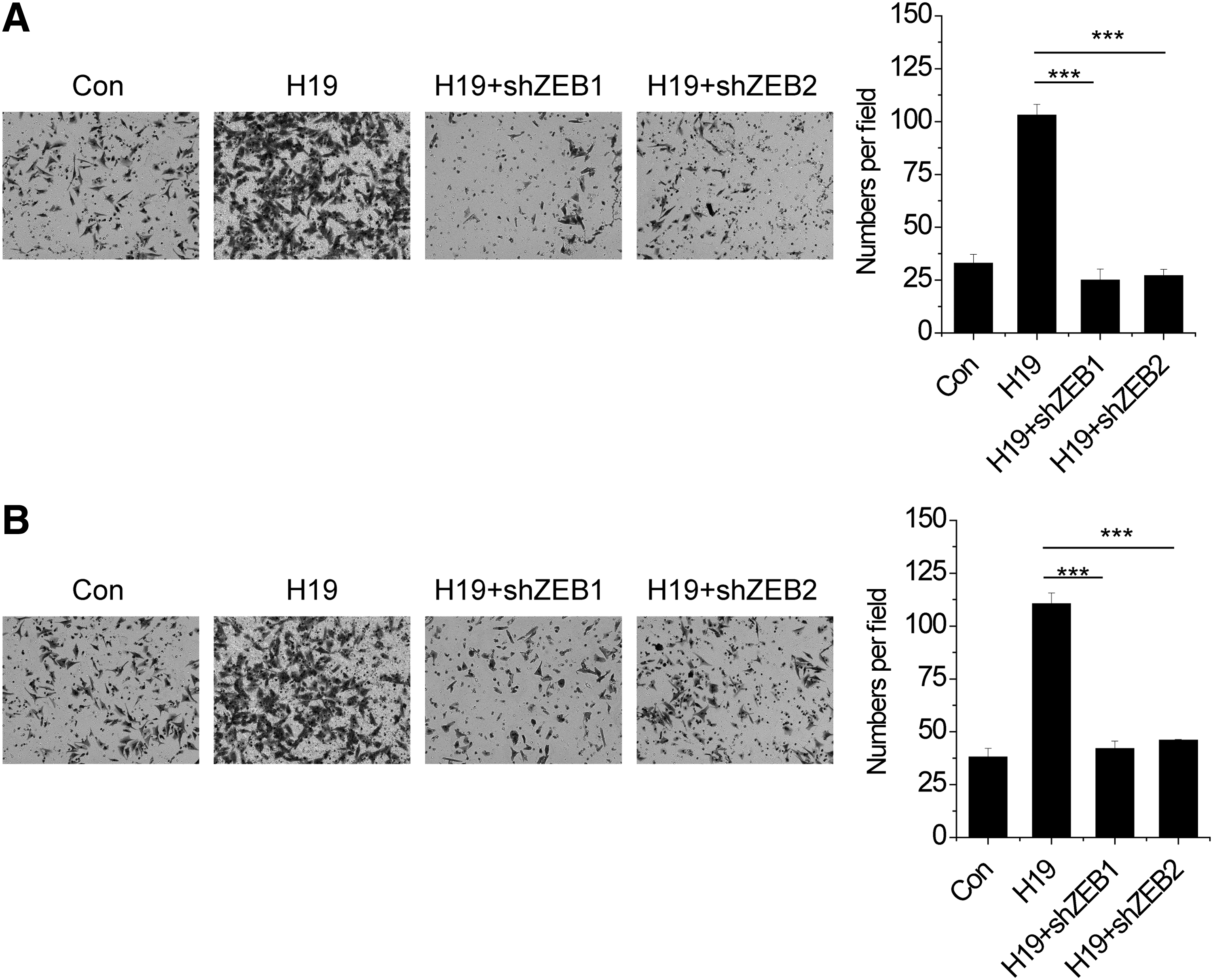
H19 promotes migration and invasion through upregulation of ZEB1 and ZEB2.
Discussion
Growing studies have demonstrated that lncRNAs participate in the initiation and progression of various cancers, including osteosarcoma, through regulation of multiple target genes involved in the progression and metastasis. Hence, identification of specific lncRNA and their target genes involved in tumorigenesis would be helpful for the diagnosis and therapy of patients with human malignancies. In this study, for the first time, we reported that H19 promotes osteosarcoma cell invasion by competitively binding the miR-200 family, upregulating ZEB1 and ZEB2, and then inducing invasion. Epithelial–mesenchymal transition (EMT) facilitates tumor invasion and dissemination (Ocana et al., 2012). Loss of E-cadherin is considered to be a fundamental event in EMT. Previous studies have reported that H19 induced EMT and inhibited E-cadherin expression through association with EZH2 (Luo et al., 2013). It is known that E-cadherin is a direct target gene of ZEB1 and ZEB2 (Galvan et al., 2015). Taken together, H19 directly or indirectly regulates E-cadherin expression through association with chromatin regulator or upregulation of ZEB1 and ZEB2.
Previous studies have reported that the aberrant regulatory network of mRNA, miRNA, and lncRNA plays a critical role in tumor progression (Cesana et al., 2011; Tay et al., 2014; Chen et al., 2015). On one hand, H19 transcriptionally regulates miRNA expression through epigenetic mechanism (Keniry et al., 2012). On the other hand, H19 acts as a molecular sponge of miRNAs, such as let-7, miR-141, and miR-106a (Kallen et al., 2013; Imig et al., 2015; Zhou et al., 2015). In this study, we demonstrated that H19 also interacts with miR-200 family and then regulates ZEB1 and ZEB2 expression. There exists a double-negative feedback loop between miR-200s and ZEB1/ZEB2 (Massague, 2008), and the upregulation of ZEB1 and ZEB2 by H19 may further augment the effects.
Taken together, this study demonstrates an important role for H19 in metastasis of osteosarcoma. H19 upregulates ZEB1 and ZEB2 by competitively binding the miR-200 family and then promotes migration and invasion. Individual therapy targeting H19 may afford more curative effects.
Footnotes
Acknowledgment
This study was supported by grants from the Support Project of Cangzhou science and technology (09ZD247).
Disclosure Statement
No competing financial interests exist.